Article
Antidermatophytic Activity of the Fruticose Lichen
Usnea orientalis
Ashutosh Pathak 1, Dalip Kumar Upreti 2 and Anupam Dikshit 1,*
1 Biological Product Lab, Department of Botany, University of Allahabad, Allahabad 211012, U.P., India;
ashupathaks@rediffmail.com
2 Lichenology Laboratory, CSIR-National Botanical Research Institute, Rana Pratap Marg, Lucknow 226001,
U.P., India
* Correspondence: anupambplau@rediffmail.com
Abstract: In the present study, the new biological sources in the form of lichen Usnea orientalis Motyka was screened for its antidermatophytic potential. Six species of dermatophytes were chosen on the basis of their prevalence for antidermatophytic assays, and the Clinical Laboratory Standard Institute (CLSI)-recommended broth microdilution procedure was used to detect the efficacy of extract against dermatophytes. Thin layer chromatography of lichen extracts reveals the presence of two secondary metabolites viz. salazinic acid and usnic acid. U. orientalis extract exhibited promising antidermatophytic activity against all tested pathogens. Amongst all tested pathogens, E. floccosum exhibited most susceptibility towards extract, whereas T. mentagrophytes exhibited the least susceptibility. Topical application of U. orientalis extract might be helpful in the cure of dermal infections.
Keywords: antidermatophytic; dermatophytes; DPPH; Lichen
1. Introduction
2. Material and Methods
2.1. Preparation and Percent Yield of Extract
Lichen thalli were collected from Koti, Chakrata district, Uttarakhand, India and identified with the help of relevant keys [16]. The voucher specimen was deposited in the Botanical Survey of India, Allahabad, India: U. orientalis (Accession No. BSA-8760). Two grams of air-dried thallus (vegetative as well as fruiting) was washed thoroughly using tap water followed by distilled water and pat-dried. Then, thalli were subjected to cold extraction of secondary metabolites in 50 mL of acetone. Subsequently, the solvent was filtered by Whatman No. 1 filter paper after 48 h.
The weight of crude extract obtained was 0.16 g after vacuum drying the filtrate via rotary evaporator. Percent yield of crude extract was calculated according to the equation below:
Percent yield (%) = (Dry weight of extract/Dry weight of sample) × 100.
Stock solution (50 mg/mL) of crude extract was prepared in dimethyl sulphoxide (DMSO) for the evaluation of antidermatophytic and free radical scavenging activity.
2.2. Thin Layer Chromatography of Extract
Solvent A (Toluene (180 mL) 1,4 dioxane (45 mL): Acetic acid (5 mL)) and Solvent C (Toluene (170 mL): Acetic acid (30 mL)) was used as mobile phase, whereas silica-coated aluminum plate (TLC Silica gel 60 F254, Merck KGaA, Germany) was used as stationary phase and Usnic acid (Tokyo, Japan, Chemical Industry Co., Ltd.) was taken as standard [17].
2.3. Test Pathogens and Inocula Preparation
Fungal cultures of Epidermophyton floccosum (MTCC No. 7880), Microsporum canis (MTCC No. 3270), M. fulvum (MTCC No. 7684), M. gypseum (MTCC No. 2867), Trichophyton rubrum (MTCC No. 296), and T. mentagrophytes (MTCC No. 7687) were procured from Microbial Type Culture Collection and Gene Bank (MTCC), Chandigarh, India, and were subcultured on Sabouraud Dextrose Agar (SDA) medium under laminar flow cabinet (Laminar flow ultra clean air unit, Micro-Filt, India). Inocula were prepared in saline media and then adjusted to a 0.5 McFarland standard, corresponding to ca 0.5 × 10⁶ CFU/mL, and transmittance of inocula prepared were 70–72% at 520 nm for each pathogen [19].
2.4. Antifungal Assay for Opportunistic Filamentous Fungi
2.4.1. Determination of Fungistatic Concentration
Antifungal susceptibility test was performed according to the Clinical Laboratory Standard Institute (CLSI)-recommended broth microdilution method in RPMI-1640 medium HEPES modification (Sigma Aldrich) supplemented with MOPS buffer (3-morphollinopropane-1-sulfonic acid) (Qualigens Fine Chemicals) [20]. Brief steps involved per plate were as follows: Inocula prepared was diluted 1:50 times in testing media, i.e., RPMI 1640; the test was performed in 96-well flat bottom microtiter plates; Column 1 was named as negative control consisting of 100 μL of RPMI-1640 broth media and 100 μL of inocula prepared in formaldehyde; Column 2 was named as broth control consisting of 200 μL of media; Columns 3 and 4, 6 and 7, and 9 and 10 were vertically diluted with extract having a final concentration of 1.25 to 0.009 mg/mL and named as treated; Column 5, 8, and 11 were taken as positive controls and contained only 100 μL of inocula and 100 μL of RPMI-1640 broth media, respectively; Column 12 was named as extract control and contained vertically diluted extract in the aforementioned concentrations. To nullify the effect of extract color, optical density (O.D.) of the extract control was subtracted from treated columns corresponding to extract treated [21]. Percent inhibition was calculated using following equation:
Minimum inhibition concentrations (MICs) was calculated based on optical density recorded with a spectrophotometer (SpectraMax Plus384, Molecular Devices Corporation, USA) at 530 nm after 96 h of incubation at 30 ± 2 °C (Figure 1).
The antifungal activity of the chemical drug Sertaconazole nitrateBP (SN) (Glenmark Pharmaceuticals, Nasik, India) was taken as the reference standard. An amount of 50 mg/mL of stock solution of SN was prepared and tested at the same concentrations as the U. orientalis extract.
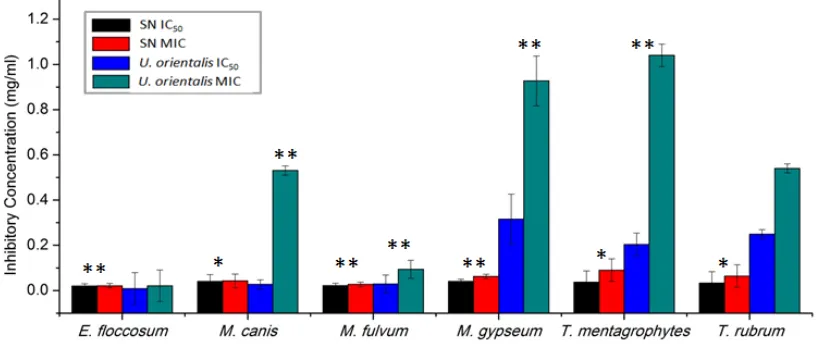
Figure 1. Antidermatophytic activity U. orientalis extract compared with Sertaconazole nitrate. Error bars show the standard error mean. ** Level of significance ≤ 0.01. * Level of significance ≤ 0.05.
2.4.2. Determination of Fungicidal Concentration
A portion of 20 μL of U. orientalis-treated columns from the above MIC wells were transferred into 7-mL tubes of a fresh RPMI 1640 medium. Tubes were incubated at 30 ± 2 °C for 4 weeks and checked for the turbidity. The aforementioned procedure was performed with SN, and the concentration at which no turbidity has been achieved was defined as the minimum fungicidal concentration (MFC) [22,23].
2.5. Statistical Analysis
An independent sample T-test was performed between the positive control and sertacoazole-treated dermatophytes. Positive control and extract-sertacoazole-treated dermatophytes for the measure of Levene’s test for equality of variances and t-test for equality of means via SPSS v20.
3. Results and Discussion
3.1. Percent Yield of Extract
Percent yield of extract obtained from U. orientalis thallus was 8%.
3.2. Thin Layer Chromatography
A light yellow turning into a green-colored spot was observed and confirmed as usnic acid in U. orientalis extract and the other compound was salazinic acid. The chromatographic study confirmed the presence of two compounds viz. usnic acid and salazinic acid in the U. orientalis extract.
3.3. Antifungal Test for Opportunistic Filamentous Fungi
drugs, there is a need for new antidermatophytic compounds. The antidermatophytic activity of SN and U. orientalis were represented graphically in the form of IC50 (50% inhibitory concentration) and MIC (minimum inhibitory concentration) values were represented graphically in Figure 1.
E. floccosum exhibited equal susceptibility towards SN and U. orientalis extract with an MIC value equivalent to 0.021 mg/mL and an MFC value equivalent to 0.039 mg/mL. The IC50 value for E. floccosum was achieved at 0.009 mg/mL against U. orientalis and 0.020 mg/mL against SN. M. gypseum was found least susceptible among Microsporum spp. with IC50 = 0.040 mg/mL; MIC = 0.062 mg/mL; MFC = 0.078 mg/mL against SN and IC50 = 0.316 mg/mL; MIC = 0.927 mg/mL; MFC = 1.250 mg/mL against U. orientalis. M. fulvum was found most susceptible among Microsporum spp. with IC50 = 0.022 mg/mL; MIC = 0.027 mg/mL; MFC = 0.039 mg/mL against SN and IC50 = 0.029mg/mL; MIC = 0.094 mg/mL; MFC = 0.156 mg/mL against U. orientalis. The IC50, MIC, and MFC for M. canis were obtained at 0.040 mg/mL, 0.043 mg/mL, and 0.078 mg/mL for SN and 0.027 mg/mL, 0.531 mg/mL, and 0.625 mg/mL for U. orientalis extract.
The IC50, MIC, and MFC for T. rubrum were achieved at 0.033 mg/mL, 0.064 mg/mL, and 0.078 mg/mL against SN and 0.249 mg/mL, 0.54 mg/mL, and 0.625 mg/mL against U. orientalis extract. T. mentagrophytes was found least susceptible amongst all tested pathogens with IC50 = 0.037 mg/mL, MIC = 0.09 mg/mL, and MFC = 0.156 mg/mL against SN and IC50 = 0.204 mg/mL, MIC = 1.04 mg/mL, and MFC = 1.25 mg/mL against U. orientalis extract. The efficacy of U. orientalis extract is equivalent to sertaconazole nitrate against E. floccosum, but was found to be less effective against all other dermatophytes.
In another study, extracts of U. florida exhibited an MIC between 0.050 and 0.100 mg/mL against M. gypseum, T. mentagrophytes, and T. rubrum [25]. In the present study U. orientalis exhibited a MIC between 0.531 and 1.04 mg/mL and was found to be less active than U. florida.The ethno-medicinal use of U. orientalis extract in swelling caused by dermatophyte infection might be cured, but in vivo efficacy and potency of the lichen extract needs to be investigated.
3.4. Statistical Analysis
The level of significance was calculated in terms of p-value. Results obtained were statistically significant except between U. orientalis treatments and control T. rubrum (p-value = 0.19) and U. orientalis treatments and control E. floccosum (p-value = 0.19); p-values calculated for SN-treated E. floccosum, M. fulvum,and M. gypseum were less than 0.01, whereas, for M. canis, T. mentagrophytes, and T. rubrum, p-values were less than 0.05. p-value calculated for U. orientalis treated all pathogens except T. rubrum, which showed a level of significance less than 0.01 (Figure 1).
4. Conclusions
U. orientalis extract exhibited broad-range antidermatophytic activity against all three genera of dermatophytes, and usnic acid (a well-known antifungal compound) was present in the lichen extract. Topical application of lichen extract might be helpful in the cure of cutaneous infections.
Acknowledgments: G.P. Sinha, Head, Botanical Survey of India, Central Regional Centre, Allahabad for accepting the voucher specimens of Lichens, and University Grant Commission (UGC) New Delhi, India, for financial assistance.
References
1. Hawksworth, D.L. Freshwater and marine lichen-forming. In Aquatic Mycology across the Millennium; Hyde, K.D., Ho, W.H., Pointing, S.B., Eds.; Fungal Diversity, Hong Kong, China, 2000; Volume 5.
2. Molnar, K.; Farkas, E. Current results on Biological Activities of Lichen Secondary Metabolites: A Review. Z. Naturforschung C 2010, 65c, 157–173.
3. Shukla, P.; Upreti, D.K.; Tewari, L.M. Secondary metabolite variability in genus Usnea in India: A potential source for bioprospection. J. Environ. Sci. Technol. 2015, 2, 44–55.
4. Wang, L.S.; Qian, Z.G. Pictorial Handbook to Medicinal Lichens in Chin; Yunnan Provincial Science and Technology Publishers: Kunming, China, 2013.
5. Dias, M.F.R.G.; Quaresma-Santos, M.V.P.; Bernardes-Filho, F.; Amorim, A.G.F.; Schechtman, R.C.; Azulay, D.R. Update on therapy for superficial mycoses: Review article part 1. An. Bras. Dermatol. 2013, 88, 764–774.
6. Peres, N.T.A.; Maranh o, F.C.A.; Rossi, A.; Martinez-Rossi, N.M. Dermatophytes: Host-pathogen interaction and antifungal resistance. An. Bras. Dermatol. 2010, 85, 657–667.
7. White, T.C.; Oliver, B.G.; Graser, Y.; Henn, M.R. Generating and testing molecular hypotheses in the dermatophytes. Eukaryot. Cell 2008, 7, 1238–1245.
8. Burzykowski, T.; Molenberghs, G.; Abeck, D.; Haneke, E.; Hay, R.; Katsambas, A.; Roseeuw, D.; van de Kerkhof, P.; van Aelst, R.; Marynissen, G. High prevalence of foot diseases in Europe: Results of the Achilles Project. Mycoses 2003, 46, 496–505.
9. Abdel-Rahman, S.M.; Simon, S.; Wright, K.J.; Ndjountche, L.; Gaedigk, A. Tracking Trichophyton tonsurans through a large urban child care center: Defining infection prevalence and transmission patterns by molecular strain typing. Pediatrics 2006, 118, 2365–2373.
10. Heidrich, D.; Garcia, M.R.; Stopiglia, C.D.O.; Magagnin, C.M.; Daboit, T.C.; Vetoratto, G.; Schwartz, J.; Amaro, T.G.; Scroferneker, M.L. Dermatophytosis: A 16-year retrospective study in a metropolitan area in southern Brazil. J. Infect. Dev. Ctries. 2015, 9, 865–871.
11. Stephenson, J. Investigators seeking new ways to stem rising tide of resistant fungi. J. Am. Med. Assoc. 1997, 277, 5–6.
12. Wingfield, A.B.; Fernandez-Obregon, A.C.; Wignall, F.S.; Greer, D.L. Treatment of tinea imbricata: A randomized clinical trial using griseofulvin, terbinafine, itraconazole and fluconazole. Br. J. Dermatol. 2004, 150, 119–126.
13. Smith, K.J.; Warnock, D.W.; Kennedy, C.T.C.; Johnson, E.M.; Opwood, V.; van Cutsem, J.; et al. Azole resistance in Candida albicans. Med. Mycol. 1986, 24, 133–144.
14. Orozco, A.; Higginbotham, L.; Hitchcock, C.; Parkinson, T.; Falconer, D.; Ibrahim, A.; Ghannoum, M.A.; Filler, S.G. Mechanism of fluconazole resistance in Candida krusei. Antimicrob. Agents Chemother. 1998, 42, 2645–2649.
15. Dias, M.F.R.G.; Quaresma-Santos, M.V.P.; Bernardes-Filho, F.; Amorim, A.G.F.; Schechtman, R.C.; Azulay, D.R. Update on therapy for superficial mycoses: Review article part I. An. Bras. Dermatol. 2013, 88, 764–774.
16. Awasthi, D.D. A compendium of the Macrolichens from India, Nepal and Sri Lanka; Bishen Singh Mahendra Pal Singh: Dehra Dun, India, 2007.
17. Orange, A.; James, P.W.; White, F.J. Microchemical Methods for Identification of Lichens; British Lichen Society: 2001.
18. Santos, D.A.; Barros, M.E.S.; Hamdan, J.S. Establishing a method of inoculum preparation for susceptibility testing of Trichophyton rubrum and Trichophyton mentagrophytes. J. Clin. Microbial. 2006, 44, 98–101.
19. Rex, J.H.; Alexander, B.D.; Andes, D.; Arthington-Skaggs, B.; Brown, S.D.; Chaturveli, V.; et al. Reference Method for Broth Dilution Antifungal Susceptibility Testing of Filamentous Fungi, Approved Standard; 2nd ed.; M38A2 28(16); Clinical and Laboratory Standard Institute (CLSI): Wayne, PA, USA, 2008.
20. Pathak, A.; Shukla, S.K.; Pandey, A.; Mishra, R.K.; Kumar, R.; Dikshit, A. In vitro antibacterial activity of ethno medicinally used lichens against three wound infecting genera of enterobacteriaceae. Proc. Natl. Acad. Sci. India Sect. B Biol. Sci. 2015, doi:10.1007/s40011-015-0540-y.
22. Pathak, A.; Mishra, R.K.; Shukla, S.K.; Kumar, R.; Pandey, M.; Pandey, M.; Qidwai, A. In vitro evaluation of antidermatophytic activity of five lichens. Cogent Biol. 2016, doi:10.1080/23312025.2016.1197472.
23. Liebel, F.; Lyte, P.; Garay, M.; Babad, J.; Southall, M.D. Anti-inflammatory and anti-itch activity of sertaconazole nitrate. Arch. Dermatol. Res. 2006, 298, 191–199.
24. Schmeda-Hirschmann, G.; Tapia, A.; Lima, B.; Pertino, M.; Sortino, M.; Zacchino, S.; Arias, A.R.; Feresin, G.E. A new antifungal and antiprotozoal depside from the Andean lichen Protousnea poeppigii. Phytother. Res. 2008, 22, 349–355.