O R I G I N A L R E S E A R C H
Evaluation of Peripheral Blood Parameters of
Pulmonary Tuberculosis Patients at St. Paul
’
s Hospital
Millennium Medical College, Addis Ababa, Ethiopia:
Comparative Study
This article was published in the following Dove Press journal: Journal of Blood Medicine
Daniel Kahase Absra Solomon Mihret Alemayehu
Department of Medical Laboratory Sciences, College of Medicine and Health Sciences, Wolkite University, Gubrie, Wolkite, Ethiopia
Background and Aim:Pulmonary tuberculosis is still among the leading cause of mor-bidity and mortality in Ethiopia. Different hematological abnormalities are commonly asso-ciated with pulmonary tuberculosis even though inconsistent results have been described. Hence, this study aimed to evaluate the hematological parameters of pulmonary tuberculosis patients visited St. Paul’s hospital millennium medical college, Addis Ababa, Ethiopia. Methods:From April to September 2018, a comparative cross-sectional study was conducted among pulmonary tuberculosis patients (n=40) and control patients (n=40). About 5 mL venous blood and 2–5 mL sputum samples were collected and examined by Cell Dyn 1800 hematology analyzer and cultured using Mycobacteria Growth Indicator Tube (BACTEC MGIT 960), respectively. Independentt-test was performed with the help of SPSS version 20 software, and p-value < 0.05 was considered as statistically significant difference. Results:The proportion of male to female in the pulmonary tuberculosis patients (PTB) and the control patients was 1.7 (25/15). Two-sample independentt-test revealed that the mean values of hemoglobin level (P=0.002), hematocrit (P=0.018), mean cell hemoglobin con-centration (P=0.001) and relative lymphocyte percentage (P=0.036) of PTB were signifi -cantly lower than the control group. Moreover, significantly higher mean values were also observed in total white blood cell count (P=0.004), platelet count (P<0.001) and erythrocyte sedimentation rate (P<0.001). Among the hematologic abnormalities detected, thrombocy-tosis and anemia presented in 65% and 25% of PTB patients, respectively.
Conclusion:Statistically significant mean differences were observed in hemoglobin, hema-tocrit (HCT), MCHC, relative lymphocyte percentage, WBC, platelet count, relative neutro-phil percentage and ESR values. So, the utilization of such data is important in providing preliminary information for diagnosis and management of pulmonary tuberculosis. In fact, a further large scale study is needed to substantiate thisfinding.
Keywords:hematologic parameter,Mycobacterium tuberculosis, thrombocytosis, anemia
Introduction
Mycobacterium tuberculosis is an intracellular pathogen that is able to cause tuberculosis and humans are the only natural reservoir.1 During M. tuberculosis
infection, host immunity plays an important role in the host–pathogen interaction. Neutrophils migrate early on the site of infection, followed by monocytes, which can be observed to differentiate into macrophages. Then, macrophages present processedMycobacterium tuberculosisantigen to T-lymphocyte which is a type of
Correspondence: Daniel Kahase Department of Medical Laboratory Sciences, College of Medicine and Health Sciences, Wolkite University, P.O. Box: 07, Gubrie, Wolkite, Ethiopia
Tel +251 9 1241 4564 Fax +251 11 322 0041 Email [email protected]
Journal of Blood Medicine
Dove
press
open access to scientific and medical research
Open Access Full Text Article
Journal of Blood Medicine downloaded from https://www.dovepress.com/ by 118.70.13.36 on 24-Aug-2020
white blood cell. These all host responses are more likely responsible for clinical manifestation of tuberculosis.2,3
Tuberculosis (TB) is one of the top ten causes of death, and the leading cause from a single infectious agent world-wide. Currently, world health organization reported the fastest regional declines of tuberculosis from 2013 to 2017 were in the WHO European Region followed by WHO African Region. Despite the decline in the WHO African Region, 2.5 million people fell ill with all forms of tuberculosis in the African region, accounting for a quarter of new TB cases worldwide in 2017.4
The severity of tuberculosis epidemics varies widely among countries. Ethiopia ranked 10th among the thirty high burden tuberculosis countries worldwide, with an esti-mated 164 incidents of all forms of TB cases per 100 000 population.4Ethiopia ministry of health also reveals that TB is the eighth leading cause of hospital admission and the third leading cause of hospital deaths in Ethiopia in 2011.5
Studies present hematological and biochemical abnorm-alities are common in pulmonary tuberculosis patients.6,7 Leukocytosis, monocytosis, lymphocytosis, thrombocytosis, lymphopenia, and anemia are among the reported hematolo-gic abnormalities.8–11Modulating a normal hematopoiesis process is mentioned as a mechanism for peripheral blood abnormalities to occur.12These hematological changes can act as important markers for diagnosis and persistent excre-tion of acid-fast bacilli, which is associated with failure of these indices to return to normal.6In addition, these changes have correlation with the severity of clinical findings of pulmonary tuberculosis.13
If clinical laboratory assays such as hematologic para-meters are interpreted carefully, it can be useful in assisting diagnosis and prognosis at a low cost. To the best of our knowledge, few studies are available in Ethiopia. Studies done in Jima and Gonder, Ethiopia stated the occurrence of hematological changes in pulmonary tuberculosis patients,14,15 even though their results are inconsistent in some of the hematological indices. As well, to the best of our knowledge, no study has been conducted in the study area. Therefore, this study planned to evaluate peripheral blood findings among PTB patients at St. Paul’s hospital millennium medical college, Addis Ababa, Ethiopia.
Materials and Methods
Study Design and Study Area
From April to September 2018, hospital-based comparative cross-sectional study was implemented at St. Paul’s hospital
millennium medical college, which is the second largest referral hospital in Ethiopia. It is located in Addis Ababa, the capital city of Ethiopia. The hospital has different depart-ments and annually offers diagnosis and treatment for approximately 100,000 up to 150,000 patients. All patients triaged in pulmonology department during the study period, with signs and symptoms suggestive of pulmonary tubercu-losis were considered as a source population for PTB group.
Study Population
From the presumptive PTB patients who provided blood and sputum samples, culture confirmed consecutive pul-monary tuberculosis (PTB) patients were taken as PTB group. The control patients were those matched for sex and age of PTB patients and who visit the hospital for other medical cases which majorly have no effect on hematological parameters such as HIV and other chronic diseases. In addition, the control groups were also negative for pulmonary tuberculosis by culture.
Inclusion Criteria
Patients who did no take anti-tuberculosis drug, no record of any other chronic disease, non-pregnant women and HIV negative patients were included in the study.
Exclusion Criteria
Patients with signs of concomitant chronic or acute infec-tion other than pulmonary tuberculosis, bleeding manifes-tations, endocrine disorders, other organ dysfunction or systemic disorders and chronic inflammatory disease on clinical examination were not included as PTB group.
Data Collection
After written informed consent was obtained from each study subjects, all subjects were asked to provide a detailed history and were subjected to a physical examination. Then, morning sputum and venous blood samples were collected.
Sputum Sample Collection, Processing and Culturing
Each collected morning sputum sample (2–5 mL) was mixed with an equal volume of N-acetyl-L-cysteine-sodium hydroxide (NALC-NaOH) solution, vortexing for 20 s was done and it was kept for 15 mins at 20–25°c for decontamination process. Then, Phosphate Buffered saline wasfilled to 50 mL mark on falcon tube and vortexing was done. After it was centrifuged at 3000 g for 15 mins, the supernatant was discarded and a portion of the deposits was used for culture.16,17
Journal of Blood Medicine downloaded from https://www.dovepress.com/ by 118.70.13.36 on 24-Aug-2020
Portion of each specimen sediment (0.5mL) was inocu-lated into BACTEC MGIT 960 (Becton Dickinson, Franklin Lakes, NJ07417, USA) media and incubated in an automated BACTEC MGIT 960TM machine (Becton Dickinson Diagnostic Instrument Systems) for a maximum of 42 days. Cultures exhibiting growth were subjected to light micro-scopy for the presence of acid-fast bacteria using Ziehl-Neelsen stain and tested by Capilia TB-Neo rapid test. Finally, both Acid-fast bacteria and Capilia TB-Neo rapid test positive isolates were considered as Mycobacterium tuberculosiscomplex.16
Blood Collection and Analysis
Five milliliter blood samples were collected aseptically from each of the study participants and correctly labeled with the patient’s identification number. Then, complete blood count (hemoglobin, MCH, hematocrit, MCV, MCHC, platelet count, RBC count, total leukocyte count, and differential leukocyte count (Lymphocyte, Neutrophil and MID cell) were measured using Cell Dyn 1800 hematology analyzer. In addition, Erythrocyte sedimentation rate measurement was performed using Westergreen method).
Quality Assurance
The validity of the studyfindings was assured by properly using established standard operating procedures (SOPs) and manufactures instruction for both hematological tests and TB culture. Control organisms Mycobacterium
tuber-culosis H37Rv (ATCC 27294) reference strain was also
used as a quality control for sputum culture. The quality performance of the hematology analyzer was checked before running the patients' sample by performing normal, low and high blood controls.
Operational De
fi
nitions
Hematological reference values were utilized from study done in Addis Ababa, Ethiopia. Accordingly, Leukocytosis, lym-phopenia and thrombocytosis were defined as WBC > 10.2 × 10³cells/µL for both sex, lymphocyte percentage < 27.4 for male and < 25.3 for female and platelet count > 337 × 103 cells/µL for both sex, respectively.18
Anemia was defined per WHO guidelines as Hgb <13 g/dl for males and Hgb <12 g/dl for females. Hemoglobin values of 9.0–11.0 g/dl for women and 9.0–12.0 g/dl for men were considered as mild while Hemoglobin values 8.0–9.9 g/dl were considered as moderate anemia.19
Data Analysis
Data were entered into Microsoft Excel, exported to SPSS version 20 for further analysis. Descriptive statistics were used to describe the distribution of age, sex among the pulmonary tuberculosis cases and the control patients. Two-sample independent t-tests were utilized for compar-ison of the hematological parameters mean values between the pulmonary tuberculosis cases and the control group. A P-value of < 0.05 was considered as statistically sig-nificant difference.
Results
Characteristics of Study Subjects
This study enrolled forty culture-positive pulmonary tuber-culosis patients and an equal number of control patients. Of the pulmonary tuberculosis patients (cases), males accounted for 25 (62.5%) and females accounted for 15 (37.5%). The proportion of male to female in the control patients was similar to the cases as shown inTable 1. The mean age of the pulmonary tuberculosis patients was 40 years (Sd. ±15). There was no statistically significant difference between the mean age (p=0.850) of the pulmonary tuberculosis patient (PTB) and the control group.
Comparison of Hematological
Parameters
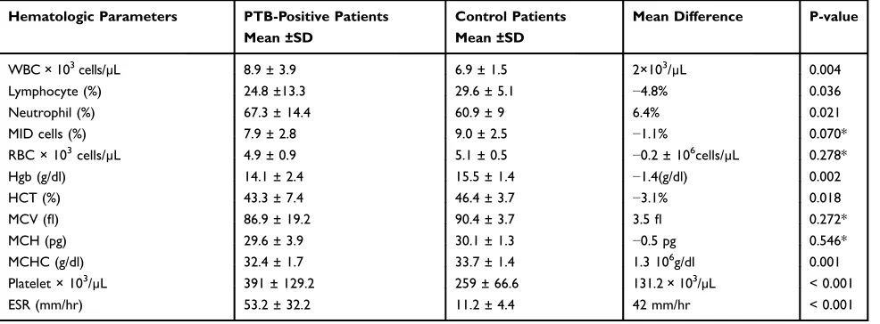
In the current study, the mean values for blood hemoglobin level (P=0.002), hematocrit (HCT) (P=0.018), mean cell hemoglobin concentration (MCHC) (P=0.001) and relative lymphocyte percentage (P=0.036) in PTB patients were found to be less than that of control patients. These differ-ences were statistically significant as seen inTable 2. Other measured hematologic parameters like absolute total white blood cell count (WBC) (P=0.004), platelet count (P<0.001) and erythrocyte sedimentation rate (ESR) (P<0.001) values found higher than the control patients, which were also statistically significant. However, there were no statistically significant differences observed in red cell (RBC) count, mean cell volume (MCV), and mean cell hemoglobin (MCH) and MID cells (%) (Table 2).
Hematologic Abnormalities in Pulmonary
Tuberculosis Patients
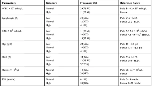
Of the total forty pulmonary tuberculosis patients (PTB), leukocytosis, thrombocytosis, and lymphopenia were observed in 27.5%, 65%, and 60% of the patients, respec-tively (Table 3). About 25% of PTB study subjects showed
Journal of Blood Medicine downloaded from https://www.dovepress.com/ by 118.70.13.36 on 24-Aug-2020
anemia. Of these, the majority (70%) were with mild and the remaining 30% were with moderate anemia.
Discussion
A range of diseases can have an effect on hematological indices including tuberculosis. Limited studies are avail-able on blood parameters of pulmonary tuberculosis patients in Ethiopia and even the results of the studies vary in some of the hematological parameters. Thus, the current study evaluated hematological values of forty
newly diagnosed pulmonary tuberculosis patients who were nonpregnant and HIV negative.
This study depicted a significant decrement of mean hemoglobin (p꞊0.002) and hematocrit values (p꞊0.018) of the pulmonary tuberculosis patients as compared to the control patients. Similar results with our findings also explained in studies done by Atomsa et al, Rohini et al, and Akpan et al.14,20,21 Scientifically different studies demonstrate that Mycobacterium tuberculosis can utilize heme as an iron source for survival22,23 which can be the reason for hemoglobin decrement.
It is known that hemoglobin measurement is important to define and classify anemia, which is an associated problem in tuberculosis patients.9,19 Anemia was found in 25% of our cases, more frequently mild anemia. This percentage is with little difference from study done by Bozóky et al, which showed as 32% of pulmonary tuber-culosis patients had anemia.12 Other studies report high proportion of anemia in tuberculosis patients as 45.8%,14 46%15and 60%.6Those differences could reflect the dif-ferences of the study participants in their nutritional status, malabsorption syndrome and stage of the disease during diagnosis. Indeed, the small sample size of the current study may have also an effect on the differences observed. Despite high percentage (72.5%) of PTB patients had normal total WBC count in this study, the mean total WBC count showed significantly higher (p=0.004) along with neutrophilia in those with PTB than the control patients. This finding is supported by previous studies done by Morris et al,6Olaniyi et al,8and Amilo et al.10
Table 1Sex and Age Group Distribution Among Eighty (n=80) Study Participants
Variables Status of Study Participants
PTB-Positive Patients (n=40)
Control Patients (n=40)
Sex
Male 25 (62.5%) 25 (62.5%)
Female 15 (37.5%) 15 (37.5%)
Total 40 40
Age groups (Years)
16–24 3 (7.5%) 2 (5%)
25–34 15 (37.5%) 16 (40%)
35–44 10 (25%) 10 (25%)
45–54 4 (10%) 4 (10%)
55–64 4 (10%) 5 (12.5%)
≥65 4 (10%) 3 (7.5%)
Total 40 40
Abbreviation:PTB, pulmonary tuberculosis.
Table 2 Two Sample Independentt-Test for Hematological Parameters Among the Pulmonary Tuberculosis Patients and Control Patients at St. Paul’s Hospital Millennium Medical College, 2018
Hematologic Parameters PTB-Positive Patients Mean ±SD
Control Patients Mean ±SD
Mean Difference P-value
WBC × 103cells/µL 8.9 ± 3.9 6.9 ± 1.5 2×10³/µL 0.004
Lymphocyte (%) 24.8 ±13.3 29.6 ± 5.1 −4.8% 0.036
Neutrophil (%) 67.3 ± 14.4 60.9 ± 9 6.4% 0.021
MID cells (%) 7.9 ± 2.8 9.0 ± 2.5 −1.1% 0.070*
RBC × 103cells/µL 4.9 ± 0.9 5.1 ± 0.5 −0.2 ± 106cells/µL 0.278*
Hgb (g/dl) 14.1 ± 2.4 15.5 ± 1.4 −1.4(g/dl) 0.002
HCT (%) 43.3 ± 7.4 46.4 ± 3.7 −3.1% 0.018
MCV (fl) 86.9 ± 19.2 90.4 ± 3.7 3.5fl 0.272*
MCH (pg) 29.6 ± 3.9 30.1 ± 1.3 −0.5 pg 0.546*
MCHC (g/dl) 32.4 ± 1.7 33.7 ± 1.4 1.3 106g/dl 0.001
Platelet × 103/µL 391 ± 129.2 259 ± 66.6 131.2 × 10³/µL < 0.001
ESR (mm/hr) 53.2 ± 32.2 11.2 ± 4.4 42 mm/hr < 0.001
Note:*Non-significant difference.
Abbreviations:PTB, pulmonary tuberculosis; MID, cells-monocyte, eosinophil and basophil.
Journal of Blood Medicine downloaded from https://www.dovepress.com/ by 118.70.13.36 on 24-Aug-2020
In fact, increased polymorphonuclear leukocytes and macrophages occur as a part of cell-mediated immunity to combat bacterial pathogen.2 However, the mean lym-phocyte percentage which is a subpopulation of WBC showed significantly lower (p=0.036) than that of control subjects in our finding. This significant decrement of lymphocyte is in agreement with previous studies done by Akintunde et al,24 and Johnson et al.25 But, studies done by Shafee et al,11and Amilo et al,10 contradict our result. These differences could reflect the variation in the study population.
Erythrocyte sedimentation rate raised with changes in plasma proteins, particularly increases in fibrinogen, immunoglobulins, and C-reactive protein in a wide range of infectious, inflammatory, degenerative, and malignant conditions.26 In this study, as expected, significantly ele-vated (p<0.001) ESR values were observed in the cases as compared to the control patients, with 85% of PTB patients had abnormal ESR values. Mandal et al also suggest that active TB is associated mostly with high ESR values.27
Based on ourfindings, thrombocytosis was detected in majority (65%) of the PTB patients, and the remaining patients had normal platelet count. One study conducted
by Rathod et al reported as 75% of tuberculosis patients were with thrombocytosis.28 Lower thrombocytosis per-centage (22%) also reported by Shafee et al.11The percen-tage difference could be due to different definition was applied in this study for thrombocytosis (>337103/µL).
This is due to platelet count of Ethiopian is lower than the commonly utilized standard values.18
The increment of platelet count in the PTB than the control group in this study was statistically significant (p<0.001) which coincide with studies done by Amilo et al,10 and Al-Omar et al.29 A study done by Rohini et al on forty pulmonary tuberculosis patients showed statistically significant decrement of platelet count20 which contradicts our result. The variation could be the early diagnosis of patients in the current study which could have an effect on platelet count. Moreover, a study hypothesizes that increased platelet count in many cases is causally related to elevated interleukin-6 (IL-6) which is known to promote mega-karyocytopoiesis during the acute phase of infection.30 This study was limited to a small number of partici-pants due to the study laboratory techniques are time-consuming and costly. Therefore, this might have an effect in our conclusion.
Table 3 The Proportion of PTB Patients (n=40) with Low, Normal and High Values of Some Hematological Profile at St. Paul’s Hospital Millennium Medical College, 2018
Parameters Category Frequency (%) Reference Range
WBC × 103cells/µL Normal 29(72.5%) Male 3–10.2× 10³cells/µL
Female
High 11(27.5%)
Lymphocyte (%) Low 24(60%) Male 24.9–45.5%
Female 25.3–47.5%
Normal 12(30%)
High 4(10%)
RBC × 103cells/µL Low 11(27.5%) Male 4.7–5.5 ×10⁶cells/µL
Female 4.1–4.9 ×10⁶cells/µL
Normal 16(40%)
High 13(32.5%)
Hgb (g/dl) Low 20(50%) Male 15–17.2 g/dl
Female 13.1–15.5 g/dl
Normal 16(40%)
High 4(10%)
HCT (%) Low 18(45%) Male 44.9–51.7%
Female 38.8–45.2%
Normal 13(32.5%)
High 9(22.5%)
Platelet × 103/µL Normal 14(35%) Male 98- 337× 103/µL
Female
High 26(65%)
ESR (mm/hr.) Normal 6(15%) Male 0–15 mm/hr.
Female 0–20 mm/hr.
High 34(85%)
Journal of Blood Medicine downloaded from https://www.dovepress.com/ by 118.70.13.36 on 24-Aug-2020
Conclusion
This study revealed mean values of hemoglobin level, hematocrit (HCT), MCHC, relative lymphocyte percentage of PTB showed statistical decrements whereas total WBC, platelet count, relative neutrophil percentage and ESR values of PTB patients showed statistically significant increments over the control patients. Moreover, PTB patients found to have lymphopenia (60%) rather than lym-phocytosis (10%) and relative to different studies low bur-den of anemia (25%) was observed in our PTB patients. So, these data are important to provide preliminary information in the diagnosis and management of pulmonary tuberculo-sis. Indeed, this study needs to be substantiated by further large-scale study.
Abbreviations
CBC, complete blood count; ESR, erythrocyte sedimenta-tion rate; MCV, mean cell volume; MCH, mean cell hemo-globin; MCHC, mean cell hemoglobin concentration; PTB, pulmonary tuberculosis; SPSS, Statistical Package for Social Sciences; RBC, red blood cells; TB, tuberculosis; WBC, white blood cell; WHO, World Health Organization.
Data Sharing Statement
The data sets used and/or analyzed during the current study are available from the corresponding author on rea-sonable request.
Ethics and Consent Statement
Ethical clearance obtained from Ethical Review Committee of Medical Laboratory Sciences, Allied Health Sciences, Faculty of Medicine; Addis Ababa University, Ethiopian Public Health Institute and St. Paulo’s Hospital Millennium Medical College. Written informed consent was also obtained from all eligible study participants, for patients aged below 18 years old from their guardian. The study was carried out in accordance with the Declaration of Helsinki. Finally, Laboratory confirmed cases were reported to and managed by the clinicians.
Acknowledgments
We would like to acknowledge the support of St. Paul’s Millennium Medical College Hospital staff and Ethiopian Public Health Institute particularly, national tuberculosis laboratory staff for their technical support. The authors are also thankful to all the study participants for their collaboration.
Author Contributions
Daniel Kahase conceived, designed the study and drafted the manuscript. All authors contributed to data analysis, drafting or revising the article, gave final approval of the version to be published, and agree to be accountable for all aspects of the work.
Funding
The study was supported by Addis Ababa University. The funder had no role in data collection, study design, data analysis, and interpretation.
Disclosure
The authors report no conflicts of interest in this work.
References
1. Murray PR, Rosenthal KS, Kobayashi GS, Pfaller MA. Medical Microbiology. 8th ed. Elsevier;2016.
2. Schluger NW, Rom WN. The host immune response to tuberculosis.
Am J Respir Crit Care Med. 1998;157(3):679–691. doi:10.1164/ ajrccm.157.3.9708002
3. Cooper AM. Cell-mediated immune responses in tuberculosis.Annu Rev Immunol. 2009;27:393–422. doi:10.1146/annurev.immunol.021908.132 703.
4. World Health Organization.WHO Report 2018 for 30 High-Burden Countries Profile. Geneva: World Health Organization;2018. 5. Federal Democratic Republic of Ethiopia, Ministry of Health.Health
and Health-Related Indicators. Addis Ababa, Ethiopia: Federal Ministry of Health;2011.
6. Morris CD, Bird AR, Nell H. The haematological and biochemical changes in severe pulmonary tuberculosis. Q J Med. 1989;73(3): 1151–1159.
7. Kurup R, Flemming K, Daniram S, Marks-james S, Roberts Martin R. Hematological and biochemistry profile and risk factors associated with pulmonary tuberculosis patients in Guyana.Tuberc Res Treat.2016;2016:6983747. doi:10.1155/2016/6983747 8. Olaniyi JA, Aken’ova YA. Haematological profile of patients with
pulmonary tuberculosis in Ibadan, Nigeria. Afr J Med Med Sci.
2003;32(3):239–242.
9. Lee SW, Kang Y, Yoon YS, et al. The prevalence and evolution of anemia associated with tuberculosis. J Korean Med Sci. 2006;21 (6):1028–1032. doi:10.3346/jkms.2006.21.6.1028
10. Amilo GI, Meludu SC, Ele PU, Ezechukwu C, Onyenekwe C, Chukwu MI. Haematologic indices in pulmonary tuberculosis with or without HV Co-infection in South Eastern Nigeria. Adv Sci Technol Res J.2013;11:1–7.
11. Shafee M, Abbas F, Ashraf M, et al. Hematological profile and risk factors associated with pulmonary tuberculosis patients in Quetta, Pakistan. Pak J Med Sci. 2014;30(1):36–40. doi:10.12669/pjms. 301.4129
12. Schlossberg D. Tuberculosis and Non-Tuberculous Mycobacterial Infection. 4th ed. Philadelphia: Saunders;2000.
13. Bozoky G, Ruby E, Goher I, Tóth J, Mohos A. Hematologic abnorm-alities in pulmonary tuberculosis. Orv Hetil. 1997;138(17): 1053–1056.
14. Atomsa D, Abebe G, Sewunet T. Immunological markers and hema-tological parameters among newly diagnosed tuberculosis patients at Jimma University Specialized Hospital.Ethiop J Health Sci.2014;24 (4):311–318. doi:10.4314/ejhs.v24i4.6
Journal of Blood Medicine downloaded from https://www.dovepress.com/ by 118.70.13.36 on 24-Aug-2020
15. Abay F, Yalew A, Shibabaw A, Enawgaw B. Hematological abnorm-alities of pulmonary tuberculosis patients with and without HIV at the University of Gondar Hospital, Northwest Ethiopia: a comparative cross-sectional study. Tuberc Res Treat. 2018;2018:5740951. doi:10.1155/2018/5740951
16. Mycobacteriology laboratory manual. A publication of the Global Laboratory Initiative a Working Group of the Stop TB Partnership. First Edition, April 2014. Available from:www.who.int/tb/laboratory/ mycobacteriology-laboratory-manual.pdf. Accessed March 20, 2018. 17. Sankar MM, Kumar P, Munawwar A, Singh J, Parashar D, Singh S.
Recovery of Mycobacterium tuberculosis from sputum treated with cetyl pyridinium chloride.J Clin Microbiol.2009;47(12):4189–4190. doi:10.1128/JCM.01295-09
18. Tsegaye A, Messele T, Tilahun T, et al. Immunohematological refer-ence ranges for adult Ethiopians.Clin Diagn Lab Immunol.1999;6 (3):410–414. doi:10.1128/CDLI.6.3.410-414.1999
19. World Health Organization. Haemoglobin Concentrations for the Diagnosis of Anaemia and Assessment of Severity. WHO/NMH/ NHD/MNM/11.1;2011.
20. Rohini K, Bhat MS, Srikumar PS, Kumar AM. Assessment of hematological parameters in pulmonary tuberculosis patients.
Indian J Clin Biochem. 2016;31(3):332–335. doi:10.1007/s12291-015-0535-8
21. Akpan PA, Akpotuzor JO, Akwiwu EC. Some haematological para-meters of tuberculosis (TB) infected Africans: the Nigerian perspective.J Natural Sci Res.2012;2:50–57.
22. Tullius MV, Harmston CA, Owens CP, et al. Discovery and characteriza-tion of a unique mycobacterial heme acquisicharacteriza-tion system.Proc Natl Acad Sci USA.2011;108(12):5051–5056. doi:10.1073/pnas.1009516108 23. Jones CM, Niederweis M. Mycobacterium tuberculosis can utilize heme
as an iron source.J Bacteriol. 2011;193(7):1767–1770. doi:10.1128/ JB.01312-10
24. Akintunde EO, Shokunbi WA, Adekunle CO. Leucocyte count, pla-telet count and erythrocyte sedimentation rate in pulmonary tuberculosis.Afr J Med Med Sci.1995;24(2):131–134.
25. Jemikalajah JD, Okogun GA. Hematological indices in human immu-nodeficiency virus and pulmonary tuberculosis infections in parts of Delta State, Nigeria.Saudi Med J.2009;30(2):253–256.
26. Cheesbrough M.District Laboratory Practice in Tropical Countries Part Two. 2nd ed. Cambridge university press;2006.
27. Mandal SK, Chavan L. Erythrocyte sedimentation rate values in cases of active tuberculosis without HIV co-infection. JMSCR.
2016;4(10):13156–13159. doi:10.18535/jmscr/v4i10.58
28. Rathod S, Samel DR, Kshirsagar P, Pokar M. Thrombocytosis: can it be used as a marker for tuberculosis?Int J Res Med Sci.2017;5 (7):3082–3086. doi:10.18203/2320-6012.ijrms20172991
29. Al-omar IA, Al-ashban R, Shah A. Hematological abnormalities in Saudis suffering from pulmonary tuberculosis and their response to the treatment.Res J Pharma.2009;3(4):78–85.
30. Hollen CW, Henthorn J, Koziol JA, Burstein SA. Elevated serum inter-leukin-6 levels in patients with reactive thrombocytosis.Br J Haematol.
1991;79(2):286–290. doi:10.1111/j.1365-2141.1991.tb04534.x
Journal of Blood Medicine
Dove
press
Publish your work in this journal
The Journal of Blood Medicine is an international, peer-reviewed, open access, online journal publishing laboratory, experimental and clinical aspects of all aspect pertaining to blood based medicine including but not limited to: Transfusion Medicine; Blood collec-tion, Donor issues, Transmittable diseases, and Blood banking logistics; Immunohematology; Artificial and alternative blood based
therapeutics; Hematology; Biotechnology/nanotechnology of blood related medicine; Legal aspects of blood medicine; Historical per-spectives. The manuscript management system is completely online and includes a very quick and fair peer-review system. Visit http://www.dovepress.com/testimonials.php to read real quotes from published authors.
Submit your manuscript here:http://www.dovepress.com/journal-of-blood-medicine-journal
Journal of Blood Medicine downloaded from https://www.dovepress.com/ by 118.70.13.36 on 24-Aug-2020