Complex Interactions of the Domains of the p85
adaptor subunit of Phosphoinositide 3-kinase.
by
Pamela Das
A thesis submitted to the University of London
for the degree Doctor of Philosophy February 1998
Ludwig Institute of Cancer Research
91, Riding House St
London W1P8BT
Department of Biochemistry
University College,
ProQuest Number: U643030
All rights reserved
INFORMATION TO ALL USERS
The quality of this reproduction is dependent upon the quality of the copy submitted.
In the unlikely event that the author did not send a complete manuscript and there are missing pages, these will be noted. Also, if material had to be removed,
a note will indicate the deletion.
uest.
ProQuest U643030
Published by ProQuest LLC(2015). Copyright of the Dissertation is held by the Author.
All rights reserved.
This work is protected against unauthorized copying under Title 17, United States Code. Microform Edition © ProQuest LLC.
ProQuest LLC
789 East Eisenhower Parkway P.O. Box 1346
To Anjali
TABLE OF CONTENTS
ACKNOW LEDGEM ENTS...1
A BBREV IA TIO NS... 3
A B S T R A C T ... 6
C hapter 1 INTRODUCTION ...7
1.1 Growth Factor Receptors and their ligands...8
1.1.1 Receptor Tyrosine kinases...9
1.1.1.1 Structural features of RTKs... 9
1.1.1.2 Function of the RPTK domains...9
1.1.1.3 Growth Factor Activation of RTK’s...11
1.1.1/4 Formation of signalling complexes...12
1.1.1.5 RPTK Substrates and Targets...13
1.1.1.6 Mechanisms of signal propagation... 14
1.1.2 Other receptor subtypes... 14
1.1.2.1 G protein-linked receptors (GPLRs) ... .14
1.1.2.1.1 Novel mechanisms of RTK signal generation... 15
1.1.2.2 Tyrosine kinase coupled receptors...16
1.2 Protein modules 17 1.2.1 SH2 Domains ...17
1.2.1.1 SH2 domain binding...17
1.2.1.2 SH2 domain structure...18
1.2.1.3 SH2 domain function...19
1.2.2 PTE domains . . . 2 1 1.2.3 The c-Met binding (MDB) domain...22
1.2.4 PDZ Domains ... 22
1.2.5 SH3 Domains ... 22
1.2.5.1 Structure of SH3 domains... 24
1.2.5.2 SH3 domain binding... 25
1.2.5.3 SH3 domain function... 29
1.2.5.4 Co-operative regulation by SH3 and SH2 domains... 32
1.2.6 The WW domain... 33
1.2.7 PH domains ...34
1.3 Signalling molecules downstream of RTK’s ... 36
1.3.1 Substrates without catalytic activity...36
1.3.1.1 Grb2 ...36
1.3.1.2 She ...37
1.3.1.3 Crk and Nek... 38
1.3.2 Docking proteins... 40
1.3.3 Substrates with catalytic activity...41
1.3.3.1 Protein tyrosine kinases (The Src Family)... 41
1.3.3.1.1 Introduction...41
1.3.3.1.2 Domain structure and regulation...42
1.3.3.1.3 Three dimensional structure of Src tyrosine kinases... 44
1.3.3.1.4 Biology of the Src tyrosine kinase family... 45
1.3.3.2 Serine /Threonine kinases... 47
1.3.3.2.1 Introduction...47
1.3.3.2.2 MAPK cascade... 47
1.3.3.3 Protein phosphatases (PTPs)... 48
1.3.3.4 Lipid modifying enzymes... 50
1.3.4 Ras and Ras-related GTP-binding proteins...50
1.3.4.1.Ras 51 1.3.4.1.1 Structure of Ras... 52
1.3.4.1.3. Ras Effectors... 54
1.3.4.2 Rho family GTPases... 56
1.3.4.2.1 Rho family GTPase function... 56
1.3.4.2.1 Rho family members and growth control...58
1.3.4.3 GEFs for the Rho family GTPases. The DH domain... 59
1.3.4.4 GAPS for the Rho/Rac-related GTPases...60
1.3.4.4.1 BCR protein... 60
1.3.4.4.2 BH domain containing proteins... 61
1.3.4.4.3 Functions of BH domain containing proteins... 62
1.3.4.4.3 Three-dimensional structure of members of the Rho family and their GAPs... 63
1.4 Lipid metabolism 64 1.4.1 Introduction ...64
1.4.2 Phospholipase C Family... 65
1.4.3 Protein kinase C ...67
1.4.4 Phosphoinositide kinases...6 8 1.4.5 Phosphatidylinositol transfer protein (PtdlnsTP)... 69
1.4.6 Lipid Phosphatases... 70
1.4.7 Phosphoinositide 3-kinase (PI3K)...72
1.4.7.1 Characterisation of the enzyme... 72
1.4.7.2 Classes of PI3Ks... 73
1.4.7.3 Class I PI3Ks...74
1.4.7.3.1 Class lA PI3Ks... 74
1.4.7.3.2 Class IB PI3Ks... 75
1.4.7.4 Class II PI3Ks... 75
1.4.7.5 Class III PI3Ks... ...7 6 1.4.7.6 PIK-related kinases...76
1.4.7.7 Inhibitors of PI3K... 77
1.4.7. 8 Upstream regulation of PI3K... 77
1.4.7.9 Downstream effectors of PI3K... 79
1.4.7.9.1 PDKl-PKB/Akt... 79
1.4.7.9.2 p70 S6 kinase (p7 0S6k )... 80
1.4.7.9.3 PKC and related kinases... 81
1.4.7.9.4 Rab GTPases... 82
1.4.7.10 PI3K and apoptosis... 82
C hapter 2 MATERIALS AND M ETHODS... 85
2.1 DNA Cloning Techniques... 85
2.1.1 Preparation of DNA fragments for ligation...85
2.1.1.1 DNA digestion with restriction enzymes...85
2.1.1.2 DNA amplification by the polymerase chain reaction (PCR) 85 2.1.1.3 End-filling of PCR products... 85
2.1.1.4 Phosphatase treatment of digested plasmids...8 6 2.1.1.5 Identification and purification of digested DNA fragments ...8 6 2.1.1.6 Synthesis and Purification of oligonucleotides...8 6 2.1.2 DNA ligation and transformation...8 6 2.1.2.1 DNA Ligation...8 6 2.1.2.2 Preparation of competent E.coli...87
2.1.2.3 Transformation of E .coli... 87
2.1.3 DNA preparation... 87
2.1.3.1 Small scale double-stranded plasmid DNA preparation... 87
2.1.3.2 Large scale double-stranded plasmid DNA preparation...8 8 2.1.4 DNA Sequencing...8 8 2.2 Cell Culture Methodology...89
2.2.1 General cell culture techniques and seeding densities for insect
2.2.1.2 Transfection of Sf9 cells... 89
2.2.1.3 Baculovirus production and amplification... 90
2.2.1.4 Identification and purification of recombinant baculovirus ...90
2.2.1.5 Protein expression in Sf9 cells...90
2.2.2 Mammmalian cell culture techniques... 90
2.2.2.1 Cell lines and culture... 90
2.2.2.2 In vivo labelling with 35Smethionine... 91
2.2.2.3 Transfection of COS cells... 91
2.2.2.4 Cell culture and scrape loading with recombinant proteins ...91
2.2.2.5 Immunofluorescence and microscopy...92
2.3 The Analysis Of Cellular Proteins...92
2.3.1 Preparation of whole cell lysates... 92
2.3.2 Triton X-100 lysis of cells...92
2.3.3 Immunoprécipitations of proteins... 92
2.3.4 SDS polyacrylamide gel electrophoresis (PAGE)... 93
2.3.4.1 Coommassie Blue Staining of SDS-PAGE gels...93
2.3.4.2 Silver Staining of SDS-PAGE gels... 93
2.3.5 Estimation of protein concentration...93
2.3.6 Electroblotting SDS-PAGE-resolved proteins...94
2.3.7 Western blotting...94
2.3.7.1 Stripping and Reprobing PVDF membranes... 94
2.4.1 Purification of native p85a...94
2.4.1.1 Preparation of phosphotyrosine affinity matiix... 94
2.4.1.2 Phosphotyrosine Affinity Purification ...9 5 2.4.2 Purification of p85 mutants... 95
2.4.5 Preparation of glutathione S-transferase-fusion proteins... 96
2.5 Size Determination Methods... 96
2.5.1 Size-Exclusion Liquid Chromatography...96
2.5.1.1 Sample preparation for size exclusion chromatography...96
2.6 Analysis Of Protein Phosphorylation And Kinase Activity... 97
2.6.1 In vitro protein kinase assay... 97
2.6.2 Immunodetection of phosphotyrosine...97
2.6.3 PI3K assay ... 97
2.7 Analysis Of Protein-Protein Interactions...98
2.7.1 Affinity Purification of p85 domain-binding proteins... 98
2.7.1.1 Biotinylation of Proteins and peptides...98
2.7.2 GTPase activity assays... 98
2.7.3 Protein interaction analysis in real-time using the BIAcore biosensor ... 99
2.7.4 In vitro activation of protein-tyrosine kinases and substrate association 100 C hapter 3 CONSTRUCTION AND EXPRESSION OF TH E p85a MUTANTS 101 3.1 Aims 101 3.2 Results 101 3.2.1 Construction and Expression of bacterial p85a GST fusion proteins 101 3.2.2 Site-directed mutagenesis of selected pro line residues... 102
3.2.3 Deletion of domains in the p85a protein... 102
3.2.3.1 p85aASH3... 102
3.2.3.2 p85aABH...103
3.2.3.3 p49a and p55y...103
3.2.4 Expression of p85 mutants in insect (Sf9) cells... 104
3.2A2 Additional constructs... 105
C hapter 4 STRUCTURAL AND FUNCTIONAL STUDIES OF THE BH DOMAIN IN THE p85 SUBUNIT OF PI3K ...106
4.1 Introduction 106 4.1.1 Aims 107 4.2 Results 108 4.2.1 Expression and Purification of the p85a BH domain... 108
4.2.1.1 Expression of B H a l... 108
4.2.1.2 Expression of BH a2... 108
4.2.1.3 Expression of BHa3 and BHp...110
4.2.2 Preliminary 1-d and 2-d proton NMR spectra of BHa3 and BHp ... 110
4.2.3 The search for GAP activity of BHa3 and BHp towards the Rho family GTPases... I l l 4.2.3.1 Scrape Loading of the p85a BH domain into Swiss 3T3 cells... I l l 4.2.4 Identification of BH domain-binding proteins...112
4.2.4.1 Binding of 35S methionine labelled mammalian cells to p85a-GST fusion proteins...112
4.2.4.2 Identification of BHa3-associated proteins in rat tissues using a far-Westem assay...113
4.3 Discussion 115 C hapter 5 THE SH3 DOMAIN AND PROLINE RICH MOTIFS OF p85 ... 120
5.1 Introduction ... 120
5.1.1 The three-dimensional structure of the p85a SH3 domain... 120
5.1.2 Aims 121 5.2 Results... ... 121
5.2.1 Affinity purification of p85a SH3 domain-binding proteins...122
5.2.2 Analysis of proteins bound to deletion mutants of p85a...123
5.2.3 Site-directed mutagenesis of internal proline residues in full length p85. ... 123
5.2.4 The PI3K complex can only be co-precipitated by exogenous SH3 domains when the endogenous p85aSH3 domain is deleted... 125
5.3 Discussion... ... 127
5.4 SRC and PI3K 131 5.4.1 Introduction ...131
5.4.1.1 Aims ...132
5.4.2 Results 132 5.4.2.1 Recombinant p85 proteins are substrates for Src family protein-tyrosine kinases... 132
5.4.2.1.1 A complex forms between the p85 proteins and Src... 132
5.4.2.2 Phosphorylation of Src is required for efficient phosphorylation of the p85a proteins... 133
5.4.2.2.1 The association of PI3K with phosphorylated Src ...134
5.4.2.2.2 The p85a carboxy-terminal SH2 domain can bind the Src tyrosine kinase... 135
5.4.3 Discussion ... 136
C hapter 6 DIMERIZATION OF p 8 5 a ... 139
6.1 Introduction... ... 139
6.1.1 Aims ...140
6.2 Results... ... 140
6.2.2 HP-SEC of p85a, p55y and p49a COST transfectants... 141 6.2.3 HP-SEC of p85 deletion mutants...142 6.3.4 HP-SEC of p85 proteins containing mutations in the p85aPl
motif 144
6.2.5 Alternative techniques to confirm HP-SEC data...145 6.2.5.1 Co-immunoprecipitations from COST cells transfected
with p85a mutants... 145
62.5.2 Determination of Molecular Weights by Sedimentation
Equilibrium ... 146 6.2.6 HP-SEC of recombinant p85ot/pl 10a complex... 146
6.3 Discussion 149
C hapter 7
GENERAL DISCUSSION...154 C hapter 8
ACKNOWLEDGEMENTS
This PhD thesis would not have been possible without the help of my colleagues, friends and family.
First and foremost, I'd like to thank my supervisor Professor Mike Waterfield for
giving me a chance to work in such a challenging and exciting field. I thank him for his
kindness and generosity when the going got tough which it did on a number of
occasions. I'd like to thank Meredith Layton and Ailsa Harpur for their stimulating
discussion and for being brave enough to pursue this project with me. A very special
thankyou to Meredith for bringing calm when there was chaos, for being a dear friend
and never giving up on me. I am gratefull to my bounty reference hunters. Miles, Katy
and Ben and all my friends who put up with the strange personality changes that
accompanies the stresses of writing any PhD. Most of all I'd like to thank my devoted
ABBREVIATIONS
Accepted abbreviations in this thesis are listed in Biochem. J. (1992) 281, 11-12. In addition the following abbreviations are used:
Abl ALL BCR BH Btk CD CDK2 OvlL CSFl c-Src DAG DMEM DOS DTT ECL EOF ERK ECS FGF FPLC Gabl GAP GAP GDI GDP GEF GH GLUT GPLR GRB-2 GST GTP HP-SEC HSQC
Abelson murine leukaemia virus
Acute lymphocytic leukaemia
Breakpoint cluster region
BCR homology
Bruton’s tyrosine kinase
Circular Dicroism
Cychn-dependent kinase 2
Chronic myelogenous leukaemia Colony stimulating factor
cellular Src
Diacylglycerol
Dulbecco’s modified Eagle’s medium Daughter of Sevenless
Dithiothreitol
Enhanced Chemiluminescence
Epidermal growth factor
Extracellular signal-regulated kinase
Fetal Calf serum
Fibroblast growth factor
Fast Phase Liquid Chromatography Grb-2 associated binder
GTPase Activating Protein
GTPase activating protein
Guanine dissociation inhibitor
Guanosine 5’-diphosphate
Guanine nucleotide exchange factor
Growth hormone
Glucose transporter
G-protein linked receptor
Growth factor receptor-bound protein 2
Glutathione S-Transferase
Guanosine 5’-triphosphate
High performance Size exclusion chromatography
IgG Ins InsPs IPTG 1RS JAKs INK Kd MAPK NGF NMR NP-40 n-Src PCR PDGF PDVF PH PI3K PtdIns4K PtdIns5K PITP PKC PLC PLL PMSF PTB PtdIns(3)P PtdIns(3,4)P2 PtdIns(3,4,5)P3 PtdIns(3,5)P2 PtdIns(4)P PtdIns(4,5)P2 PtdlnsTP PTK PTK PTP pTyr RPTK RPTP Immunoglobulin Inositol
Inositol 1,4,5-trisphosphate
Isopropyl-2-D-thiogalactopyranoside Insulin receptor substrate
Janus Kinases
Jun-N-terminal kinase
Equilibrium Dissociation constant
Mitogen-activated protein kinase
Nerve growth factor
Nuclear magnetic resonance
NonidetP-40
neuronal Src
Polymerase Chain Reaction
Platelet-derived growth factor PolyvinyHdene fluoride
Pleckstrin homology
Phosphoinositide 3-Kinase
Phosphoinositol 4-Kinase
Phosphoinositol 5-kinase
Phosphatidylinositol Transfer Protein Protein kinase C
Phospholipase C
Poly-L-lysine
Phenylmethylsulphonylfluoride
Phosphotyrosine binding domain
Phosphatidylinositol-3-phosphate Phosphatidylinositol-3,4-bisphosphate Phosphatidylinositol-3,4,5-trisphosphate Phosphatidylinositol-3,5-bisphosphate Phosphatidylinositol-4-phosphate Phosphatidylinositol-4,5-bisphosphate
Phosphatidylinositol transfer protein
Protein tyrosine kinase
Protein tyrosine kinase
Protein tyrosine phosphatase
Phosphotyrosine
Receptor protein tyrosine kinase
RU
RP-HPLC
SAPK
SE-AUC
SH Ship
SHP
80S
SPR
STAT
TCA
TGPP
TLC
TM
TNF
TRTTC
WASP
1-d 2-d
Resonance Units
Reversed Phase High Performance Liquid
Chromatography
Stress activated protein kinase
Sedimentation equilibrium analytical ultracentrifugation Src homology
SH2-containing inositol 5-phosphatase
SH2-containing protein tyrosine phosphatase
Son of Sevenless
Surface Plasmon Resonance
Signal Transducers and Activators of Transcription
Trichloroacetic acid
Transforming growth factor-p
Thin layer Chromatography
Transmembrane
Tumour necrosis factor
Tetramethylrhodamine B isothiocyanate
Wiskott Aldrich syndrome protein
1-dimensional
Abbreviations for amino acids
Amino Acid Three Letter
Abbreviation
One-letter Symbol
Alanine Ala A
Arginine Arg R
Asparagine Asn N
Aspartic acid Asp D
Cysteine Cys C
Glutamine Gin Q
Glutamic acid Glu E
Glycine Gly G
Histidine His H
Isoleucine De I
Leucine Leu L
Methionine Met M
Phenylalanine Phe F
Proline Pro P
Serine Ser S
Threonine Thr T
Tryptophan Trp W
Tyrosine Tyr Y
ABSTRACT
This thesis describes studies of signal transduction in mammalian systems which are
designed to help understand how molecules interacting at the cell surface regulate
intracellular responses. In recent years, it has become clear that phosphoinositides (PI)
are key components of growth factor triggered signal transduction cascades, and this
thesis describes work to elucidate the structure and function of members of the
Phosphoinositide 3-kinase (PI3K) family of enzymes. The p85 adaptor subunit of
PI3K contains several domains including SH3 (Src homology domain 3), SH2 (Src
homology domain 2) and BH (Breakpoint cluster region homology). This thesis
describes work which examines the relationship between the structure and function of
these domains. As part of the study, a number of deletions and point mutations were
introduced into the p85 cDNA. The encoded proteins were expressed either in bacteria
(E.coli), insect (Sf9) and mammalian (COST) cells, and the structure and function of the intact p85 and its isolated domains were examined using biochemical and
biophysical techniques.
To investigate the effects of the mutations on the regulation of the enzymatic complex,
specific studies involving size-exclusion chromatography of the recombinant proteins, identification of associated proteins using affinity chromatography, biosensor analysis
to establish binding affinities, and lipid and protein kinase assays of the recombinant
enzymatic complex were used.
This study presents data and describes proposals which clarify the possible
contributions of the SH3 and BCR domains to the function of the p85 adaptor subunit
of PI3K. The results presented here show how the SH3 and BCR domains associate
with other regions of p85 in vitro, and mechanisms are proposed to explain interactions of p85 with other proteins involved in signal transduction in vivo . Finally based on the data presented, mechanisms by which this enzyme is activated and regulated
Chapter 1: INTRODUCTION
The term signal transduction is used to describe the conversion of an input signal
received at the extracellular surface of the plasma membrane of a cell, into an intracellular signal that ultimately alters gene expression in the nucleus. The pathways
involved in signal transduction are thought to consist of a series of biochemical events
that alter cell metabolism. The study of these signalling events is of vital importance in
understanding the highly complex systems that control cell metabolism and gro'Ath.
Pathways controlling cell growth and differentiation in multicellular organisms are, in
part, regulated by polypeptide growth factors, differentiation factors and hormones.
Many of these ligands cannot diffuse across the plasma membrane, but instead interact
with cell surface receptors, which is the first event in signal transduction. The binding
of a ligand to its receptor then elicits a cascade of events including protein
phosphorylation, stimulation of second messenger generating systems, ion fluxes and
enhanced DNA synthesis. Given this fundamental role in normal patterns of cellular regulation, it is perhaps not unusual to find that breakdown, or subversion, of
intercellular signalling pathways is implicated in several disease states including cancers.
Following agonist stimulation, an emerging theme suggests that these signalling
pathways may actually be regulated by stoichiometric complexes of cellular proteins. A
convergence of genetic, biochemical and structural data has focused attention on
conserved protein modules that regulate signal transduction through their ability to
mediate protein-protein interactions. These conserved modules represent common regulatory features of many distinct signalling pathways which are used to build up
complex networks of interacting proteins.
1.1 Growth Factor Receptors and their ligands
Most receptors serve to link extracellular ligand signals to the cytoplasm of target cells.
They are composed of three major domains; an extracellular domain connected via a
single membrane-spanning domain to a cytoplasmic domain (reviewed in Ullrich and
Schlessinger, 1990). The extracellular domain is responsible for ligand binding and
transmission of the biological signal to the cytoplasmic domain, whose role is to
receptor superfamily which are entirely cytoplasmic and the G-protein linked receptor
superfamily which span the membrane seven times.
All known growth factor receptors are classified both by the classes of ligands they
bind and their own structural organisation. Known receptor superfamilies include: the
steroid receptors, which function essentially as ligand-dependent transcription factors
and have lipid soluble ligands (Evans, 1988), the tumour necrosis factor (TNF)
receptor superfamily, which are involved in regulation of cytotoxicity, apoptosis and
proliferation reviewed in (Bazan, 1989; Smith et a i, 1994), and the serine/threonine kinases superfamily of receptors which interact with a family of structurally related, dimeric ligands that includes the three isoforms of transforming growth factor p
(TOPP). Ligand stimulation, activates the SMAD family of signal transducer proteins
which relay signals from activated serine/threonine kinase receptors to the nucleus (Massague et a i, 1997). TGFp and related growth factors are involved in growth inhibition, induction of differentiation of cartilage and adipocytes, tumour suppression
and chemotaxis. The immunoglobulin superfamily of receptors have a common fold,
containing immunoglobulin domains and are generally involved in cell surface
recognition. Finally the cytokine receptor superfamily which includes receptors for
many interleukins, colony-stimulating factors and interferons and are characterised by the presence in their extracellular domains of one or two copies of a conserved domain
of about 200 amino acids (Bazan, 1989) Of relevance to this thesis, are two other
receptor superfamilies, the Receptor tyrosine kinases (RTKs) and the G-protein-
coupled receptors (GPLRs), (Figure 1.1) which will be discussed in more depth in the following sections.
1.1.1 Receptor Tyrosine kinases
1.1.1.1 Structural features of RTKs
Although the members of the receptor tyrosine kinase (RTK) superfamily can be
classified into at least 14 different subgroups based on the details of their structural
organisation (Figure 1.2), all share several structural and functional features. The
extracellular domain, which interacts with the polypeptide ligand, is connected via a
single hydrophobic membrane-spanning domain to an intracellular region that contains a
protein tyrosine kinase (PTK) catalytic domain (reviewed in Ullrich and Schlessinger,
1990). The RTK superfamily is sub classified according to the structural motifs present
in the extracellular domain and the presence or absence of an insert within the kinase
1.1.1.2 Function of the RPTK domains
The extracellular domain is responsible for the binding of the receptor to its
corresponding growth factor. The variety of structural motifs that are found in this
region reflect the different strategies adopted by different family members for the specific and high-affinity recognition of structurally different ligands.
The primary role of the transmembrane domain is to anchor the receptor in the plasma
membrane, thereby making a link between the extracellular environment and the internal
compartment of the cell. However, there is evidence that the transmembrane domain
can also influence receptor function. For example, a single amino acid substitution of
Val 664 to Glu in the transmembrane domain of the c-Neu/c-ErbB2 RTK produces an
oncogenic receptor (Bargmann and Weinberg, 1988) which dimerises in the absence of
ligand and is constitutively activated (Sternberg and GuUick, 1990).
The juxtamembrane sequences separate the transmembrane region of RTKs from the catalytic domain. This region differs most between RTK subclasses but is conserved
within members of the same subclass. The juxtamembrane domain is thought to be
involved in a phenomenon termed transmodulation, which is the ability of one type of receptor to affect the activity of a second receptor of another type (Schlessinger, 1988; Ullrich and Schlessinger, 1990). For example, phosphorylation of the epidermal
growth factor receptor (EGF-R) receptor, in particular on threonine 654 in the
juxtamembrane region by protein kinase C (PKC), results in the loss of high affinity
EGF binding (Livneh et al., 1987) and attenuation of the kinase activity of the EGF-R (Davis, 1988).
The catalytic domain is the best conserved domain within the RTK superfamily and
contains several residues that form a typical ATP-binding site motif, which is conserved
in all protein kinases (Hanks et al., 1988). The kinase domain catalyses the transfer of the y-phosphate of ATP to tyrosine residues on substrate proteins. Mutation of a
conserved lysine residue in the ATP-binding site motif has been shown to abolish
kinase activity in the EGF, insulin and platelet derived growth factor (PDGF) receptors
both in vivo and in vitro. Deletion mutants of RTK’s have revealed that the intrinsic kinase activity was dispensable for their expression and targeting to the cell surface
(reviewed in Schlessinger and Ullrich, 1992; van der Geer and Pawson, 1994),
however, the kinase activity is absolutely required for signal transduction of both early
and delayed responses to growth factors including mitogenesis and transformation.
The kinase domains of the PDGF and FGF subclass receptors are separated into two
as the kinase insert region. Although it has been demonstrated that the kinase insert
region is not required for catalysis (Taylor et al., 1989), it contains a number of autophosphorylation sites, for example tyrosines 740, 751 and 771 in the human PDGPP receptor (Fantl et al., 1992; Kashishian et al., 1992; Kazlauskas and Cooper, 1989). These phosphorylated tyrosines are known binding sites for downstream
effectors (reviewed in Pawson and Schlessinger, 1993 and section 1.2), thus it appears
that the role of the kinase insert region is to modulate RTK interactions with various
cytoplasmic substrate and effector proteins.
The carboxy-terminal tail sequences are among the most divergent between all known
receptor tyrosine kinases (Yarden and Ullrich, 1988). Several autophosphorylation
sites have been mapped to this region of neu and the EGF receptor (Hazan et al., 1990; Margolis et al., 1989), the insulin receptor (Tomqvist and Avruch, 1988), the nerve growth factor (NGF) receptor, (Obermeier et al., 1993; Obermeier et al., 1993) and the PDGF receptor (Ronnstrand et al., 1992). As with the kinase insert region, many of these autophosphorylation sites appear to be required for interactions with downstream
effectors (section 1.2.1 and 1.2.2). Several studies also suggest that the carboxy-
terminal tail sequences may negatively regulate receptor tyrosine kinase signalling
functions (Haley etal., 1989; Roussel etal., 1987; Velu et al., 1989).
1.1.1.3 Growth Factor Activation of RTK’s
Growth factors and cytokines exert their effects by binding to cell surface receptors,
resulting in the transmission of this signal across the plasma membrane and activation of signalling pathways inside the cell. The currently accepted model proposes that
ligand binding drives the dimérisation or oligomerization of receptor molecules
(Schlessinger, 1988; Ullrich and Schlessinger, 1990; Williams et al., 1988) (Figure 1.3). A steady-state equilibrium is thought to exist at the cell surface between inactive
receptor monomers and active dimers. Ligand binding to the extracellular domain
stabilises receptor dimers, thus shifting the equilibrium in favour of dimer formation.
There are a number of mechanisms by which receptor dimers are formed. For example,
dimerization may be driven by a dimeric ligand, which would therefore contain two
receptor binding sites, or by ligand-induced conformational change in the extracellular
domain of the receptor that expose interaction sites for other receptor monomers. Also
two receptor binding sites may exist on one ligand as in the case of growth hormone
(Cunningham et al., 1991; de Vos et al., 1992).
The EGF receptor was the first RTK to be shown to dimerize upon ligand binding
(Yarden and Schlessinger, 1987). EGF-induced receptor dimerization is probably
domain that stabilises the interaction of two occupied receptor molecules. PDGF is a
dimeric molecule, and the dimerization of its receptor is thought to be mediated by
ligand-induced bridging of two PDGF receptors (Hart et al., 1988; Heldin et al., 1988; Heldin and Westermark, 1989; Heldin and Westermark, 1989). The two known forms
of PDGF (A and B types) are able to form A A or BB homodimers, or AB heterodimers. Similarly, there are two forms of the PDGF receptor (a and P) that are
displayed on the cell surface as either a a or PP homodimers or a p heterodimers.
Quantitative binding studies indicate that the various dimeric forms of the PDGF
receptor exhibit differential binding affinities for the different dimeric forms of the
PDGF ligands. Hence, receptor dimerization can expand the repertoire of receptor
ligand interactions, and may even extend the diversity of intracellular signals generated.
The insulin receptor, on the other hand, is displayed on the cell surface as a disulphide- bridged homodimer of a and p insulin receptor subunits. Insulin appears to activate its
receptor by inducing an conformational change within a pre-existing dimeric structure
(White etal., 1984).
One consequence of receptor dimérisation is the juxtaposition of the cytoplasmic domains of the dimerised receptors that results in a transient activation of the tyrosine
kinase catalytic domain, possibly induced by a conformational change. In turn, this
allows transphosphorylation of specific tyrosine residues within the intracellular domain
of the RTK, most of which lie outside the catalytic domain. Activation of kinase
activity appears to be essential for a biological response. A variety of mutagenesis
studies on several receptors have shown that abolition of this kinase activity, does not
affect ligand binding, but results in the loss of many downstream signalling events
(Rozengurt, 1986; Ullrich and Schlessinger, 1990), Unlike the PDGF receptor, dimérisation of the EGF receptor is sufficient to activate its intrinsic tyrosine kinase
activity, but autophosphorylation of a tyrosine residue within the kinase domain of the
PDGF receptor is required for its activation. Intermolecular phosphorylation may also
occur between two related receptors, such as the transphosphorylation of c-erbB2 by
the EGF receptor (King et al., 1988) and the transphosphorylation between FGF or PDGF receptor subtypes (Bellot et al., 1991; Kelly et al., 1991).
1.1.1.4. Formation of signalling complexes
The tyrosine residues that become phosphorylated following growth factor stimulation
of RTKs are located at many positions within the intracellular domain, and are specific
for each receptor. Distinct phosphotyrosine-containing motifs, comprising the
autophosphorylation site and amino acid residues immediately carboxy-terminal to the
phosphotyrosine (pTyr), act as specific and high-affinity recruitment sites for
protein-protein recognition events is the presence of distinct domains in the downstream signalling molecules, which mediate the recognition of specific tyrosine-phosphorylated
sequences (Cantley et û/., 1991; Cohen et al., 1995).
The first class of protein domains to be recognised that specifically bind tyrosine-
phosphorylated targets were the SH2 domains (section 1.2.1). Receptor
autophosphorylation serves as a switch for assembling SH2 domain-containing
molecules into receptor-bound signalling complexes (Pawson and Schlessinger, 1993).
(Figure 1.3). SH2 domains are present in many intracellular proteins, and more than
one SH2 domain can be present in a single protein (Figure 1.4 and Table 1.1). Another
recently identified pTyr binding domain is the PTB domain (section 1.2.2), which is
thought to represent an alternative mechanism for mediating the assembly of signalling
complexes during tyrosine kinase signalling (van der Geer and Pawson, 1995). Other conserved recognition modules have been identified that mediate complex formation
between signalling molecules. One of the best studied is the SH3 domain (reviewed in
Musacchio et al., 1992), which mediates protein-protein interactions through the recognition of proline-rich motifs (Gout et ai, 1993; Ren et al., 1993) (section 1.2.5). Many SH2 domain-containing proteins also contain SH3 domains (Table 1.1 and Figure 1.4.). Another domain, known as the pleckstrin homology (PH) domain
(Musacchio et al., 1993), often co-exists in the same protein with SH2 and SH3 domains. The amino acid sequence of PH domains is less well conserved than those for SH2 and SH3 domains, which may be reflected in their broader binding specificity.
PH domains are believed to be involved in both protein-protein and protein-lipid
interactions (Figure 1.4) (section 1.2.7).
1.1.1.5 RPTK Substrates and Targets
There are two main classes of RTK targets. Firstly, there are enzymes whose activity
can be altered directly by phosphorylation by the receptor kinase or which can gain
access to their substrates by the translocation to the plasma membrane which is a consequence of receptor binding. Examples include phospholipase C-y (PLC-y)
(section 1.4.2), the Ras GTPase activating protein (RasGAP) (section 1.2.5.5), the
non-receptor tyrosine kinase c-Src, (section 1.3.3.1) and the tyrosine-specific
phosphatase SH-PTP2 (Syp) (section 1.3.3.3). Secondly, there are proteins that lack
an obvious catalytic domain. These proteins commonly contain SH2 and SH3 domains
and have become known as adapters because they are believed to serve as
intermediaries between RTKs and downstream signalling molecules. Examples include
Nek, c-Crk, SHC and Grb2 (section 1.3.1). In this category, there are also docking
proteins such as the insulin receptor substrate 1 (IRS-1) (Yenush and White, 1997)
proteins following phosphorylation and thus act as an RPTK surrogate, triggering
signalling via SH2 domain containing protein-mediated pathways. Binding of SH2
domain-containing proteins to activated RTKs can result in activation of an associated activity, therefore not all the proteins that bind to RTKs are necessarily for the kinase
activity. An example is Grb2 which is not tyrosine phosphorylated upon stimulation of
cells with growth factors that bind to RTKs. Instead, receptor binding induced
translocation of Grb2 from the cytosol to the plasma membrane appears to be sufficient for the transmission of downstream signals (section 1.3.1.1). Proteins such as Grb2
can be called targets of RTKs, rather than substrates.
1.1.1.6 Mechanisms of signal propagation
There are two general mechanisms by which signalling molecules are regulated by
interaction with RTKs (Panayotou and Waterfield, 1993; Pawson and Schlessinger,
1993): Firstly, enzymes may undergo a change in activity when they interact with the
receptor. This usually arises through a conformational change in the enzyme upon receptor binding, or phosphorylation of specific tyrosine residues in the enzyme by the
receptor tyrosine kinase activity (Backer et a l, 1992; Shoelson et al., 1993). Secondly, cytosolic enzymes can be physically juxtaposed with their membrane-bound substrates,
upstream activators, or downstream effectors as a result of receptor binding and
translocation to the plasma membrane (Aronheim et al., 1994; James et al., 1993; Leevers et al., 1994). The specific set of intracellular molecules that binds to an activated receptor mediates which signal transduction pathways will be stimulated in a
given cell type. Cross-talk between these pathways may augment or desensitise their
respective signals, presumably in order to finely tune cellular responses.
1.1.2 Other receptor subtypes
1.1.2.1 G protein-linked receptors (GPLRs)
G protein-linked receptors are characterised by having seven membrane-spanning
domains and are coupled to intracellular responses through a direct interaction with
heterotrimeric GTPases or G proteins (Bourne et a i, 1990; Kaziro et al., 1991) (Figure 1.1). Despite the variety of the stimuli for GPLR’s ,which include light, taste and
smell, and common intercellular messengers like aromatic amines, proteins, peptides,
lipids and others, the receptors that regulate G proteins are both structurally and
functionally homologous. They are of similar size (40-50kDa) and show considerable
amino acid sequence similarity. The seven membrane-traversing regions are linked by
three cytoplasmic and three extracellular loops (Figure 1.1). The extracellular amino-
terminal tail may contain one or more glycosylated residues. The third intracellular loop
sites of phosphorylation. Ligand recognition is frequently defined by conserved,
charged residues in the transmembrane segments, which form a ligand binding pocket buried deeply in the membrane (reviewed in Baldwin, 1994).
G proteins are composed of three polypeptide subunits an a subunit that binds and
hydrolyses GTP, and a Py dimer which forms a stable, non-covalently bound complex
that associates with membranes (Hamm and Gilchrist, 1996). To date, twenty different a subunits, five P subunits and twelve y subunits have been identified. The G a
subunits are the most diverse in structure and function and can be divided into 4
subtypes according to their amino acid sequence homology; these are Gas, G ai, Goq
and G ai2 . This family of G proteins is distinct from the family of small G proteins
whose most notable member is the proto-oncogene Ras (Bourne et a i, 1990).
At present, it is not yet fully understood how ligand-binding activates GPLRs and
transduces a signal. Heterotrimeric G proteins act as molecular switches that can be turned on and off via the GTPase cycle. When GDP is bound, the G a subunit
associates with the GPy dimer to form an inactive heterotrimer. Following ligand
stimulation, the heptahelical receptor becomes activated by a mechanism that seems to involve a conformational change. Receptor activation leads to a decreased affinity of G a for GDP by an unknown mechanism, then G a binds GTP and consequently
dissociates from both the receptor and the Py dimer. This activated state of G a persists until the GTP is hydrolysed to GDP by the intrinsic GTPase activity of the G a subunit,
and the G a and Gpy subunits reassociate in what has been proposed to be a two-step process (Neubig et a i, 1994). Thus, binding and hydrolysis of GTP drive the transition between the activated GTP-bound state and the inactive GDP-bound state. The rate of GTP hydrolysis may be regulated by the interaction of the G a subunit with
its effector or target molecules (Bourne et a i, 1991). Examples of such effectors are phosphodiesterases, adenylyl cyclase, phospholipase A2, ion channels, and
phospholipase C (PLC) isoforms (reviewed in Bimbaumer, 1992). While prominence was originally given to the a-subunits of G proteins as the pathway for downstream
signalling, roles for py subunits have also emerged. It has been suggested that a and
with PLCpi. Thus PLC pi acts as a GTPase activating protein that inactivates Gotq in
a similar manner to the GTPase activating protein for Ras (RasGAP) (Berstein et al.y
1992).
1.1.2.1.1 Novel mechanisms of RTK signal generation
As well as activation by a specific ligand, RTKs have recently been shown to be
involved in signal integration of a variety of stimuli, including calcium-dependent
responses in neuronal cells (Rosen and Greenberg, 1996) or cellular stress responses to
stimuli such as UV radiation (Knebel et al., 1996). There is also evidence for a role for RTKs in signalling via G-protein coupled receptors. It has been reported that EGFR
and the neu oncoprotein become rapidly tyrosine-phosphorylated upon stimulation of Rat-1 cells with the GPLR agonists endothelin-1, lysophosphatic acid and thrombin,
suggesting there is an intracellular mechanism for transactivation (Daub et al., 1996). These results demonstrate a role for RTKs as downstream mediators in GPLR-derived mitogenic signalling, and suggest the existence of a ligand-independent mechanism of
RTK activation through intracellular crosstalk.
1.1.2.2 Tyrosine kinase coupled receptors
The cytokine receptor superfamily includes receptors for many interleukins, colony
stimulating factors, interferons and certain other factors and hormones (reviewed in (Daub et al., 1996; Kishimoto et al., 1994; Mui and Miyajima, 1994). Like the RTKs, cytokine receptors comprise an extracellular ligand binding domain, a single
transmembrane domain and an intracellular domain. However, unlike RTKs the
intracellular domains of cytokine receptors lack intrinsic enzymatic activities. However,
despite this structural difference between cytokine receptors and RTKs, their
mechanism of activation appears to be similar. In many cases, the ligand-binding
subunit(s) form signalling complexes with transmembrane signal-transducing molecules
that are structurally related to cytokine receptors but that are themselves unable to bind
ligands. Although receptor subunit heterodimerisation appears to be the most common
mechanism of cytokine receptor activation, there are examples of cytokine receptors that
are activated by homodimerisation, for example the receptor for growth hormone (GH).
Analysis of co-crystals of GH in complex with the extracellular domain of the GH
receptor revealed that each ligand binds two receptor molecules simultaneously
(Cunningham et al., 1991; de Vos et a i, 1992; Ultsch et al., 1991). As for RTKs, activation of cytokine receptors by homo or heterodimerisation leads to receptor
phosphorylation and activation of downstream signal transduction pathways. Because
cytokine receptors have no intrinsic kinase activity, they employ constitutively bound
Phosphorylation of cytokine receptors by activated JAKs creates binding sites for the SH2 domains of the STAT transcription factors (Boulton et al., 1994; Yi et al., 1993).
The T cell receptor is composed of several transmembrane proteins with very short
cytoplasmic sequences, which are associated with a large number of invariant subunits
also lacking intrinsic enzymatic activities, but capable of interacting with cytoplasmic
tyrosine kinases (Cambier and Jensen, 1994; Cunningham et al., 1991; de Vos et a i,
1992; Weiss and Littman, 1994) (Figure 1.1). Tyrosine kinases of the Src family bind
to the T cell receptor even in its resting state (Iwashima et a i, 1994; Letoumeur and Klausner, 1992). The precise mechanism that triggers antigen receptor activation and
subsequent phosphorylation of the invariant subunits is unclear. However it has been
suggested that antigen binding causes receptor aggregation that allows intersubunit
interactions and cross-phosphorylation of tyrosine kinases in the Src families.
1.2 Protein modules
Numerous proteins involved in signal transduction possess protein modules which are
found within the same polypeptide chain and allow the formation of a network of
protein-protein and protein-phosphoHpid complexes that can, in principle, disseminate
signalling information to a wide range of cellular processes. The resulting interactions are important for signalling from the cell surface to the nucleus, for protein trafficking
and subcellular localisation, for the control of cell architecture and cell-cell interactions
and for cellular responses to infection. The evolutionary conservation of these domains
gives clues to the function of the protein which contains them. Genetic analysis
provided initial evidence for a biological function of these modules. Biochemical and
structural studies have indicated that they mediate an extremely complex and versatile
network of interactions which may co-ordinate cellular responses to the environment.
How these domains attain specificity in a cell awaits further studies.
1.2.1 SH2 Domains
Src homology 2 (SH2) domains are modules of approximately 100 amino acids, which
were originally identified in the cytoplasmic tyrosine kinases v-fps and v-Src, by virtue
of their effects on both catalytic activity and substrate phosphorylation (Pawson, 1988;
Sadowski et al., 1986). SH2 domains are found in many intracellular signalling proteins and have been shown to recognise distinct phosphotyrosine (pTyr)-containing
1.2.1.1 SH2 domain binding
SH2 domain interactions with pTyr containing motifs are both transient and dynamic,
and are characterised by fast rates of both association and dissociation. This has
considerable implications for the regulation of complex formation, as it facilitates
competition between different domains, and/or access for specific tyrosine
phosphatases that may down-regulate the response. These interactions can be
mimicked using isolated SH2 domains and short pTyr containing peptides of 5-10
amino acids in length in vitro. SH2 domains bind to their cognate pTyr containing peptides with relatively high affinity (KD=10-100nM), and to pTyr containing peptides
of random sequence with an approximately 1,000-fold lower affinity (Felder et al.,
1993; Panayotou et al., 1993; Piccione et al., 1993). They have no detectable affinity for non-phosphorylated peptides. Most of the binding energy therefore derives from
interactions of the SH2 domain with pTyr, although the amino acids surrounding the
pTyr can increase the affinity by up to three orders of magnitude (Piccione et al., 1993).
Mapping of SH2 binding sites has revealed that SH2 specificity is largely determined by the three residues immediately carboxy-terminal to the pTyr, (pTyr-X-X-X, where
X-X-X varies for different SH2s). For example, the consensus binding site for the two
SH2 domains of the regulatory subunit of phosphoinositide 3’-kinase (PI3K) is p-Tyr- (Val/Met)-X-Met. Site-directed mutagenesis of the platelet-derived growth factor P
receptor (PDGFpR) at sites containing the consensus sequences Y^^^MDM and
y751 VPM severely disrupts the binding of PI3K to PDGFpR mutants. Mutation of the
two tyrosine residues (Y) to phenylalanine (F) results in the abolition of PDGF-
stimulated DNA synthesis (Cantley et al., 1991; Fantl et al., 1992), however this mutant retains considerable mitogenic activity when expressed in some cell types (Kazlauskas et al., 1992). In addition, the binding site for GAP, another SH2 domain containing protein, can be mutated in the PDGFpR with no apparent effect on PDGF
stimulated mitogenesis (Fantl et al., 1992; Kazlauskas et al., 1992), suggesting that SH2-mediated interactions are not absolutely required for all cellular responses, and it is
likely that some may be redundant
1.2.1.2 SH2 domain structure
The structure of SH2 domains from several proteins have been determined by either x-
ray crystallography or NMR, and reveal an overall similarity in three dimensional structure, in which a central anti-parallel P sheet is flanked by two a helical regions
with loops of variable sizes linking these secondary structural elements (Kuriyan and
Cowbum, 1993)and Cowburn 1993). The amino-and carboxy-termini are close
be inserted into a protein with a minimum of structural alteration and with its functional
site exposed towards the solvent. The three dimensional structure of several SH2 domains in complex with pTyr-containing peptides has also been determined, and
showed that the phosphopeptide binding site is bipartite. The pTyr residue is buried
within a deep pocket in the SH2 domain (Eck et a i, 1993; Waksman et a i, 1992; Waksman et a i, 1993) which is lined by basic residues (Figure 1.5). Residues within this pocket are among the most conserved between various SH2 domains and includes
an arginine residue which forms hydrogen bonds with two oxygen atoms within the
phosphate moiety of pTyr, and which is part of a conserved amino acid motif
(FLVRES) that is often used to identify SH2 domains.
In some cases, the specificity of SH2 domains may not be absolute for pTyr. There is a
report of high-affinity binding of an SH2 domain to a phosphoserine or
phosphothreonine containing peptide (Pendergast et al., 1991). The Abl SH2 domain has been shown to bind to peptides derived from the amino terminal region of the BCR protein (Pendergast et al., 1991). In addition, the SH2 domains of PLCy, RasGAP and c-Src have been shown to interact with phosphoserine residues, although with a
reduced affinity compared to phosphotyrosine residues (Muller et al., 1992).
The specificity of SH2 domain binding has been examined by screening a combinatorial
peptide library to determine the optimal peptide ligand for various SH2 domains
(Songyang et al., 1993; Songyang et al., 1994). Most SH2 domains fall into one of two broad categories. The first group, known as group I ligand binders, are typified by
the SH2 domains of Src and Ick. These SH2 domains bind preferentially to
phosphopeptides containing hydrophilic amino acids at the +1 and +2 positions, and
have a small hydrophobic pocket which accommodates a hydrophobic residue at the +3
position. Binding of the group I SH2 domains to phosphopeptides has been described
as “a two-pronged plug engaging a two-holed socket” (Waksman et al., 1993). In contrast, the SH2 domains PLC-yl and the Syp/SH-PTP2 protein-tyrosine phosphatase
(PTP) belong to the group II ligand binders. These domains recognise at least five,
mainly hydrophobic residues following the pTyr, which fit into an extended
hydrophobic groove running across the ligand-binding surface (Lee et al., 1994; Pascal
et al., 1994). The Grb2 SH2 domain is unusual in that an asparagine residue at the +2 position has been shown to be critical for binding (Songyang et al., 1993). The three dimensional structures of the Grb2 SH2 domains in complex with a pTyr containing
residue at Tyr+2 in close contact to the specificity pocket (Rahuel et al., 1996; Wang et al., 1996).
1.2.1.3 SH2 domain function
Activation and autophosphorylation of the EGFR and other receptors creates a binding
site for the SH2 domain of Grb2 and recruits the Grb2-SOS complex to the receptor, bringing SOS into proximity with its membrane-bound substrate, Ras. Translocation is also important for the pathways that activate PLC-yl, PI3K and RasGAP because like
SOS, these molecules are cytosolic, and their substrates, phospholipids and Ras
respectively, are membrane associated.
Phosphorylation of SH2-containing molecules may recruit other molecules to the
activated complex. For example, the phosphorylation of Syp by the PDGFR creates a
new binding site for Grb2 on Syp (Lechleider et al., 1993; Li et al., 1994; Valius and Kazlauskas, 1993). Alternatively, phosphorylation by the activated receptor may alter
the intrinsic catalytic activity of the SH2 domain-containing protein. For example, phosphorylation of PLC-yl by the EGFR increases its activity towards profllin-bound
phosphatidylinositol-4,5-bisphosphate (PtdIns(4,5)P2). Hydrolysis of PtdIns(4,5)P2 frees profilin, which then regulates actin polymerisation (Aderem, 1992).
SH2 domain-containing proteins that are downstream of non-RTK receptors function in
a similar manner. Most cytokine receptors are constitutively bound to JAK kinases (Darnell et al., 1994), JAKs are members of a family of non-receptor tyrosine kinases, and associate either constitutively or inducibly with membrane-bound receptors,
particularly those of the cytokine superfamily. Ligand-binding induces receptor
oligomerization and concomitant JAK activation, possibly by cross-phosphorylation.
JAK kinases also phosphorylate activated receptors, creating binding sites for a range
of SH2 domain-containing proteins, which may also become phosphorylated by JAKs
(Boulton et al., 1994; Yi et al., 1993). One class of these SH2 domain-containing proteins is the ST AT transcription factors, (Schindler et al., 1992) which become phosphorylated at a tyrosine immediately carboxy-terminal to the SH2 domain, thus
inducing dimerization by mutual interactions between this pTyr and the endogenous
SH2 domain (Schindler et al., 1992; Shuai et al., 1994). ST AT dimers can then translocate to the nucleus where they bind specific promoter elements and induce gene expression (Damell etal., 1994).
For some SH2 domain-containing proteins, binding to a phosphorylated receptor is the
only event required for their activation. For example, the phosphatase activity of Syp is
binding site in the PDGF receptor (Sugimoto et a/., 1994). Little structural alteration is seen within SH2 domains upon ligand binding (Lee et al., 1994), however ligand binding may induce conformational changes within the rest of the protein, thus altering
internal interactions of the SH2 domains with the rest of the protein. In Src, when
tyrosine 527 in the carboxy-terminal tail becomes phosphorylated, it interacts with the
SH2 domain of Src and down regulates the kinase (Superti Furga et at., 1993) (section 1.3.3.1). A somewhat similar mechanism is apparently utilised by the Syp
phosphatase, as limited protease digestion of the carboxy-terminal tail of Syp activates
phosphatase activity and mitigates the increase in activity seen upon phosphopeptide binding (Sugimoto et al., 1994).
SH2 domain function can be inhibited by binding to a substrate other than pTyr. A
phosphoinositide product of PI3K, PtdIns(3,4,5)P3, has been shown to bind to the
SH2 domains of the p85 regulatory subunit of PI3K (Rameh et al., 1995). The amount of PI3K associated with tyrosine-phosphorylated proteins inversely correlates with the
amount of PtdIns(3,4,5)P3 in the cell, suggesting that the product of activated PI3K is able to down-modulate PI3K activity by competing for pTyr binding, thus creating a
negative feedback loop. In the same study, the SH2 domain of Src has also been
shown to bind PtdIns(3,4,5)P3, and this binding was competed for by a
phosphopeptide specific for the Src SH2 domain.
1.2.2 PTB domains
Recently, a distinct protein module also capable of binding to pTyr was found in several proteins, including the adaptor protein She, and the insulin receptor substrates IRS-1
and IRS-2 (Blaikie et al., 1994; Kavanaugh and Williams, 1994; van der Geer and Pawson, 1995). This domain has been identified not only in proteins involved in
tyrosine kinase signalling, but also in the neuronal proteins Fe65 and X I 1, and a
membrane-associated protein. Numb. This module, termed the phosphotyrosine binding (PTB); (van der Geer et al., 1996) or phosphotyrosine interaction (PI) domain differs from the SH2 domain in that it recognises phosphotyrosine in the amino acid
sequence context NPXpY, in contrast to SH2 domains, which recognise residues
carboxy-terminal to pTyr. Different PTB domains exhibit selectivity for residues at
positions -5 to -8 to the pTyr (Li et al., 1997; van der Geer et al., 1996). The three dimensional structure of the She PTB domain in complex with a phosphopeptide
derived from the TrkA receptor determined by NMR (Zhou et al., 1995), and an x-ray crystal structure and NMR derived three dimensional structure of the IRS-1 PTB domain in complex with phosphopeptides derived from the insulin receptor (Eck et al.,
connecting loops, and have very similar folds despite their low sequence similarity. Both have a carboxy-terminal amphipathic a-helix capping one end of the p-sandwich.
The amino-terminal residues of the peptide ligand form an additional anti-parallel p
strand to the second p-sheet. Interestingly, determination of these structures showed
them both to have exactly the same topology as PH domains (Lemmon et al., 1996). Indeed, like the PH domains, the She PTB domain has been shown to bind phospholipids (Harlan et al, 1994; Zhou et a l, 1995), suggesting that both the PTB and PH domains despite their lack of sequence homology, share a structural fold that is
capable of binding to either phospholipids or phosphopeptides.
1.2.3 The c-Met binding (MDB) domain
Recently, another new domain capable of interacting with pTyr has been identified (Weidner et a l, 1996). The c-Met receptor was shown to bind an 83 amino acid region in Gabl, a multiple pTyr-containing docking protein that serves as an intermediary in
EGF and insulin receptor signalling (section 1.3.2) (Holgado Madruga a/., 1996)
Mutation of the two tyrosine residues that become phosphorylated when c-Met is
activated prevented Gabl binding. Unlike SH2 and PTB domains, this domain is rich
in proline residues and is the smallest of the phosphotyrosine binding domains.
Subsequent sequence analysis and database searches have identified an MDB domain in
SICl, a protein in yeast (Nugroho and Mendenhall, 1994) and in Pikl-p30, a cyclin- dependent kinase inhibitor (HP.Parek in press). A three-dimensional structure of a Met
domain has not yet been determined. Despite the small number of known Met domains,
two conserved arginine residues have been identified that may be important for pTyr binding.
1.2.4 PDZ Domains
PDZ domains (also known as DHR domains or GLGF repeats) are repeat domains
comprising approximately 90-residues that are found in a number of proteins implicated
in ion-channel formation and receptor clustering, and the linking of receptors to effector
enzymes (Doyle et a l, 1996; Gomperts, 1996). PDZ domains are thought to be protein-recognition modules. Some PDZ domains recognise proteins containing the
consensus tripeptide motif S/TXV near their carboxy-termini with high specificity
(Saras and Heldin, 1996; Songyang et a l, 1997). Other PDZ domains form homotypic dimers. The PDZ domain of a neuronal enzyme, nitric oxide synthase, binds to the
PDZ domain of a 95kDa post synaptic density protein (PSD-95), and is implicated in its
by three a-helices (Cabral et aL, 1996). A groove runs over the surface of the domain, ending in a conserved hydrophobic pocket and a buried arginine, thought to be the
binding site for the carboxy-terminal peptide.
1.2.5 SH3 Domains
Many of the proteins involved in the transmission of signals from RTKs contain, in
addition to SH2 domains, another conserved motif of 50-75 residues, the SH3 domain
(Cohen et al., 1995; Musacchio et al., 1992; Pawson and Schlessinger, 1993) This domain was originally identified through sequence comparison of protein tyrosine kinases such as Src, the oncogene product Crk, phospholipase Cy, a -sp e c trin , myosinIB and yeast proteins CDC25 and FUSl (Lehto et al., 1988; Mayer et al., 1988; Rodaway et al., 1989; Stahl et al., 1988). Unlike SH2 domains or Src-related tyrosine kinases, SH3 domains have been identified in yeast. This may suggest that SH3
domains have evolved independently of tyrosine kinases and SH2 domains, but have been subsequently been acquired by the SH2 domain containing-signalling proteins of
metazoan organisms to allow coupling of tyrosine kinase signalling pathways to SH3
domain regulated pathways.
The first indications of the importance of SH3 domains were from mutagenesis studies
of the c-Abl and c-Src tyrosine kinases. Mutations and deletions in the SH3 domain of
these kinases (Franz et al., 1989; Hirai and Varmus, 1990; Hirai and Varmus, 1990; Jackson and Baltimore, 1989; Kato etal., 1986; Nemeth etal., 1989) can activate their transforming ability, suggesting that the normal function of these particular SH3
domains in vivo is to negatively regulate kinase activity.
SH3 domains are present in a wide variety of proteins (Table 1.1), including many which associate with the cytoskeleton or membranes (Rodaway etal., 1989; Rodaway
et al., 1990) such as myosin 1 (Jung et al., 1987) and the a chain of spectrin (Wasenius et al., 1989). SH3 domains are also present in signal transduction enzymes, such as non-receptor tyrosine kinases and PLCy, two neutrophil NADPH oxidase-
associated proteins, p47phox and p67phox (Leto etal., 1990; Rodaway etal., 1989; Rodaway et al., 1990; Volpp et al., 1989) which are absent in patients with distinct forms of chronic granulomatous disease (section 1.2.5.3), and in the transcriptional
regulators Vav and Hsl(Leto et al., 1990; Volpp et al., 1989). The location of the SH3 domains within their host protein varies, as does the number of SH3 domains. Proteins with enzymatic activities, such as Src, PLCy or Ras-GAP often contain only a single
contain two SH3 domains, while the adaptor protein Nek contains three SH3 domains
(Figure 1.4).
SH3 domains have a low degree of sequence similarity at the amino acid level. Amino
acid sequence alignments are generally hmited to approximately 60 amino acid residues which constitute the core of the SH3 domain. The highest degree of conservation
within the SH3 domain is localised at the amino- and carboxy-termini and in some parts
of the central region. There is one amino acid motif, ALYD, that is characteristic of
SH3 domains and which lies within residues in the amino-terminal part of the domain
(Figure 1.7(D)).
1.2.5.1 S tru ctu re of SH3 domains
SH3 domains with known three dimensional structure all have the same overall
topology, despite their limited sequence similarity, and other SH3 domains are
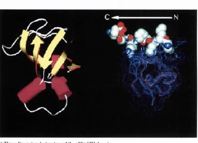
predicted to have the same fold. The first three-dimensional structure of an SH3 domain determined was that of chicken a-spectrin, (Musacchio et al., 1992), which revealed a compact |3-barrel of five anti-parallel P-strands. The five P strands,
designated a to e form two orthogonal p-sheets of three strands each, with one P strand
shared by both sheets (Figure 1.6A). The amino-and carboxy-termini of the domain are close together, as are those of several other protein modules, including SH2 domains.
This allows the domain to be incorporated into any protein as an independently folding
module, with little effect on the structure of the host protein.
The residue numbering adopted in this section is according to (Musacchio et al., 1992). Beta strand a in the SH3 domain of spectrin and Fyn is composed of four amino acids
(residues 3-6), and is connected to strand b (residues 25-30) by a long loop (RT-loop), which resembles an irregular anti-parallel P-hairpin loop. Mutating residues R13 and
T14 in the RT loop of the Src SH3 domain causes cell transformation (Kato et al.,
1986). In the three-dimensional structures of the Src and PLC-y SH3 domains (Kohda
et al., 1993; Yu et al., 1992), the hairpin structure formed by strands a and b is extended beyond the P sheet. An extra p-strand is also observed around positions 17-
20 in these SH3 domains.
Strand b in the SH3 domains of spectrin, Fyn, Src and PLCy can be divided into two
halves, designated b l and b2, that each participate in one of the P-sheets that makes the
SH3 domain structure. The neuronal variant of Src (n-Src) has a six-residue insert
within the loop connecting strands b2 and c, and this loop has been named the n-Src
loop. This insert increases the tyrosine kinase activity and alters the transforming
Pyper and Bolen, 1990) and it has been shown that a different set of proteins which
binds to the n-Src SH3 domains (Cicchetti et a i, 1992; Ren et a/., 1993; Weng et al.,
1993). A 15-residue insert in the n-Src loop of the SH3 domain of the p85a subunit of
PI3K completely disrupts strand b2. Two different three-dimensional structures have been reported for this region of the p85a SH3 domain. In one NMR derived structure,
the n-Src loop forms a large loop on the surface of the domain which is stabilised by
hydrophobic interactions (Booker etal., 1993). In another NMR structure, this insert forms a short a helix and two 3io helices (Koyama et al., 1993). As the experimental conditions and numbers of nuclear Overhauser effect and hydrogen-bond restraints
were very similar, the reason for this discrepancy remains unclear. The recently determined x-ray crystal structure of the p85a SH3 domain is in better agreement with
the latter NMR structure (Liang et al., 1996).
Strands c and d (36-41 and 46-50) are connected by a tight turn in the spectrin SH3
domain but an insert of two amino acids is present in the same loop in the Fyn and Src
SH3 domains. This loop was designated the distal loop (Noble et al., 1993) because it is on the opposite face of the SH3 domain to the putative ligand binding site. A turn of
a 31 0 helix, starting at the conserved P51 (positions), connects strand d and strand e
(residues 55-58).
In summary, the overall fold of an SH3 domain, therefore, is a |3-sandwich, composed
of two three-stranded p-sheets. The first sheet is composed of strands a, b l and e,
while the second comprises strands b2, c and d (Fig). In the p85aSH3 domain,
strand b2 is completely absent. In the Src and PLCy SH3 domains strand a is longer,
and a short additional P strand at positions 17-20 seems to be present.
1.2.5.2 SH3 dom ain binding
Mutagenesis studies of two SH3 domain binding proteins, 3BP1 and 3BP2, mapped
their Abl SH3 domain binding site to a 10 amino acid proline-rich region (Ren et al.,
1993) with a potential consensus amino acid motif XPXXPPPZXP (where X is any
amino acid and Z is a hydrophobic amino acid). A number of SH3 domain binding proteins subsequently have been identified, including 80S, the p85a subunit of PI3K,
and p47phox, and the binding sites have also been mapped to proline-rich sequences
(Finan et al., 1994; Musacchio et al., 1994) Proline-rich regions are a relatively common feature in proteins (Reviewed in Williamson, 1994), so it was important to
establish whether specific rules exist which define proline-rich sequences constitute
high-affinity SH3 domain binding sites, and the basis of ligand binding specificity