PEDIATRIC
S
Aa 1992
VON.. #{149}o: . 2:
Head
Injury
in
Very
Young
Children:
Mechanisms,
Injury
Types,
and
Ophthalmologic
Findings
in 100
Hospitalized
Patients
Younger
Than
2
Years
of Age
A. C. Duhaime, MD*; A. J. Alano, MDII; W. J. Lewander, MDII; L. Schut,
MD*; L. N. Sutton, MD*; T. S. Seidi, MSW; S. Nudelman RN; D. Budenz,
MD
II;
R. Hertle, MD0;
W. Tsiaras, MD#; and S. Loporchio, MD#ABSTRACT. Head injury in the youngest age group is
distinct from that occurring in older children or adults because of differences in mechanisms, injury thresholds, and the frequency with which the question of child abuse is encountered. To analyze some of these characteristics in very young children, the authors prospectively studied
100 consecutively admitted head-injured patients 24
months of age or younger who were drawn from three
institutions. Mechanism of injury, injury type, and as-sociated injuries were recorded. All patients underwent
ophthalmologic examination to document the presence
of retinal hemorrhages. An algorithm incorporating in-jury type, best history, and associated findings was used to classify each injury as inflicted or accidental. The
results confirmed that most head injuries in children
younger than 2 years of age occurred from falls, and
while different fall heights were associated with differ-ent injury types, most household falls were neurologi-cally benign. Using strict criteria, 24% of injuries were presumed inflicted, and an additional 32% were suspi-cious for abuse, neglect, or social or family problems.
Intradural hemorrhage was much more likely to occur
from motor vehicle accidents and inflicted injury than
from any other mechanism, with the latter being the
most common cause of mortality. Retinal hemorrhages
were seen in serious accidental head injury but were
most commonly encountered in inflicted injury. The
presence of more serious injuries associated with
partic-ular mechanisms may be related to a predominance of
rotational rather than translational forces acting on the
From the Departments of *Neurongey, C1inica1 Social Work, §Nursing,
and flOphthahnology, Children’s Hospital of Philadelphia, University of Pennsylvania School of Medicine; and Departments of #{182}Pediatrics and #Ophthalmology, Rhode Island Hospital, Brown University School of
Med-icine, Providence.
Received for publication Aug 26, 1991; accepted Jan 21, 1992.
Reprint requests to(A.C.D.) Dept of Neurosurgery, Children’s Hospital, 34th and Civic Center Blvd. Philadelphia, PA 19104.
PEDIATRICS (ISSN 0031 4005). Copyright C 1992 by the American
Acad-emy of Pediatrics.
head. Pediatrics 199290:179-185; head injury, infants, child abuse, retinal hemorrhage.
It has been well established that head injury in
children differs in several important ways from that
seen in the adult population. While various recent
studies have focused on incidence,”2 mechanism,35
various injury types,9 and outcome10’3 of head
trauma in the pediatric population, most efforts have grouped all children together for purposes of com-parison with adults. Because of our clinical impression
that head injuries in babies have many features
dis-tinct from those seen in older children, and because
of the relatively frequent question of nonaccidental
injury in these very young children,’4’15 we
prospec-tively
studied
all patients
aged
24 months
or
youngerwho were admitted with a diagnosis of head injury
in three
institutions.
The
circumstances
and
mecha-nisms of the trauma, injury type and severity, and
the ophthalmologic findings were analyzed in each
case to establish both relative frequencies of different injury types and mechanisms and their associated
findings.
MATERIALS AND METHODS
All consecutively admitted children 24 months of age or younger with a primary diagnosis of head injury determined at discharge or death were included in the study protocol; in this way, children in whom the diagnosis of head trauma was not apparent at the time of admission were not excluded. Patients were drawn from three teaching hospitals, of which two were located in urban seftings. Criteria for hospital admission after head injury included history of loss of consciousness, abnormal level of consciousness or other neurologic abnormality, skull fracture, orintracranial hem-orrhage. Children were identified for inclusion into the study by admission diagnosis or by regular surveillance of all trauma diag-noses apparent after neurosurgical, neurologic, or trauma surgery consultation.
at Viet Nam:AAP Sponsored on September 1, 2020
www.aappublications.org/news
Skull fracture +1-CpidUral
Multiple,stellate, or basilar skull fracture
180 HEAD INJURY
A biomechanical profile (Table I) was administered at the time of admission to the hospital or upon entry into the study by the evaluating physician and amended as additional information be-came available. Fall heights were judged by the distance through which the patient’s head moved, so that children falling from a standing or sitting position had distances adjusted accordingly. In general, couches were estimated at 11/2 to 2 feet in height, beds at
2to 2#{189}feet, and changing tables at 3 feet. Falls from an adult’s arms were classified as falls greater than 4 feet. When available, specific measurements were used to assign fall distances. Police
reports, emergency medical technician interviews, and accounts of
additional witnesses were sought to corroborate injury details. When applicable in unwitnessed accidents, distance estimates were based on the child’s position before and when first seen after the accident. In patients with clinical or radiographic evidence of trauma but with no history of trauma on admission, information was obtained regarding mechanism of injury by multiple interviews with caretakers by the appropriate members of the medical and social service teams. Injuries in which no history of trauma could
be obtained after such efforts were designated with respect to
mechanism as ‘no history of trauma.’
Children underwent detailed physical examination by a pedia-trician, neurosurgeon, and, in most cases, a trauma surgeon. Radio-logic evaluation included skull films, computed tomographic scan, and/or magnetic resonance imaging scans as clinically indicated as well as skeletal survey in all children in whom additional injury was suspected on dinical or historical grounds. Patients with equiv-ocal skeletal surveys underwent delayed, subsequent surveys or radioisotope bone scans for documentation of concomitant trauma.
All patients underwent fundoscopic examination withIn 36
hours of admission by ophthalmologists or emergency department pediatricians familiar with the diagnosis and detection of retinal hemorrhages. The majority (82%) were performed using short-acting mydriatic agents.
The families or caretakers of each patient were interviewed by atrauma social worker or pediatric emergency department attend-ing physician experienced in the evaluation of child abuse. Physi-dams and social workers from the child maltreatment evaluation team at each hospital assessed all patients in whom a suspicion of inflicted injury or inadequate supervision was raised. To determine whether a given patient’s injury would be dassified as inflicted or accidental for purposes of statistical analysis, an algorithm was developed which incorporates the patient’s specific injury type, final best history attainable, and the associated physical and
radio-TABLE 1. Biomechamcal Profile
1. Narrative description of accident by witnesses.
2. What time did the accident occur? 3. Was the accident witnessed? By whom?
4. What position was the baby in prior to the fall/accident? 5. Through what distance did the baby move?
6. What position was the baby in after the fall/accident? 7. Did the baby strike the head? If so, where and against what?
8. If the baby was struck by a moving object, how was the object moving and how did it strike the baby?
9. If this was an auto accident, please note speed of car, point of impact to the car, speed of other involved vehicles, and other pertinent information.
10. Was there pressure on or compression of the chest or abdomen at or after the accident? Describe.
1 1. If the accident was unwitnessed, what is the estimated time between the accident occurring and when the baby was first seen?
12. What did the baby do immediately after the accident (or, if unwitnessed, when first seen)? Check all that apply:
Unresponsive
Eyes Opened Closed
Tone ____Flaccid ____Increased... Normal
Time until responsivenesss returned:
_____
Seizure How long after accident and duration? Cry or other sound_____
Other13. What did the caretaker do on reaching the baby and how did the baby respond?
14. Was the baby manipulated in any way after the accident (eg, shaking, resuscitation, etc)? If yes, please describe.
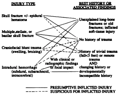
graphic findings. The scheme was designed to use objective and readily available data and to be independent of ophthalmologic findings and family social circumstances (Fig. 1). Such a scheme provides a conservative estimate of the overall incidence of child abuse, focusing only on injuries believed by the authors to clearly result from a deliberate and intended assault. Determination is based on combinations of findings indicating the presence of either (1) unexplained injuries such as healing long-bone fractures; (2) injuries unequivocally caused by mechanical trauma with no his-tory of trauma obtainable; or (3) a history of forces considered by the authors to be mechanically insufficient to cause particular injury
types, when seen in association with a changing or developmentally incompatible history. These latter features of the history have been described as typical in child abuse.’6”7 Falls clearly described as less than 3 feet in height were designated as ‘trivial’ trauma, and when given as an explanation for a high-force injury along with variability in the history or a developmentally incompatible sce-narlo, nonaccidental injury was presumed. For example, an
other-wise healthy 1-month-old baby with multiple skull fractures who
‘rolled off the center of a mattress’ meets criteria, as does a 3-month-old with acute bilateral subdural hematomas, a fresh per-iorbital bruise, and no history of trauma. Conversely, a 6-month-old child with a linear skull fracture, who may have fallen off a bed or may have been struck by a falling object, is ‘suspicious’ but not ‘presumptive’ for inflicted injury according to the algorithm,
and this would be dassed as accidental. Such children in our
experience may have been poorly supervised, rather than targets of intended assault. Likewise, patients with intradural hemorrhage and no history of trauma must also have dinical or radiographic findings of blunt impact to the head, unexplained long-bone frac-tures, or other soft-tissue inflicted injury, in order to completely eliminate the possibility of a spontaneous intracranial hemorrhage such as might rarely occur from a vascular malformation or a bleeding disorder. Thus, the scheme as shown provides a means to classify injuries as presumptively inflicted which is deliberately biased to reduce false positives and thus may underestimate the true incidence of child abuse.
In addition to injuries dassified as inflicted according to the algorithm, admitted assaults were also classified as inflicted inju-ties. Instances of neglect resulting in accidental injury, inadequate supervision, accidents involving intoxicated caretakers, or suspi-cious circumstances not reaching criteria for presumptive infliction according to the algorithm were classified for purposes of statistical
INJURY TYPE BFSTHLSTORY OR
ASSOQATED NDD1GS
Unexplained long-bone
fractures or old fractures; inflicted
soft-tissue y
histcuy of trauma
.---,:-:: ::2i::;::History of trivial trauma
7
(falk3feet)orremote- A,. With clinical or trauma radiographic findings AND Intradural hemorrhage ,,.-lf focal impact changing history or
(subdural, subarachnoid,
.
developmentallyintracerebral) incompatible history
-
PRESUMPTIVE INFLICTED INJURY---- - -- SUSPICIOUS FOR INFLICI’ED INJURY Fig 1. Scheme for determination of inflicted injury in very young children. The algorithm is used by finding the patient’s injury type in the left-hand column, then matching it with the appropriate best history or associated findings in the right-hand column. If the columns are connected by a solid line, the injury is classified as ‘presumed inflicted injury.’ If the columns are connected by a dashed line, the injury is suspicious for inflicted injury but does not meet criteria for a presumption of inflicted injury, and so it is Classified as accidental in this study. (Note that concussive injuries are assigned to a category in the left-hand column according to their associated objective clinical or radiographic findings, such as bruising, fracture, or hemorrhage.) For more details please see text.
at Viet Nam:AAP Sponsored on September 1, 2020
www.aappublications.org/news
analysis as accidental injuries, but referrals to the appropriate child protection agencies were made in all cases of suspected child abuse or neglect independent of the study classification.
Patients were categorized according to reported mechanism of
head injury, head injury type (Table 2), presence or absence of retinal hemorrhages, inflicted or noninflicted injury, and whether the child died. Patients sustaining more than one injury type (eg, skull fracture and epidural hematoma) were classified for purposes of statistical analysis by the more severe injury type. The perform-ance of a formal social service or child maltreatment evaluation team consult was also analyzed separately as an additional marker for suspicion for family or social problems, neglect, or abuse, to identify the frequency with which these issues were present in the population studied.
Data were analyzed using univariate analysis of variance to assess distribution of demographic variables and the x2statistic for contingency tables to assess the relationship between variables.
RESULTS
Of the 100 children studied, 65 were male and 35
were female. Mean age was 9.0 months (standard
deviation 7.1 months), with a range of 11 days to 24
months. The distribution of reported mechanisms of
injury is given in Table 3. Grouping similar
mecha-nisms, there were 73 reported falls, 9 motor vehicle
accidents, 14 patients with no history, 2 patients with
admitted assault, and 2 impacts by moving objects.
The distribution of injury types is shown in Table
2. Thirty-two children had a soft-tissue injury and/or
concussion only. Twenty-seven children had a linear
skull fracture with or without loss of consciousness.
There were eight depressed fractures and eight
in-stances of multiple, stellate, or basilar skull fracture.
This study population had no cases of ‘ping-pong’
fracture (in which the bone is indented but not
frac-TABLE 2. Distribution of Injury Types in 100 Consecutively
Admitted Head-Injured Children 24 Months of Age or Younger
Injury Type No.
Concussion/soft tissue injury only 32
Skull fracture (± concussion) 43
Unear 27
Depressed 8
Multiple 3
Stellate 1
Basilar 4
Epidural hematoma 3
Intradural hemorrhage 22
Subdural hematoma ± subarachnoid hemorrhage, 16
contusion
Subarachnoid hemorrhage only 3
Focal contusion only 3
Total 100
TABLE 3. Reported Mechanisms of Injury
Mechanism of Injury No.
Falls 73
<4ft 34
>4ft 21
Down stairs 10
Down stairs in walker 8
Motor vehicle accidents 9
Passenger-unrestrained 6
Passenger-restrained 2
Pedestrian 1
Impact by moving object 2
Assault (admitted) 2
No history 14
Total 100
hired). Three children had epidural hematomas (all
with fractures), and 22 children had intradural
hem-orrhages (subarachnoid or subdural hemorrhage or
parenchymal contusion). In the latter category, 10
patients also had skull fractures.
Twenty-four patients were classified as having
in-flicted injuries. All of these children were also iden-tified by the child maltreatment teams using
hospital-specific protocols, and reports to child protection
agencies were filed in each case. An additional 32
patients had social service or child maltreatment
eval-uation team consults because of social problems or
suspicion of a nonaccidental injury or neglect but did
not meet criteria for presumed inflicted injury as
defined in this study. Of the 24 patients meeting
criteria for presumed inflicted injury, there were 15
boys and 9 girls, with a mean age of 8.7 months
(standard deviation 6.3 months), which did not differ
from the overall study population. Reported history
in this group was a fall less than 4 feet in 8 patients, admifted assault in 2 patients, and no history in 14 patients. Two children with presumed inflicted injury
had craniofacial soft-tissue injury; 7 had linear or
depressed skull fractures without intradural
hemor-rhage; 2 had multiple, basilar, or stellate fractures;
and 13 had intracranial hemorrhage with or without
fracture. All 13 of these latter patients had subdural
hemorrhage. Subarachnoid hemorrhage and focal
contusions were also seen, particularly in those
pa-tients who underwent magnetic resonance imaging.
Five of the 13 patients with subdural hemorrhage also
had skull fractures diagnosed radiographically. Nine
of the 24 patients with inflicted injury also had acute
long-bone fractures or healing fractures. The
relation-ship between inflicted injury and the presence of an
intradural hemorrhage is statistically significant (P <
.0002).
Exduding those children with inflicted injury, an
analysis can be made of the association between
mechanism of injury and injury type. Linear skull
fractures were as likely to occur from a fall less than
4 feet as from falls greater than 4 feet, falls down
stairs, or falls down stairs in walkers. However, more
complex skull injuries were associated with the
greater
mechanical impact forces generated from the higher falls. All noninflicted depressed skull fractures occurred from falls greater than 4 feet, falls down stairs, or impact from a moving object. Basilar (n = 3)or bilateral (n = 3) fractures without intracranial
hemorrhage were all associated with falls greater than
4 feet or falls down stairs. The only instance of stellate
fracture in this series was in a child with an admitted
assault who was struck on the head by her caretaker. While all three epidural hematomas occurred from
falls less than 4 feet, no other types of intracranial
hemorrhages were associated with this mechanism of
injury when the trauma was accidental. In contrast, more significant falls resulted in focal parenchymal contusions (n = 4) or focal subarachnoid hemorrhage
(n = 2); these children typically had a benign clinical
course. Motor vehicle accidents (n = 7) were
associ-ated with a
high
incidence of subdural (n = 3) ordiffuse subarachnoid hemorrhage (n = 2) and
paren-chymal contusions and a more severe neurologic
at Viet Nam:AAP Sponsored on September 1, 2020
www.aappublications.org/news
182 HEAD INJURY
jury. The difference in injury types seen among
pa-tients with accidental injuries sustained in motor
ye-hide accidents compared with those from all other
mechanisms (excluding inflicted injury) is significant,
with the former having a higher incidence of
intra-dural hemorrhage (x2 contingency tables, P < 0002).
Retinal hemorrhages were found in 10 patients.
Mean age was 10.9 months (range 3 to 24 months).
Nine of the 10 were inflicted injuries (P < .0005),
with 2 of these patients reporting a history of a trivial
fall, 1 having an admitted assault, and the remaining
6 patients having no history of trauma. One patient
had a history of unexplained apnea, shaking to
resus-citate, and cardiopulmonary resuscitation but was
later found to have multiple long-bone fractures. No
other patients had a history of shaking. The patient
with an accidental injury and retinal hemorrhages
was a passenger in a high-speed motor vehicle
acci-dent who subsequently died of his head injury. All
10 patients with retinal hemorrhages also had
sub-dural hemorrhage; these were all small and were not
treated surgically. Seven of the 9 patients with
in-flicted injury and retinal hemorrhages had seizures as
part of their acute course. Two had skull fractures
and 5 had associated long-bone or old fractures.
There were four deaths in the overall series. One
of these was accidental and three were inflicted; all
had subdural hemorrhage and three of the four (two
inflicted and one accidental) had retinal hemorrhages.
DISCUSSION
The most common mechanisms of head injury in
very young children involve falls from short distances
which impart a predominantly translational (linear)
force to the head. While such forces may result in
local skull deformations sufficient to cause simple
skull fractures, translational forces of this degree
ap-plied to the brain are not of great acute clinical
consequence, except when an epidural hematoma
occurs.’82#{176} In our experience, ping-pong fractures
also frequently occur from short falls against a
pointed object such as a table corner, though there
were none found in the present series. Greater degrees
of translational force as occur in falls from greater
heights appear to result in focal impact forces
asso-ciated with multiple, depressed, comminuted, or
bas-ilar skull fractures as well as focal brain contusions
and subarachnoid hemorrhage.6’7’9’21’22 However,
ex-cept when translational forces are extreme, damage
is predominantly focal and recovery of global
neuro-logic function is usually rapid. In contrast, it has been
shown experimentally and clinically that more
signif-icant diffuse brain injury results from the introduction
of a significant angular component to the head’s
deceleration. Angular (rotational) deceleration leads
to much more brain deformation and shear strain
than is seen in translational events, and at
progres-sively greater angular decelerations the phenomena
of concussion, subdural hematoma, and diffuse
ax-onal injury will occur.18’19’23’24
The results of this study confirm the widely noted
observation that simple falls from low heights rarely,
if ever, result in significant primary brain injury.4’5
Secondary brain injury from the mass effect and
herniation resulting from an arterial epidural
hema-toma, which requires only that a fracture or skull
displacement occurs across and ruptures a dural
ar-tery, may result from such falls; however, when the
hematoma is evacuated before secondary damage
occurs the outcome is usually very good.25 While the
exact frequency and biomechanics are unknown,
cal-varial, linear, depressed, and ping-pong fractures
oc-cur fairly commonly from falls in young infants as
the requisite skull deformation can be produced with
relatively little impact force in this age group. Under-lying brain injury is almost always minimal when the
mechanism is a low-height fall.
The present results also confirm the intuitive notion
that greater biomechanical forces produce more
sig-nificant injuries, but here too the type of force
deter-mines the particular type of associated injury.
Trans-lational forces of greater magnitudes (eg, falls from
greater heights or focal impact from a heavy moving
object) produce more extensive skull fractures. These
include basilar fractures which occur through bone of
greater stress tolerance than that of the convexity,
depressed fractures, stellate fractures, or multiple
fractures. These forces may also be associated with
focal contusions and usually localized subarachnoid
hemorrhage. Such forces may also be generated when
a child acts as a missile in a motor vehicle or
pedes-trian accident if the head and brain move in a line
rather than rotate when they decelerate. These
chil-then often have a relatively benign clinical course
even when fractures and focal contusions are
exten-sive (Fig. 2). Contusional brain swelling and focal
neurologic deficit may complicate recovery or increase mortality when large forces are involved.
Subdural hematomas result from displacement of
the brain relative to the dura sufficient to cause
rup-Fig 2. Computed tomographic scan of an 11-month-old child who was an unrestrained passenger in a motor vehicle accident. Despite extensive fractures (arrows) and focal contusions and extraaxial hemorrhage (asterisk), the child made a good recovery.
at Viet Nam:AAP Sponsored on September 1, 2020
www.aappublications.org/news
ture of the bridging veins which course from the
brain’s surface to the overlying venous sinuses.228
Thus, unlike epidural hematomas which occur from
focal impact injuries, subdural hematomas almost
always result from angular deceleration of the head
in which the brain continues to rotate relative to the
more stationary skull and dura. This explains the high
incidence of subdural hematoma seen in adults
in-volved in motor vehicle accidents in which the head
rotates around an axis in the lower cervical spine,
often decelerating abruptly as it impacts against a
surface. The same shear forces involved in rupturing
the bridging veins are also applied to the brain
paren-chyma itself and, depending on the velocity and
duration of the deceleration, may give rise to diffuse
axonal injury. This type of brain damage often results
in severe and permanent neurologic sequelae.18’2931
While an unusual focal impact could theoretically tear
a cortical vessel or bridging vein and result in
hem-orrhage into the subdural space, such an injury would
be expected to produce the appropriate focal
neuro-logic deficits, rather than diffuse neurologic
dysfunc-tion. Such an injury is rarely encountered and was
not seen among the patients studied.
In this series, subdural hematomas were
uncom-mon in accidental injuries, occurring in only three
children involved as passengers in motor vehide
ac-cidents. None were treated surgically. One child died
from uncontrollable diffuse brain swelling and the
other two children made gradual recoveries. The
small size and less erect sitting posture in very young
children probably contribute to the uncommon
oc-currence of sufficient angular rotation to cause
sub-dural hemorrhage in the clinical setting of accidental
injury.
In contrast, subdural hemorrhage and diffuse
sub-arachnoid hemorrhage are common in inflicted injury
in very young children, occurring in 13 of 24 patients
in this series. The mechanism of injury of inflicted
trauma is rarely clear, as an accurate history is almost
always lacking and the mechanism varies among
patients. In most cases, an extrapolation of forces
required to cause a given injury type can be
approxi-mated; this is true for both fractures and for
intracra-nial hemorrhages. While subdural hematomas in child
abuse classically have been attributed to shaking,32’33
it is our belief that the high incidence of subdural
hemorrhage in inflicted injury in infants results from
the application of a rapid angular deceleration to the
brain which requires an impact to occur. The
mech-anism postulated is that of a child being held by the
perpetrator who shakes, swings, or throws the child,
the head thus moving through an arc, stopping
ab-ruptly against a surface. Previous autopsy studies and
biomechanical analysis using infant models suggest
that shaking alone does not generate sufficient
decel-eration forces to cause the subdural hemorrhages and
brain injuries seen in these children.34 The frequent
radiologic or clinical findings of blunt impact in series
of shaking’ injuries corroborate this conclusion, as
does the rarity of an unsolicited history of
shak-ing.3439 For these reasons, we now refer to this
syn-drome as the “shaken impact syndrome.’36 The
pres-ence of fractures or bruises will be determined by the
surface against which the rotating head decelerates.
Thus, if the head strikes a soft padded surface, contact
forces will be dissipated over a broad area and
exter-nal or focal injuries may be undetectable while
intra-cranial rotational shear forces can be sufficient to
result in subdural hemorrhage and severe brain
in-jury.18’34
In young children with malleable skulls and patent
sutures, the possibility of dural sinus tears resulting
from bony displacement associated with impact is
also a possible source of subdural hemorrhage. Such
a mechanism might explain the preponderance of
hemorrhages in the occipital region and posterior
interhemispheric fissure, where the lambdoid and
sagiftal sutures overlie the confluence of venous
si-nuses. The diffuse brain injuries often associated with these hemorrhages in nonaccidental trauma are likely
to result from large angular deceleration forces and
shearing of the brain parenchyma. The degree to
which superimposed hypoxia, possibly related to
sei-zures, cervicomedullary compromise, or concomitant
strangulation, exacerbates the brain injury and
sub-sequent swelling in these patients remains
Un-known.4042 In some patients, particularly those with
a more unilateral injury, the entire affected
hemi-sphere appears swollen or infarcted, though in our
experience magnetic resonance angiography
per-formed within 24 hours of admission demonstrates
patency of the circulation. Whether a transient func-tional effect of the injury on vasomotor tone or a local
toxic effect of subdural blood plays a role in this
phenomenon remains to be studied.43
Retinal hemorrhages are also overrepresented in
nonaccidental trauma (9 of 24, P < .000002), being
found in only one patient with accidental trauma, the
victim of a fatal motor vehide accident associated
with subdural hemorrhage. The mechanism of retinal
hemorrhage which is found so often in child abuse
remains speculative. Historically this also had been
attributed to violent shaking.333446 Other
postu-lated mechanisms include increased pressure to the
central retinal vein from increased intracranial or
increased intrathoracic pressure or some effect related
to direct head trauma.47’48 Studies linking
cardiopul-monary resuscitation to retinal hemorrhages have
been inconclusive, and they further complicate
at-tempts to determine mechanism of injury in cases of
suspected abuse with a history of resuscitation.49’5#{176}
Nonetheless, it is dear that retinal hemorrhages can
occur under a variety of circumstances, including
vaginal delivery, spontaneous subarachnoid
hemor-rhage, systemic hypertension, intracranial
hyperten-sion, thoracic or abdominal trauma, and in-hospital
resusdtation.4955 Whether superimposed hypoxia or
ischemia with reflow exacerbates the finding remains
unknown.
Traumatic retinoschesis resulting from
accelera-tion/deceleration forces applied to the eye has also
been postulated as a mechanism for retinal
hemor-rhages.56 The latter may be particularly relevant in
very young children because of the more solid
con-sistency of the vitreous body in the infant and the stronger adhesions at the vitreoretinal interface.5759
Threshold values for the degree of deceleration
at Viet Nam:AAP Sponsored on September 1, 2020
www.aappublications.org/news
* Injury types preceded by question marks are uncommonly asso-ciated with the given mechanism.
184 HEAD INJURY
quired to result in retinal hemorrhage have not been
established. Since this study was completed, we have
seen three additional patients with well-witnessed
accidental head injuries who had acute retinal
hem-orrhages. Mechanisms included a nonfatal motor
ye-hide accident, a fall down stairs in a walker, and a
fatal three-story fall. Thus, because of the variable
etiologies and unclear biomechanical thresholds for
retinal hemorrhages, it is at the present time
impos-sible to extrapolate a specific mechanism of injury for
a given patient with
this finding.
Deciding whether a head injury in a very young
child is accidental or nonaccidental has always be
problematic for clinicians and is complicated by the
fact that there is often no history given by the
care-takers who bring the child to medical
atten-tion.21’34’#{176}’61Most determinations of nonaccidental
injury are based on the notion of 1history insufficient
to explain injuries.’ Social factors and certain specific
associated injuries such as long-bone fractures or
injuries of different ages have also been used. The
presence of retinal hemorrhages is believed by some
authors to be virtually pathognomonic for child
abuse,3944 but for the reasons stated above this is
unreliable. From the data generated in this study and
in our larger experience over many years, the expected
injuries associated with various accidental
mecha-nisms can be approximated, although some
uncer-tainty remains (Table 4). Further biomechanical
stud-ies are needed to determine specific threshold
condi-tions for various injury types in this age group.
The scheme used in the present study for
determi-nation of inflicted injury incorporates our current
understanding of the biomechanical forces associated
with a given injury type as well as reliance on those
features of the history, physical examination, and
radiographic findings which are most objectively
ver-ifiable. The scheme was designed to eliminate
de-pendence on the presence of retinal hemorrhages or
psychosocial factors to determine nonaccidental
in-jury. In addition, the possibility of a rare medical
condition mimicking trauma is maintained. The
scheme also includes strict criteria for a presumption
of inflicted injury, with lesser injuries with no history
TABLE 4. Expected Injury Types Mechanisms in Very Young Children*
Associated With Accidental
Mechanism Injury Types
Concussion/soft tissue injury Linear fracture
Epidural hematoma Ping-pong fracture ? Depressed fracture
Fall >4 ft Injuries listed above plus the following: Depressed fracture
Basilar fracture Multiple fractures Subarachnoid hemorrhage Contusion
?Subdural hematoma ?Stellate fracture
Motor vehicle accident Injuries listed above plus the following: Subdural hematoma
Diffuse axonal injury Fall <4 ft
of trauma or very trivial trauma, which may occur
when children are unsupervised, classified as
suspi-cious but not presumptive for inflicted injury. The
conclusions reached by using the algorithm correlated
extremely well with those reached by the trauma
social workers and child abuse team, who often had
additional social data available.
In light of these strict criteria for determining
in-flicted injury, it is of interest that nearly one quarter
(24%) of all admissions for head injury in this age
group can be classified as inflicted. This figure is
higher than that of more general seriesU1 and is cause
for grave concern. Compared to accidental injuries,
inflicted injuries have a disproportionately high
mci-dence of intracranial hemorrhage (13 of 24, P < .0002)
and mortality (3 of 24, or 125%). An additional 32%
of all patients in this series required further
investi-gation because of a suspicion of abuse or neglect.
Thus, the majority of head injuries in very young
children appear to be preventable, with social or
family problems a major risk factor for their
occur-rence.
CONCLUSIONS
Accidental blunt head injuries in children younger
than 2 years of age are very common, but except for
those caused by motor vehicle accidents and falls
from extreme heights, they are almost always benign
in their acute clinical consequences. Inflicted injury is
extremely common among children admitted for head
injury, accounting for nearly one quarter of
admis-sions in this series. This mechanism is associated with
more severe brain damage and was the most common
cause of mortality. The determination of inflicted
injury can be assisted by using a scheme incorporating
biomechanical information and related physical and
radiographic findings independent of ophthalmologic
findings and social history. Finally, while retinal
hem-orrhages can occur in severe accidental head trauma
and in other benign circumstances, they were never
seen in trivial accidental head injuries. Retinal hem-orrhages are highly associated with inflicted injuries
in very young children and, like subdural
hemor-rhages and diffuse axonal injury, may result from
rotational, rather than translational, forces applied to
the head.
ACKNOWLEDGMENTS
Statistical reviewer was Wayne Alves, PhD, from the Depart-ment of Neurosurgery, University of Pennsylvania. He is currently at the Department of Neurosurgery, University of Virginia Medical School, Charlottesville, VA.
REFERENCES
1. Krause if, Fife D, Cox P, Ramstein K, Conroy C. Incidence, severity and external causes of pediatric brain injury. AJDC 1986;140:687-693 2. Brookes M, MacMillan R, Cully S, et al, Head injuries in accident and
emergency departments: how different are children from adults? I
Epidemiol Community Health. 1990;44:147-151
3. Joffe M, Ludwig S. Stairway injuries in children. Pediatrics. 1988;82(pt
2):457-461
4. Heifer RE, Slovis IL, Black M. Injuries resulting when small children
fallout of bed, Pediatrics. 1977;60:533-535
5. Nimityongskul P, Anderson L The likelihood of injuries when children
fall out ofbed, JPediatr Orthop. 1987;7:184-186
at Viet Nam:AAP Sponsored on September 1, 2020
www.aappublications.org/news
6. Chin K, Yue CP, Kirpal SM. The risk of intracranial complications in
pediatric head injury: results of multivariate analysis. Childs Nerv Syst. 1990;6:27-29
7. Steinbok P, Flodmark 0, Martens D, Cermann ET. Management of simple depressed skull fractures in children. INeurosurg. 1987;66:506-510
8. Scarfo GB, Mariottini A, Tomacdni D, Palma L Growing skull fractures: progressive evolution of brain damage and effectiveness of surgical treatment. Childs Nero Syst. 1989;5:163-167
9. Bonadio WA, Smith DS, Hillinan S. clinical indicators of intracranial lesion on computed tomographic scan in children with parietal skull
fractures. AJDC. 1989;143:194-196
10. Alberico AM, Ward JD, Choi SC, Marmarou A, Young HF. Outcome after severe head injury: relationship toniasslesions, diffuse injury, and ICP course in pediatric and adult patients INeurosurg. 1987;67:648-656
11. Luerssen TG, Klauber MR, Marshall LF. Outcome from injury related to patient’s age: a longitudinal prospective study of adult and pediatric head injury. INeurosurg. 1988;68:409-416
12. Kriel RL, Krach LE, Panser LE. Gored head injury: comparison of children younger and older than 6 years of age. Pediatr Neurol.
1989;5:296-300
13. Costeff H, Groswasser Z, Goldstein R. Long-term follow upreview of 31 children with severe closed head trauma. INeurosurg. 1990;73:684-687
14. Billmire ME, Myers PA, Serious head injury in infantu accident or abuse? Pediatrics. 1985;75:340-342
15. Newton RW. intracranial haemorrhage and non-accidental injury. Arch Dis Child. 1989;64:188-190
16. Kempe CH, Silverman FN, Steele BF, Droegemueller W, Silver IlK. The battered-child syndrome. JAMA. 1962;181:105-112
17. Rekate HL, McClelland CW, Rekate MW. The neurosurgical implications ofchild abuse. In: Shapiro K, ed. Pediatric Head Trauma Mt. Kisco, NY:
Futura Publishing 1983:195-211
18. Gennarelli TA, Thibault LE. Bioniechanics of head injury. In: Wilkins RH, Rengachary 55, eds. Neurosurgery. New York, NY: McGraw Hilb
1985:1531-1536
19. Ornmaya AK, Gennarelli TA. Cerebral concussion and traumatic uncon-sciousness: correlation of experimental and clinical observations on blunt head injuries. Brain. 197497:633-654
20. Stalhanimer D. Experimental models of head injury. Acta Neurochir Suppi (Wien). 1986;36:33-46
21. Meservy CJ,Towbin R, McLaurin RL, Myers PA, Ball W. Radiographic characteristics of skull fractures resulting from child abuse. AIR
1987;149:173-175
22. Liu-Shindo M, Hawkins DB. Basilar skull fractures in children. hit J Pediatr Otorhinolaryngol. 1989;17:109-117
23. Hanigan WC, Peterson RA, Njus C. Tin ear syndrome: rotational accel-eration inpediatric head injuries. Pediatrics. 1987;80:618-622
24. Margulies 55, Thibault LE. An analytical model of traumatic diffuse brain injury. IBiochem Eng. 1989;111:241-249
25. Bricolo AP, Pasut LM. Extradural hematoma: toward zero mortality. A prospective study. Neurosurgery. 1984;14:8-12
26. Yamashima T, Friede RL Why dobridging veins rapture into thevirtual subdural space? INeurol Neurosurg Psychiatry. 1984;47:121-127
27. Guthkelch AN. Infantile subdural hematoma and its relationship to whiplash injuries. Br MedJ. 1971;2:430-431
28. Vance BM. Ruptures of surface blood vessels on cerebral hemispheres
as a cause of subdural hemorrhage. Arch Surg. 1950;61:992-1006 29. Vowles GH, Scholtz CL, Cameron JM. Diffuse axonal injury in early
infancy. I clin Pathol. 1987;40:185-189
30. Yashon D, Jane JA, White RJ, Sugar 0. Traumatic subdural hematoma ofinfancy:long-term followup of92 patients.Arch Neurol.
1968;18:370-377
31. Gennareffi TA, Spielman GH, Langfitt TW, et al Influence of the type ofintracranial lesion on outcome from severe head injury: a multicenter study using a new dassification system. JNeurosurg. 1982;56:26-32 32. Caffey J. On the theory and practice of shaking infantu its potential
residual effects of permanent brain damage and mental retardation. AJDC 1972;124:161-169
33. CaffeyJ. The whiplash shaken infant syndrome: manual shaking by the extremities with whiplash-induced intracranial and intraOcUlar bleed-tags, linked with residual permanent brain damage and mental retar-dation. Pediatrics. 1974;54:396-403
34. Duhaime AC, Gennarelli TA, Thibault LE, et al. The shaken baby syndrome: a clinical, pathological and biomechanical study. JNeurosurg.
1987;66:409-415
35. Hahn YW, Raimondi AJ, McLone DC, et al. Traumatic mechanisms of head injury in child abuse. Chills Brain 1983;10:229-241
36. Bruce DA, Zimmerman RA. Shaken impact syndrome. Pediatr Ann.
1989;18:482-494
37. McQelland cQ, Rekate H, Kaufman B, et al. Cerebral injury In child
abuse: a changing profile. Childs Brain. 1980;7:225-235
38. Alexander R, Sato Y, Smith W, Bennett T. Incidence of impact trauma
with cranial injuries ascribed so shaking. AJDC 1990;144:724-726 39. Elner SC, Elner VM, Arnall M, Albert DM. Ocular and associated
systemic findings in suspected child abuse: a necropsy study. Arch Ophthalmol. 1990;108:1094-1101
40. Hadley MN, Sonntag VK, Rekate HL, Murphy A. The infant whiplash-shake injury syndrome: a clinical and pathological study. Neurosurgerg. 1989;24:536-540
41. Gilles FH, Bira M, Sotrel A. Infantile atlantooccipital inStabiIity the potential danger ofextreme extension. AJDC. 1979;133:30-37
42. Bird CR, McMahan JR, Gilles PH, Senac MD, ApthorpJS. Strangulation in child abuse: CT diagnosis. Radiology. 1987;163:373-375
43. Mifier JD, Bullock R, Graham DI, Chen MH, Teasdale GM. Ischemic brain damage in a model of acute subdural hematoma. Neurosurgery.
1990;27:433-439
a.
Eisebey AB. Retinal hemorrhage in the battered child. Chills Brain. 1979;5:40-4445. Wilkinson WS, Han DP, Rappley MD, Owings CL Retinal hemorrhage predicts neurologic injury in the shaken baby syndrome. Arch
Ophthal-mol. 1989;107:1472-1474
46. Lambert SR, Johnson it, Hoyt CS. Optic nerve sheath and retinal hemorrhages associated with the shaken baby syndrome. Arch
Ophthal-mol. 1986;104:1509-1512
47. Gilkes MJ, Mann TP. Fundi of battered babies. Lancet 1967;2:468-469 48. Harcourt B,Hopkins D. Ophthalmic manifestations of the battered-baby
syndrome. Br Med J.1971;3:398-401
49. Kanter RD. Retinal hemorrhage after cardiopulmonary resuscitation or
child abuse. IPediatr. 1986;108:430-432
50. Coetting MG, Sowa B. Retinal hemorrhage after cardiopulmonary re-suscitation in children: an etiologic reevaluation. Pediatric& 1990;85:585-588
51. Baum JD, Bulpitt CJ.Retinal and conjunctival hemorrhage in the new-born. Arch Dis Child 1970;45:344-349
52. Besio R, Caballero C, Meerhoff E, Schwarcz R.Neonatal retinal hemor-chages and influence of perinatal factors. Am JOphthalmoL
1979;87:74-76
53. McClellan NJ, Prasa R, Punt J.Spontaneous subhyaloid and retinal haemorrhages in an infant. Arch Dis Child. 1986;81:1130-1132
54. Muller PJ, Deck JH. IntraOcUlar and optic nerve sheath hemorrhage in cases of sudden intracranial hypertension. I Neurosurg.
1974;41:160-166
55. Hayrek 55, EdwardsJ. Ophthalmic arterial and venous pressure: effects of acute intracranial hypertension. BrJ Ophthalmol. 1971;55:649-663 56. Greenwald MJ, Weiss A, Oesterle CS, Friendly DS. Traumatic
retinas-chisis in battered babies. Ophthalmology. 198693:618-625
57. WolterJR. Coup-contracoupmechanism of ocular injuries.
AmJOphthal-mol. 1963;56:785
58. Fine BS, Tonsimis AJ. The structure of the vitreous body and the suspensory ligaments of the lens. Arch Ophthalmol. 1961;65:95 59. Hogan MJ. The vitreous, its structure and relation to the ciliary body
and retina. Invest Ophthalmol Via Sci 1963;2:418
60. Ludvig S. Shaken baby syndrome: a review of 20 cases. Ann Emerg Med. 1984;13:104-107
61. Duhaime AC, Gennarelli TA, Sutton LN, Schut L The ‘shaken baby syndrome’: amisnomer? IPediatr Neurosci 1988;4:77-86
at Viet Nam:AAP Sponsored on September 1, 2020
www.aappublications.org/news
1992;90;179
Pediatrics
Nudelman, D. Budenz, R. Hertle, W. Tsiaras and S. Loporchio
A. C. Duhaime, A. J. Alario, W. J. Lewander, L. Schut, L. N. Sutton, T. S. Seidl, S.
Ophthalmologic Findings in 100 Hospitalized Patients Younger Than 2 Years of Age
Head Injury in Very Young Children: Mechanisms, Injury Types, and
Services
Updated Information &
http://pediatrics.aappublications.org/content/90/2/179
including high resolution figures, can be found at:
Permissions & Licensing
http://www.aappublications.org/site/misc/Permissions.xhtml
entirety can be found online at:
Information about reproducing this article in parts (figures, tables) or in its
Reprints
http://www.aappublications.org/site/misc/reprints.xhtml
Information about ordering reprints can be found online:
at Viet Nam:AAP Sponsored on September 1, 2020
www.aappublications.org/news
1992;90;179
Pediatrics
Nudelman, D. Budenz, R. Hertle, W. Tsiaras and S. Loporchio
A. C. Duhaime, A. J. Alario, W. J. Lewander, L. Schut, L. N. Sutton, T. S. Seidl, S.
Ophthalmologic Findings in 100 Hospitalized Patients Younger Than 2 Years of Age
Head Injury in Very Young Children: Mechanisms, Injury Types, and
http://pediatrics.aappublications.org/content/90/2/179
the World Wide Web at:
The online version of this article, along with updated information and services, is located on
American Academy of Pediatrics. All rights reserved. Print ISSN: 1073-0397.
American Academy of Pediatrics, 345 Park Avenue, Itasca, Illinois, 60143. Copyright © 1992 by the
been published continuously since 1948. Pediatrics is owned, published, and trademarked by the
Pe