R E S E A R C H A R T I C L E
Open Access
Head and neck paragangliomas: diffusion
weighted and dynamic contrast enhanced
magnetic resonance imaging characteristics
Ying Yuan
†, Huimin Shi
†and Xiaofeng Tao
*Abstract
Background:To determine the feature values of head and neck paragangliomas on diffusion-weighted imaging (DWI) and dynamic contrast enhanced (DCE) magnetic resonance imaging (MRI).
Methods:Patients with primary head and neck paraganglioma who underwent both DWI and DCE-MRI before treatment between January 2010 and June 2013 were identified. Two radiologists assessed apparent diffusion coefficient (ADC) values on DWI and kinetic characteristics on DCE-MRI. The time intensity curves (TICs) and dynamic parameters, including peak height (PH), maximum enhancement ratio (ERmax), time to peak enhancement (Tpeak) and maximum rise slope (Slopemax), were generated and evaluated.
Results:Ten patients with head and neck paraganglioma were retrospectively analyzed. On conventional MRI, the tumors demonstrated as well-circumscribed, strongly enhanced lesions. Mean ADC value of the lesions was 1.12 ± 0.15 × 10⁻³mm2/s. The TICs demonstrated washout pattern (type-III) in all lesions. The mean PH, ERmax, Tpeakand Slopemaxvalue was 121.24 ± 63.99, 193.79 ± 67.18, 8.16 ± 3.29 and 25.42 ± 7.91, respectively.
Conclusions:Head and neck paragangliomas demonstrate distinctive DWI and DCE-MRI results than for other benign tumors which should be taken into account in further evaluation of MRI on head and neck lesions.
Background
Head and neck paragangliomas are rare tumors originat-ing from the parasympathetic nervous system. They mostly occur at the carotid bifurcation (carotid body tu-mors), jugular bulb (jugular paraganglioma), vagus nerve (vagal paraganglioma), middle ear mucosa (tympanic paraganglioma) and larynx (laryngeal paraganglia). Less common sites include the sella turcica, pineal gland, cav-ernous sinus, larynx, orbit, thyroid gland, nasopharynx, mandible, soft palate, face, and cheek [1]. Computed tomography (CT) and magnetic resonance imaging (MRI) have evolved as effective non-invasive imaging modalities for detection and evaluation of paraganglio-mas. MRI has advantages over CT in better soft tissue contrast, higher intrinsic flow sensitivity and non-ionizing. As reported, characteristic imaging findings
were described as well-circumscribed, strongly enhanced masses with“salt-and-pepper”appearance [2–5].
MRI methods, such as diffusion-weighted imaging (DWI) and dynamic contrast enhanced (DCE) MRI, as well as other functional imaging modalities have been showing great potential in oncologic applications of head and neck tumor [6–9]. DWI depicts the brownian mo-tion of water molecules within the tissue, quantified with apparent diffusion coefficient (ADC). DCE-MRI allows in vivo imaging of the physiology of microcirculation and provides information related to the vascularity [10]. However, previous MRI studies of head and neck para-gangliomas were mainly focused on conventional MRI and rarely assessed the application of DWI and DCE-MRI. Therefore, the aim of the current study was to de-termine the MRI characteristics of head and neck para-gangliomas on: (1) the range of ADC value; and (2) the pattern of time intensity curves (TICs) and dynamic pa-rameters generated from TICs, including peak height (PH), maximum enhancement ratio (ERmax), time to
* Correspondence:cjr.taoxiaofeng@vip.163.com
†Equal contributors
Department of Radiology, Shanghai Ninth People’s Hospital, Affiliated to JiaoTong University School of Medicine, 639 Zhizaoju Road, Shanghai 200011, China
peak enhancement (Tpeak) and maximum rise slope (Slopemax).
Methods Patients
A retrospective review of MRI findings was performed in patients with histopathologically proved head and neck paragangliomas between January 1, 2010 and June 30, 2013. All included patients underwent both conven-tional MRI, DWI and DCE-MRI within 7 days before surgery and biopsy for histopathological confirmation. Patients were excluded in any of the following condi-tions: (1) with artifacts interfering the diagnosis; (2) a head and neck cancer was previously diagnosed; or (3) with previous surgical procedures or radiation in head and neck region. This retrospective study was approved by institutional review board of Shanghai Ninth People’s Hospital.
MRI acquisition
All MRI examinations were performed using a 1.5-T scanner (Signa Excite; GE Medical Systems, Milwaukee, WI, USA) with a head and neck array coil. All patients were placed in the magnet in the supine position. The conventional MRI consisted of axial T1-weighted (T1W) sequence, axial and coronal T2-weighted (T2W) se-quences. DWI was performed using a single-shot spin echo echo planar imaging (SE-EPI) sequence. Sensitizing diffusion gradients were applied sequentially in the x, y, and z direction with b values set at 0 and 1000 s/mm2. The DCE-MRI was obtained with fast spoiled gradient echo (FSPGR) acquisition technique. The sections were acquired in the axial plane, centered on the mass. Gado-pentetate dimeglumine (Magnevist, Schering, Berlin, Germany) was intravenously bolus injected via a power injector with a flow rate of 2.0 mL/s at the dose of 0.1 mmol/kg of body weight, followed by a 20 mL saline flush. DCE-MRI was sequentially obtained for 40 dy-namic phases for each investigation. Conventional
post-contrast T1W sequences were also performed, with fat suppression in coronal sequences. DWI and DCE-MRI images were post-processed and statistically analyzed in the current study. Details of the MRI protocol are pro-vided in Table 1.
MRI interpretation
All MR images were processed by two radiologists (Y.Y., H.-M.S.; with 8 and 25 years of experience in head and neck MR imaging, respectively). Blinded to clinical infor-mation, imaging results from other modalities and histopathologic results, two radiologists selected repre-sentative regions of interest (ROIs) on DWI and DCE-MRI by consensus. Using the post processing software Functool from GE, ADC maps were automatically gener-ated. ADC values were extracted from ADC maps. DWI was evaluated by drawing freehand ROI in lesions over the axial slice with the largest tumor area, taking care to exclude obvious necrotic or non-perfused areas by visual correlation with pre- and post-contrasted T1W images. ADC values were manually measured three times in each lesion, and average ADC value was obtained. ROIs of DCE-MRI were acquired with the following method. After a color-coded mapping of the mass, we placed 2 mm × 2 mm ROIs over the entire lesion on multiple slices. The TIC of each ROI was generated. By compar-ing the TIC from each ROI, the ROI that demonstrated the greatest degree of enhancement was selected as rep-resentation for the lesion. The detail method was de-scribed in our previous studies [11, 12]. The TICs (axis coordinate, time; vertical, signal intensity) were catego-rized as three patterns: (1) the persistent pattern (type-I) with straight or curved line and continuous enhance-ment over the entire dynamic study; (2) the plateau pat-tern (type-II) with a relatively prominent increase slope and a final intensity 90–100 % of peak grade; (3) the washout pattern (type-III) with a rapid increase slope and a final intensity lower than 90 % of peak grade. From each TIC, the signal intensity (SIpre, SImax) and
Table 1MR imaging protocol
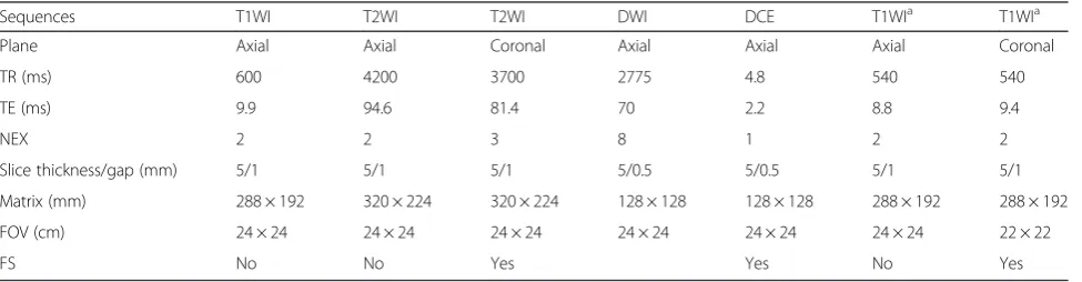
Sequences T1WI T2WI T2WI DWI DCE T1WIa T1WIa
Plane Axial Axial Coronal Axial Axial Axial Coronal
TR (ms) 600 4200 3700 2775 4.8 540 540
TE (ms) 9.9 94.6 81.4 70 2.2 8.8 9.4
NEX 2 2 3 8 1 2 2
Slice thickness/gap (mm) 5/1 5/1 5/1 5/0.5 5/0.5 5/1 5/1
Matrix (mm) 288 × 192 320 × 224 320 × 224 128 × 128 128 × 128 288 × 192 288 × 192
FOV (cm) 24 × 24 24 × 24 24 × 24 24 × 24 24 × 24 24 × 24 22 × 22
FS No No Yes Yes No Yes
DCEdynamic contrast-enhanced,DWIdiffusion weighted imaging,FOVfield of view,NEXnumber of excitations,T1WIT1-weighted imaging,T2WIT2-weighted imaging,TEecho time (msec),TRrepetition time
a
time (Tpre, Tpeak) were derived. SIprewas defined as the pre-contrast signal intensity. SImaxwas the signal inten-sity at maximal contrast enhancement. Tpre and Tpeak were the time corresponding to the SIpre and SImax. Other parameters, such as PH, ERmaxand Slopemax, were calculated upon the following formulas [11, 12]:
PH¼SImax—SIpre
ERmax¼SImaxSI−SIpre
pre 100
Slopemax¼ SImax−SIpre
SIpre Tpeak−Tpre100
Statistical analysis was carried out using STATA ver-sion 12.0 (CollegeStation, TX). P< 0.05 was considered as statistically significant.
Results
Ten patients with untreated head and neck paraganglio-mas (three male, seven female; age 36.6 ± 15.02 years) were included. DWI and DCE-MRI were performed in all patients. Carotid body tumors were diagnosed in eight patients (8/10, 80 %). Other locations included the jugular fossa (n= 1) and orbit (n= 1). The tumor size at diagnosis was 4.42 ± 1.65 cm. The radiological findings of each patient are listed in Table 2. On conventional MRI, the tumors demonstrated as well circumscribed and intensely enhanced masses with marked internal vascularity that appeared as multiple signal voids. The mean ADC value was 1.12 ± 0.15 × 10⁻³ mm2/s, signifi-cantly higher contrasted to adjacent muscle (0.74 ± 0.06 × 10⁻³mm2/s,P= 0.0006). The TIC curve unexcep-tionally demonstrated washout pattern (type-III) in the ten lesions. The mean PH, ERmax, Tpeak and Slopemax value was 121.24 ± 63.99, 193.79 ± 67.18, 8.16 ± 3.29 and
25.42 ± 7.91, respectively. MRI images of representative cases are shown in Figs. 1 and 2.
Discussion
Head and neck paragangliomas are hypervascular benign neoplasms derived from the embryologic neural crest. They account for 0.6 % of all neoplasms in the head and neck region. The most common locations are carotid space and jugular fossa. In the current study, eight of the ten paragangliomas were from the carotid body. The other two lesions were respectively in the jugular fossa and the left orbit, which is a rare location to our know-ledge [1, 13]. As reported, 10 % tumors may be multi-centric in origin and approximately 6 to 19 % are malignant, as evidenced by the presence of regional and/ or distant metastases [14, 15]. In the current study, there was no multifocal or metastatic lesion, probably due to the small amount of patients. The patients were all spor-adic cases without a familial predisposition.
Contrast-enhanced CT and MRI have evolved as ef-fective imaging modalities for detection of paraganglio-mas. The lesions have conventional MRI findings of well-circumscribed and strongly enhancing masses indi-cating abundant blood supply, which was also proved in our study. Salt-and-pepper appearance of the tumor, ex-pansive growth and remodeling of adjacent bony struc-tures are also diagnostic [16]. DWI has shown promise in the diagnosis, staging and prognostic evaluation of head and neck tumors [17]. Previous studies have con-firmed a significant difference in ADC values between benign and malignant lesions [18, 19]. Lower ADC values have been reported in the head and neck region for most malignant lesions, compared with those of benign lesions. Optimal ADC threshold values of 1.3 × 10⁻³ mm2/s (b= 800 s/mm2, 3-T MR unit) [18] and 1.25 × 10⁻³mm2/s (b= 500, 1000 s/mm2, 1.5-T MR unit) [19] were suggested to help distinguish benign from
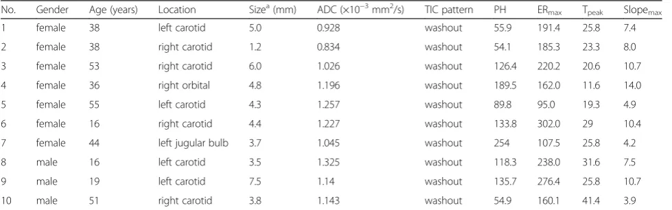
Table 2Radiological findings of patients with head and neck paragangliomas
No. Gender Age (years) Location Sizea(mm) ADC (×10−3mm2/s) TIC pattern PH ER
max Tpeak Slopemax
1 female 38 left carotid 5.0 0.928 washout 55.9 191.4 25.8 7.4
2 female 38 right carotid 1.2 0.834 washout 54.1 185.3 23.3 8.0
3 female 53 right carotid 6.0 1.026 washout 126.4 220.2 20.6 10.7
4 female 36 right orbital 4.8 1.196 washout 189.5 162.0 11.6 14.0
5 female 55 left carotid 4.3 1.257 washout 89.8 95.0 19.3 4.9
6 female 16 right carotid 4.4 1.227 washout 133.8 302.0 29 10.4
7 female 44 left jugular bulb 3.7 1.045 washout 254 107.5 25.8 4.2
8 male 16 left carotid 3.5 1.325 washout 118.3 238.0 31.6 7.5
9 male 19 left carotid 7.5 1.14 washout 135.7 276.4 25.8 10.7
10 male 51 right carotid 3.8 1.143 washout 54.9 160.1 41.4 3.9
ADCapparent diffusion coefficient,TICthe time intensity curve
a
malignant lesions. The mean ADC value was of the head and neck paragangliomas under study 1.12 ± 0.15 × 10⁻³ mm2/s. Eight tumors showed ADC value lower than 1.25 × 10⁻³ mm2/s, which could have suggested a malignant nature according to abovementioned criteria
[18, 19]. Other DWI results here! The relatively lower ADC value detected in the current study than other be-nign tumor as previously reported might be attributed to the inner texture of the lesion. Solid tumors without apoptosis or necrosis show higher signal intensity and
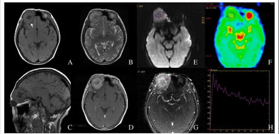
Fig. 1Left carotid body tumor in a 38-year-old female.a,bNon-enhanced axial T1W and T2W images.cContrast enhanced axial T1W image.d Contrast enhanced coronal T1W image with fat suppression.e,fROI on DWI (bvalue = 1000 s/mm2) and corresponding ADC map. The average ADC value of this lesion was 0.928 × 10⁻³mm2/s.g,hDCE-MRI and corresponding TIC, which showed a washout pattern
lower ADC value [17]. Therefore, although DWI shows the potential in characterizing and differentiating head and neck lesions, given the heterogeneous of benign and malignant lesions, there will clearly be exceptions and overlaps. When dealing with lesions with lower ADC value in head and neck region, radiologists should care-fully exclude the possibility of paraganglioma before giv-ing it a malignant diagnosis, especially for those lesions without typical conventional MRI characteristics.
DCE-MRI has been employed for tumor detection and characterization as well [20]. Malignant tumors tend to exhibit strong initial signal increase followed by washout effect, which is equivalent to washout pattern (type-III) in our study, whereas benign lesions mostly demonstrate slow initial signal enhancement combined with continu-ous signal increase (the persistent pattern). A plateau curve can be seen in both benign and malignant lesions. However, some malignant processes can mimic benign contrast kinetics and vice versa [21]. It is to be noted in the current study that the TICs unexceptionally demon-strated washout pattern in all cases. In a previously con-ducted study for visualization of paragangliomas with magnetic resonance projection angiography (MRPA), an early and rapid enhancement after contrast administra-tion and a washout effect after maximum of intensity was also detected [22]. The “washout” of intensity is usually attributed to malignant arteriovenous anasto-moses, neovascular capillary hyperpermeability and the high flow rate in the newly formed capillaries [23, 24]. Correspondingly, in the current study, the TIC pattern might be related to the typical angiographic appearance of a paraganglioma as a hypervascular mass with en-larged feeding arteries, intense tumor blush and early draining veins [16, 25], which is distinctive and not com-monly seen in benign tumors.” DWI and DCE-MRI could indicate the functional characteristics of tumor with regard to the texture and blood supply; however, these methods cannot be used exclusively with the same standard and threshold for differentiate benign from ma-lignant tumors, as also reported in a systemic review of salivary gland tumors [26]. The washout TIC together with conventional MRI characteristics of well-defined margin and strong enhancement as well as a lower ADC value (mostly lower than 1.25 × 10⁻³ mm2/s in the current study) would direct to a diagnosis of paragan-glioma in head and neck region, which is especially dis-tinct in ADC and TIC results than other benign tumors.
Our study has some limitations. First, we included a relatively small number of cases, not only due to the low incidence rate of the disease, but also the inclusion cri-teria of the study. Major treatments for head and neck paragangliomas include observation and clinical follow-up, surgical excision, embolism and radiotherapy. We only included those histopathologically confirmed by
specimens obtained at biopsy or surgery. Moreover, the inlcuded patients should undergo preoperative DWI and DCE-MRI. In spite of the small amount of cases, the re-sults of the current study could still deliver a message that head and neck paragangliomas demonstrate dis-tinctive DWI and DCE-MRI results compared to other benign lesions. Second, the selection of ROIs would affect the reproducibility of ADC values and kinetic curves. We chose to acquire a mean ADC by drawing three times of freehand ROIs over the axial slice with the largest tumor area, taking care to exclude obvious necrotic or non-perfused areas, in order to acquire ADC values most representative of the solid components in tumors. More sophisticated techniques for quantitative evaluation of DWI have been introduced, including histogram analysis and voxel-by-voxel changes; however, these techniques are mostly not yet available to clinical radiologists [17]. As for DCE-MRI, as described in our previous studies [11, 12], representative ROIs were de-fined as those demonstrated the greatest degree of peak enhancement. Two readers interpreted the images in consensus and inter-observer reliability could not be cal-culated, which might be another limitation of the study. Intravoxel incoherent motion (IVIM) MRI is a technique with the potential for simultaneously assessing both tis-sue perfusion and diffusion by using a single DWI. Re-cently, several attempts have been made to determine the feasibility of IVIM for differential diagnosing head and neck tumors [27, 28] and evaluating head and neck squamous cell carcinoma (HNSCC) [29], by comparing and combining with DWI and DCE-MRI results.
Conclusions
Head and neck paragangliomas demonstrate distinctive features on DWI and DCE-MRI than other benign tu-mors. In addition to the morphological features, a rela-tively lower ADC value and washout TIC would support the diagnosis, which is otherwise more supportive for a malignant nature. Therefore, the interpretation of MRI in head and neck lesions should take into account the possible exceptions. Further studies are needed to be performed in larger group of patients and to compare the differential ability of DWI and DCE-MRI between paragangliomas and other head and neck lesions.
Abbreviations
ADC:apparent diffusion coefficient; CT: computed tomography;
DCE: dynamic contrast enhanced; DW: diffusion-weighted; ERmax: maximum enhancement ratio; FSPGR: fast spoiled gradient echo; IVIM: intravoxel incoherent motion; MR: magnetic resonance; PH: peak height; ROI: regions of interest; SE-EPI: single-shot spin echo echo planar imaging;
Slopemax: maximum rise slope; TIC: time intensity curve; Tpeak: time to peak enhancement; T1W: T1-weighted; T2W: T2-weighted.
Competing interests
Authors’contributions
HS contributed with sequence programming and collected the data. YY performed the data analysis and drafted the manuscript. XT conceived of the study, and helped to draft and revise the manuscript. All authors read and approved the final manuscript.
Authors’information
YY: MD; HS: MD, associate professor; XT: MD; PhD; professor.
Acknowledgement
This study was supported by grants from National Natural Science Foundation of China (81402461); Subject Chief Scientist of Shanghai, Science and Technology Commission of Shanghai Municipality (13XD1402400).
Received: 1 July 2015 Accepted: 22 January 2016
References
1. Hodge KM, Byers RM, Peters LJ. Paragangliomas of the head and neck. Arch Otolaryngol Head Neck Surg. 1988;114(8):872–7.
2. Quint LE, Glazer GM, Francis IR, Shapiro B, Chenevert TL.
Pheochromocytoma and paraganglioma: comparison of MR imaging with CT and I-131 MIBG scintigraphy. Radiology. 1987;165(1):89–93.
3. van Gils AP, Falke TH, van Erkel AR, Arndt JW, Sandler MP, van der Mey AG, et al. MR imaging and MIBG scintigraphy of pheochromocytomas and extraadrenal functioning paragangliomas. Radiographics. 1991;11(1):37–57. 4. Sahdev A, Sohaib A, Monson JP, Grossman AB, Chew SL, Reznek RH. CT and
MR imaging of unusual locations of extra-adrenal paragangliomas (pheochromocytomas). Eur Radiol. 2005;15(1):85–92.
5. Amin MF, El Ameen NF. Diagnostic efficiency of multidetector computed tomography versus magnetic resonance imaging in differentiation of head and neck paragangliomas from other mimicking vascular lesions: comparison with histopathologic examination. Eur Arch Otorhinolaryngol. 2013;270(3):1045–53.
6. Jansen JF, Parra C, Lu Y, Shukla-Dave A. Evaluation of Head and Neck Tumors with Functional MR Imaging. Magn Reson Imaging Clin N Am. 2016; 24(1):123–33.
7. Payne KF, Haq J, Brown J, Connor S. The role of diffusion-weighted magnetic resonance imaging in the diagnosis, lymph node staging and assessment of treatment response of head and neck cancer. Int J Oral Maxillofac Surg. 2015;44(1):1–7.
8. Bernstein JM, Homer JJ, West CM. Dynamic contrast-enhanced magnetic resonance imaging biomarkers in head and neck cancer: potential to guide treatment? A systematic review. Oral Oncol. 2014;50(10):963–70.
9. Shah GV, Wesolowski JR, Ansari SA, Mukherji SK. New directions in head and neck imaging. J Surg Oncol. 2008;97(8):644–8.
10. Hylton N. Dynamic contrast-enhanced magnetic resonance imaging as an imaging biomarker. J Clin Oncol. 2006;24(20):3293–8.
11. Yuan Y, Yue XH, Tao XF. The diagnostic value of dynamic contrast-enhanced MRI for thyroid tumors. Eur J Radiol. 2012;81(11):3313–8. 12. Yuan Y, Kuai XP, Chen XS, Tao XF. Assessment of dynamic
contrast-enhanced magnetic resonance imaging in the differentiation of malignant from benign orbital masses. Eur J Radiol. 2013;82(9):1506–11.
13. Makhdoomi R, Nayil K, Santosh V, Kumar S. Orbital paraganglioma–a case report and review of the literature. Clin Neuropathol. 2010;29(2):100–4. 14. Mendenhall WM, Amdur RJ, Vaysberg M, Mendenhall CM, Werning JW.
Head and neck paragangliomas. Head Neck. 2011;33(10):1530–4. 15. Benn DE, Robinson BG1, Clifton-Bligh RJ. 15 YEARS OF PARAGANGLIOMA:
Clinical manifestations of paraganglioma syndromes types 1-5. Endocr Relat Cancer. 2015;22(4):T91-103.
16. Rao AB, Koeller KK, Adair CF. From the archives of the AFIP. Paragangliomas of the head and neck: radiologic-pathologic correlation. Armed Forces Institute of Pathology. Radiographics. 1999;19(6):1605–32.
17. Thoeny HC, De Keyzer F, King AD. Diffusion-weighted MR imaging in the head and neck. Radiology. 2012;263(1):19–32.
18. Srinivasan A, Dvorak R, Perni K, Rohrer S, Mukherji SK. Differentiation of benign and malignant pathology in the head and neck using 3 T apparent diffusion coefficient values: early experience. AJNR Am J Neuroradiol. 2008; 29(1):40–4.
19. Abdel Razek AA, Gaballa G, Elhawarey G, Megahed AS, Hafez M, Nada N. Characterization of pediatric head and neck masses with diffusion-weighted MR imaging. Eur Radiol. 2009;19(1):201–8.
20. Choyke PL, Dwyer AJ, Knopp MV. Functional tumor imaging with dynamic contrast-enhanced magnetic resonance imaging. J Magn Reson Imaging. 2003;17(5):509–20.
21. Padhani AR, Husband JE. Dynamic contrast-enhanced MRI studies in oncology with an emphasis on quantification, validation and human studies. Clin Radiol. 2001;56(8):607–20.
22. Arnold SM, Strecker R, Scheffler K, Spreer J, Schipper J, Neumann HP, et al. Dynamic contrast enhancement of paragangliomas of the head and neck: evaluation with time-resolved 2D MR projection angiography. Eur Radiol. 2003;13(7):1608–11.
23. Bisdas S, Seitz O, Middendorp M, Chambron-Pinho N, Bisdas T, Vogl TJ, et al. An exploratory pilot study into the association between microcirculatory parameters derived by MRI-based pharmacokinetic analysis and glucose utilization estimated by PET-CT imaging in head and neck cancer. Eur Radiol. 2010;20(10):2358–66.
24. Kitamoto E, Chikui T, Kawano S, Ohga M, Kobayashi K, Matsuo Y, et al. The application of dynamic contrast-enhanced MRI and diffusion-weighted MRI in patients with maxillofacial tumors. Acad Radiol. 2015;22(2):210–6. 25. Weber AL, McKenna MJ. Radiologic evaluation of the jugular foramen.
Anatomy, vascular variants, anomalies, and tumors. Neuroimaging Clin N Am. 1994;4(3):579–98.
26. Assili S, Fathi Kazerooni A, Aghaghazvini L, Saligheh Rad HR, Pirayesh IJ. Dynamic Contrast Magnetic Resonance Imaging (DCE-MRI) and Diffusion Weighted MR Imaging (DWI) for Differentiation between Benign and Malignant Salivary Gland Tumors. J Biomed Phys Eng. 2015;5(4):157–68. 27. Sumi M, Nakamura T. Head and neck tumours: combined MRI assessment
based on IVIM and TIC analyses for the differentiation of tumors of different histological types. Eur Radiol. 2014;24(1):223–31.
28. Sakamoto J, Imaizumi A, Sasaki Y, Kamio T, Wakoh M, Otonari-Yamamoto M, et al. Comparison of accuracy of intravoxel incoherent motion and apparent diffusion coefficient techniques for predicting malignancy of head and neck tumors using half-Fourier single-shot turbo spin-echo diffusion-weighted imaging. Magn Reson Imaging. 2014;32(7):860–6.
29. Fujima N, Yoshida D, Sakashita T, Homma A, Tsukahara A, Tha KK, et al. Intravoxel incoherent motion diffusion-weighted imaging in head and neck squamous cell carcinoma: assessment of perfusion-related parameters compared to dynamic contrast-enhanced MRI. Magn Reson Imaging. 2014; 32(10):1206–13.
• We accept pre-submission inquiries
• Our selector tool helps you to find the most relevant journal
• We provide round the clock customer support
• Convenient online submission
• Thorough peer review
• Inclusion in PubMed and all major indexing services
• Maximum visibility for your research
Submit your manuscript at www.biomedcentral.com/submit