International Journal of Medical Science and Current Research (IJMSCR)
Available online at: www.ijmscr.com
Volume1, Issue 4,Page No: 127-134
November-December 2018
127
The Utility of MRI in Evaluation of Spinal Injuries: Revisited
Dr. Sanjay Bhikarilal Shahu, Dr. Avinash Parshuram Dhok, Dr.Pulak Bhagwat Bansal, Dr. Kajal Ramendranath Mitra, Dr. Raunak Ravindra Thakare, Dr.Vikrant Vishnuji Bhende, Dr. Madhura Vijay Bayaskar
1 M.D. (Radiodiagnosis), Associate Professor, 2M.D. (Radiodiagnosis), Professor and Head of Department,
3MBBS, Junior Resident 4 M.D. (Radiodiagnosis), Professor and Dean 5 MBBS, Junior Resident 6 MBBS, Junior Resident 7 MBBS, Junior Resident
Department Of Radiodiagnosis and Imaging,
NKP Salve Institue of Medical Sciences and Lata Mangeshkar Hospital, Digdoh Hills, Hingna Road, Nagpur, Maharashtra, India
Corresponding Author:
Dr.Pulak Bhagwat Bansal
Radiology Department, NKP Salve Institue of Medical Sciences and Lata Mangeshkar Hospital, Digdoh Hills, Hingna Road, Nagpur, Maharashtra, India
440019
Type of Publication: Original Research Paper Conflicts of Interest: Nil
ABSTRACT
Since the introduction of Magnetic Resonance Imaging (MRI) in the field of imaging, it has been of paramount importance in ca ses of spinal trauma and plays a decisive role in picking up subtle abnormalities which are not possible on other modalities. Though X-rays and Computed Tomography (CT) scans are fundamental in diagnosing a case of spinal trauma, an early MRI scan in cases of spinal trauma results in accurate diagnosis and extension of cord injury, which is not possible on other imaging modalities.
The aim of this study is to emphasize the utility of MRI in cases of acute spinal trauma and to describe the various soft tissue, ligamentous and cord injuries observed in these patients.
MATERIALS AND METHODS
An observational study of 57 patients with acute spinal trauma was carried out in the department of Radio-diagnosis, NKP SIMS Nagpur. These patients were assessed for various cord abnormalities and soft tissue injuries using a 1.5 Tesla MR scanner.
RESULTS
In this observational study, we enumerated and discussed various post-traumatic spinal cord injuries and their appearance on MRI along with other commonly seen adjoining soft tissue injuries. The abnormal cord was seen in 66.6 % of cases with a most common finding of cord edema more than 3 cm (29.82 %). Cord hemorrhage was noted in 14.03 % with trauma to ligaments in 15.78 % cases. CONCLUSION
This study primarily suggested that MRI is an excellent diagnostic modality for patients with spinal cord trauma. Though CT has been the modality of choice to determine the stability of the spine, the role of MRI cannot be denied in diagnosing the cord involvement and ligamentous injuries.
Keywords: Cord Edema, Cord Haemorrhage, Ligamentous Injury, MRI, Spinal Cord Trauma.
INTRODUCTION
Spinal cord injury is a routine cause of severe disability and patient mortality(1). Extensive spinal cord trauma could lead to paraplegia, quadriplegia and other neurological deficits. MRI provides a high resolution, multi-planar images without any bony artifacts. More accurate diagnosis is possible, considering the vast number of sequences at disposal. All these factors are responsible for providing information about the need for surgical intervention
for any cord trauma and its adjacent structures. MRI findings are often used as a predictive index in cases of severe disc protrusions, epidural collection, spinal cord edema and its damage(2).
Pag
e
128
Pag
e
128
Pag
e
128
Pag
e
128
Pag
e
128
Pag
e
128
Pag
e
128
Pag
e
128
Pag
e
128
Pag
e
128
Pag
e
128
Pag
e
128
Pag
e
128
Pag
e
128
Pag
e
128
Pag
e
128
Pag
e
128
Pag
e
128
Pag
e
128
Pag
e
128
Pag
e
128
findings of myelopathy, ligamentous injury and mechanically unstable spine is suspected with criteria for National Emergency X-Radiography Utilization Study (NEXUS) should be met(4).
Spinal instability is one of the major concerns following trauma. MRI allows a radiologist to directly assess the patency of ligaments which is accountable for diagnosing the stability of the spine(5). Spinal instability can cause further injury to the cord if left undiagnosed. Multiple classifications have been proposed over the years to determine spinal stability but most of them are complex leading to high inter-reader variability. The term ‘stability’ is hard to comprehend as it could be a bony, neurological or ligamentous stability(6). An optimal time for an MRI scan after the injury has not been defined yet, however it should be likely less than 72 hours(7). The sensitivity of MRI to pick up edema and haemorrhage reduces with time due to its resorption(7). Generally ligaments give a low signal on all sequences, however, they are more apparently seen when there is a surrounding T2 hyperintensity in the form of edema or extravasation of blood in the extradural space(8).
Following are the various indications for an MRI in a case of traumatic spine(9–11):
1. To look for cord irregularities in patients
with neurological deficit.
2. Initial X-ray and CT scan suggesting
dislocations, vertebral subluxation, and increased disc space, displaced fracture fragments, hematoma formation and injury to the ligaments.
3. To differentiate between cord edema and
cord haemorrhage, which in turn can predict the prognosis of the patient (haemorrhage is associated with poor outcome).
4. In cases where radiograph suggests locked
facets, disc herniation and epidural haemorrhage needs to be ruled out to perform a closed reduction.
5. To look for marrow edema in patients with
negative x-rays and also assess the stability in cervical spine trauma.
MRI is the only imaging modality that provide direct visualization of the changes in the ligaments in a post-traumatic patient. The ligaments which could be easily visualized on a routine MRI sagittal image are Anterior Longitudinal Ligament (ALL), Posterior
Longitudinal Ligament (PLL), Interspinous
Ligament (ISL), Supra-spinous Ligament,
apophyseal joint capsule and Ligamentum
Flavum(12). They are relatively avascular structures composed primarily of strong fibro elastic tissue with very short T2 relaxation properties. Therefore, ligaments appear relatively hypo intense to other structures on all MRI pulse sequences. When overstretched or ruptured, a gap in the ligament may be identified, and the surrounding tissues may increase in signal intensity on T2- weighted or GE images because of an increase in free water content
from extracellular fluid and/or adjacent
haemorrhage(13).
Patients with spinal cord edema and cord haemorrhage suffer with severe neurological deficits and are associated with poor outcome(14) (15).
Cord Edema is described as high signal intensity area in the spinal cord on T2-weighted imaging. It basically depicts the abnormal focal accumulation of interstitial and intracellular fluid followed by spinal cord injury(16,17).
Cord Haemorrhage is presence of haemorrhagic
focus within the substance of the spinal cord followed by injury. The usual location is in the central gray matter of the cord, at the point of the impact. In an acute phase of injury deoxy-haemoglobin is produced, it is depicted as area of hypo-intensity on T2-weighted and GRE images.
In this study we focused on presence of abnormal cord findings including cord haemorrhage and edema along with ligamentous trauma and calculate their frequency.
MATERIALS AND METHODS
e
129
e
129
e129
e
129
e129
e
129
e129
e
129
e129
e
129
e
129
e
129
e
129
e129
e
129
e129
e
129
e129
e
129
e129
e
129
A total of 57 patients were taken up for this hospital based observational study. A 1.5 Tesla GE MR Scanner was used and combination of pulse sequences were used to acquire images in axial, sagittal and coronal planes. Patient position was kept supine during the scan with respective dedicated coil in place with quiet breathing to avoid artifacts.
MR Imaging of spine was performed with 1.5 Tesla MR Scanner in the axial, coronal and sagittal planes using a combination of pulse sequences. The study was performed with patient in supine position (with respective coil in place) with quiet breathing to avoid artifacts. Acquisition of sagittal T1-weighted (T1W) and T2 –weighted (T2W) fast spin echo sequence, sagittal and coronal Short Tau Inversion Recovery (STIR) sequences, and axial TIW and T2W fast spin echo images and axial Gradient Recalled Echo (GRE) sequences was done.
Sagittal images were kept 5.0 mm thick with a 0.5 mm slice gap. The field of view (FOV) of the area of interest was at 24 cm in cervical spine and at 32 cm in lumbosacral spine. However, in the dorso-lumbar spine, a large FOV was needed (34/36 cm).
T2-weighted image was obtained using a single FSE acquisition using a split echo train, resulting in an intermediate T2WIs sequence. For the short TEeff image, an echo train of three with two excitations was used, whereas for the long TEeff image an echo train of 15-30 with single excitation was used. For every sequence, 256- 448 steps were followed in both the frequency and phase axes. To improve visualization of oedema in the Posterior ligamentous complexes fat suppression was employed on the long TR sequences (STIR - short tau inversion recovery). Axial images were obtained using FSE or gradient-echo (GRE) pulse sequences. Other technical parameters included were 16° flip angle, minimum TR/TE, 224 × 320 matrix and two excitations in T1WI and one excitation in T2WIs. The TE used was up to 100 m/s in T2WIs and less than 15 m/s in T1WI in order to minimize unwanted susceptibility effects that might give an impression of bony stenosis or haemorrhage.
Inclusion criteria: All the patients of acute spinal
trauma undergoing MR Imaging formed the study group.
Exclusion criteria: Patients of spinal trauma
undergoing MRI of spine after 4 weeks of injury and patients of spinal trauma with contraindications for MRI like non- cooperation, in situ metallic implants, cochlear implants, pacemakers and claustrophobia were excluded from the study.
RESULTS
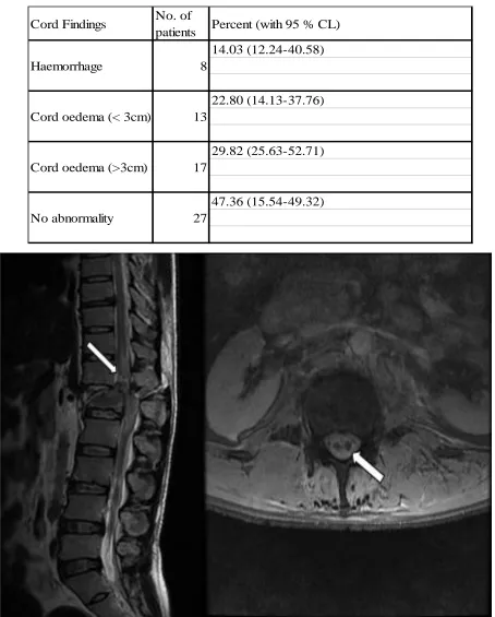
In this study 57 cases with spinal trauma were observed. Of these 48 (84.21 %) were males and 9 (15.78%) were females. Most of the patients of spinal trauma in our study were in the age group of 21-40 years (43.8%) followed by 41-60 years (38.5%). Patient below 20 years and above 60 years constituted of only 5 % each. MRI examination revealed the cord abnormalities in 38 out of 57 patients, i.e. in 66.66% of patients. Rest 33.34% of patients had no cord findings. Cord oedema more than 3 cm was there in 17 out of 57 patients (29.82%) with (95 % CL, 12.24-40.58). Cord oedema less than 3 cm was noted in 13 out of 57 patients (22.80%) with (95 % CL, 14.1-37.7). 8 of 57 patients (14.03%) showed haemorrhagic focus within the cord and (95 % CL, 12.24-40.58).
Epidural hematoma was seen in 3 patients only i.e. 5.2 %. Marrow edema was picked up on STIR sequence, 40 out of 57 patients (70.17 %) had marrow edema. 6 patients had disc herniation (10.52 %). We came across 9 patients with ligament injuries (15.78 %). Out of these 22.2 % patients suffered injury to anterior longitudinal ligament, 33.3 % had injured ligamentum flavum and 44.4 % came with posterior longitudinal ligament injury.
DISCUSSION
This study was carried out with a sample size of 57 patients who presented with spinal trauma and underwent an MRI examination. MRI serves as an essential tool to determine the site and nature of spinal cord injury with ability to pick up entities like cord edema, haemorrhage and marrow edema in addition to disc herniation, extra medullary hematoma, vertebral and ligamentous injuries.
Pag
e
130
Pag
e
130
Pag
e
130
Pag
e
130
Pag
e
130
Pag
e
130
Pag
e
130
Pag
e
130
Pag
e
130
Pag
e
130
Pag
e
130
Pag
e
130
Pag
e
130
Pag
e
130
Pag
e
130
Pag
e
130
Pag
e
130
Pag
e
130
Pag
e
130
Pag
e
130
Pag
e
130
in these cases cannot be denied. Importance of an MRI scan in decision making by assessing the degree of soft tissue involvement(11) has been stressed upon in various studies.
S. Gaudino et al.(19) Emphasized on the dependence on an MRI scan in decision making and assessing the severity of the injury and concluded that however, MDCT has wide indication in bony evaluation in traumatic cases, MRI has utmost importance in picking up cord injuries without bony lesions. Similarly, D. Hayati et al.(20) Conducted a study depicting various findings in traumatic spine and came to concordance that MRI is more sensitive as compared to any other modality in diagnosing cord injuries. Whereas, CT is limited to limited to diagnosing bony trauma and stability of the spine. G. Gamal et al.(21) Compared evaluation of patients with spinal trauma on CT and MRI, came to a conclusion that MRI is superior to MDCT in diagnosing bone marrow edema, ligamentous complex injuries, cord findings including edema and hematoma.
Out of 57 patients, 48 were males and 9 were females. The age group commonly affected were those in the age group of 21 – 40 years (43.8 %). This was in concordance with G. Gamal et al.(21) Which showed that the spinal trauma was more common in men (79.6 %) at age group of 21-30 years.
Further, we assessed the abnormal cord findings in these 57 patients. Out of these patients 38 patients i.e. 66.6 % had spinal cord abnormalities after trauma. S. Bhadury et al.(22) Came across similar results which showed 75.8 % patients with abnormal cord findings. In a previous study conducted by Kulkarni et al.(23) There was presence of cord abnormalities in 70 % of spinal injuries.
Mechanism of the trauma is important to determine the type of ligamentous injury. Hyperextension injuries to the spine generally cause injuries to the anterior column or both anterior and posterior columns. This leads to involvement of ALL and PLL(24). Similarly, injuries causing hyper flexion of
the spine can also result in posterior column or combined posterior and middle columns injuries further causing damage to facet joint capsules,
ligamentum flavum, supraspinous ligaments,
interspinous ligaments and PLL(25). Ligaments being essential components of spinal columns, their injury can transform a single column injury to a two column injury. Hence, turning a stable injury into an unstable one(26).
Disc herniation causing compression of the spinal cord could lead to central cord syndrome in few patients(27). MRI is an excellent modality to assess a traumatic disc herniation due to a brilliant contrast between the disc, CSF and vertebra. It is essential to evaluate any disc sequestration, herniation or extrusions before closed reduction in cases of spinal dislocation(28). If a disc herniation is left undetected, it might lead to a worse outcome with progressive neurological deficits at the time of follow up. At times there might be just an abnormal signal intensity with asymmetry of the disc which characterize edema. It might signify the rupture of annulus fibrosus with hematoma formation(29).
Extramedullary hematomas appears hyperintense on T1W and T2W images. It is better appreciated on GRE sequence. Similarly subarachnoid haemorrhage and subdural hematoma will show varying signal intensities(30,31).
CONCLUSION
MRI is far more superior to CT in diagnosing and categorizing the various cord and soft tissue injuries. Cord edema was the most significant abnormality seen in our study followed by cord haemorrhage. A sizeable focus of cord haemorrhage has been proven
to cause poor prognosis in various studies. In the era
e
131
e
131
e131
e
131
e131
e
131
e131
e
131
e131
e
131
e
131
e
131
e
131
e131
e
131
e131
e
131
e131
e
131
e131
e
131
TABLES AND FIGURES
Table 1. Various cord findings seen in our study
Cord Findings No. of
patients Percent (with 95 % CL)
14.03 (12.24-40.58)
22.80 (14.13-37.76)
29.82 (25.63-52.71)
47.36 (15.54-49.32)
No abnormality 27
Haemorrhage 8
Cord oedema (< 3cm) 13
Cord oedema (>3cm) 17
Pag
e
132
Pag
e
132
Pag
e
132
Pag
e
132
Pag
e
132
Pag
e
132
Pag
e
132
Pag
e
132
Pag
e
132
Pag
e
132
Pag
e
132
Pag
e
132
Pag
e
132
Pag
e
132
Pag
e
132
Pag
e
132
Pag
e
132
Pag
e
132
Pag
e
132
Pag
e
132
Pag
e
132
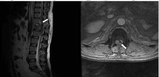
Figure 2: A 25 year old male with history of fall presented with inability to move bilateral lower limbs. T2 sagittal (a) image shows anterior wedge compression of T12 vertebra with spinal canal narrowing and hyper intensity in the cord from T10 to L1 level, suggestive of cord oedema with injury to ALL and PLL. Axial GRE (b) image shows a fluid level in the posterior thecal sac suggestive of epidural haemorrhage
e
133
e
133
e133
e
133
e133
e
133
e133
e
133
e133
e
133
e
133
e
133
e
133
e133
e
133
e133
e
133
e133
e
133
e133
e
133
BIBLIOGRAPHY
1. Chabok SY, Safaee M, Alizadeh A, Dafchahi
MA, Taghinnejadi O, Koochakinejad L. Epidemiology of traumatic spinal injury: A
descriptive study. Acta Med Iran.
2010;48(5):308–11.
2. Parashari UC, Khanduri S, Bhadury S, Kohli
N, Parihar A, Singh R, et al. Diagnostic and prognostic role of MRI in spinal trauma, its comparison and correlation with clinical profile and neurological outcome, according to ASIA impairment scale. J craniovertebral junction spine. India; 2011 Jan;2(1):17–26.
3. Daffner RH, Hackney DB. ACR
Appropriateness Criteria on suspected spine trauma. J Am Coll Radiol. United States; 2007 Nov;4(11):762–75.
4. Hoffman JR, Mower WR, Wolfson AB, Todd
KH, Zucker MI. Validity of a set of clinical criteria to rule out injury to the cervical spine in patients with blunt trauma. National Emergency X-Radiography Utilization Study Group. N Engl J Med. United States; 2000 Jul;343(2):94–9.
5. Katzberg RW, Benedetti PF, Drake CM,
Ivanovic M, Levine RA, Beatty CS, et al. Acute cervical spine injuries: prospective MR imaging assessment at a level 1 trauma center.
Radiology. United States; 1999
Oct;213(1):203–12.
6. Vaccaro AR, Lehman RAJ, Hurlbert RJ,
Anderson PA, Harris M, Hedlund R, et al. A new classification of thoracolumbar injuries: the importance of injury morphology, the
integrity of the posterior ligamentous
complex, and neurologic status. Spine (Phila
Pa 1976). United States; 2005
Oct;30(20):2325–33.
7. Selden NR, Quint DJ, Patel N, d’Arcy HS,
Papadopoulos SM. Emergency magnetic resonance imaging of cervical spinal cord injuries: clinical correlation and prognosis.
Neurosurgery. United States; 1999
Apr;44(4):783–5.
8. Benedetti PF, Fahr LM, Kuhns LR, Hayman
LA. MR imaging findings in spinal
ligamentous injury. Am J Roentgenol. 2000;175(3):661–5.
9. Hogan GJ, Mirvis SE, Shanmuganathan K,
Scalea TM. Exclusion of unstable cervical spine injury in obtunded patients with blunt trauma: is MR imaging needed when multi-detector row CT findings are normal?
Radiology. United States; 2005
Oct;237(1):106–13.
10.Benzel EC, Hart BL, Ball PA, Baldwin NG,
Orrison WW, Espinosa MC. Magnetic resonance imaging for the evaluation of patients with occult cervical spine injury. J
Neurosurg. United States; 1996
Nov;85(5):824–9.
11.Saifuddin A. MRI of acute spinal trauma.
Skeletal Radiol. Germany; 2001
May;30(5):237–46.
12.Kliewer MA, Gray L, Paver J, Richardson
WD, Vogler JB, McElhaney JH, et al. Acute spinal ligament disruption: MR imaging with anatomic correlation. J Magn Reson Imaging. United States; 1993;3(6):855–61.
13.Kerslake RW, Jaspan T, Worthington BS.
Magnetic resonance imaging of spinal trauma. Br J Radiol [Internet]. The British Institute of Radiology; 1991 May 1;64(761):386–402. Available from: https://doi.org/10.1259/0007-1285-64-761-386
14.Flanders AE, Schaefer DM, Doan HT,
Mishkin MM, Gonzalez CF, Northrup BE. Acute cervical spine trauma: correlation of MR imaging findings with degree of neurologic deficit. Radiology. United States; 1990 Oct;177(1):25–33.
15.Andreoli C, Colaiacomo MC, Rojas Beccaglia
M, Di Biasi C, Casciani E, Gualdi G. MRI in the acute phase of spinal cord traumatic lesions: Relationship between MRI findings and neurological outcome. Radiol Med. Italy; 2005;110(5–6):636–45.
16.Schaefer DM, Flanders A, Northrup BE,
Doan HT, Osterholm JL. Magnetic resonance imaging of acute cervical spine trauma. Correlation with severity of neurologic injury. Spine (Phila Pa 1976). United States; 1989 Oct;14(10):1090–5.
17.Hackney DB, Asato R, Joseph PM, Carvlin
Pag
e
134
Pag
e
134
Pag
e
134
Pag
e
134
Pag
e
134
Pag
e
134
Pag
e
134
Pag
e
134
Pag
e
134
Pag
e
134
Pag
e
134
Pag
e
134
Pag
e
134
Pag
e
134
Pag
e
134
Pag
e
134
Pag
e
134
Pag
e
134
Pag
e
134
Pag
e
134
Pag
e
134
compression: demonstration by MR imaging.
Radiology. United States; 1986
Nov;161(2):387–90.
18.Rajasekaran S, Vaccaro AR, Kanna RM,
Schroeder GD, Oner FC, Vialle L, et al. The value of CT and MRI in the classification and surgical decision-making among spine surgeons in thoracolumbar spinal injuries. Eur spine J Off Publ Eur Spine Soc Eur Spinal Deform Soc Eur Sect Cerv Spine Res Soc. Germany; 2017 May;26(5):1463–9.
19.Guarnieri G, Izzo R, Muto M. The role of
emergency radiology in spinal trauma. Br J Radiol [Internet]. 2016/03/04. The British
Institute of Radiology.; 2016
May;89(1061):20150833. Available from: https://www.ncbi.nlm.nih.gov/pubmed/26612 468
20.Kumar Y, Hayashi D. Role of magnetic
resonance imaging in acute spinal trauma: a pictorial review. BMC Musculoskelet Disord
[Internet]. BioMed Central; 2016 Jul
22;17:310. Available from:
https://www.ncbi.nlm.nih.gov/pubmed/27448 661
21.Gamal GH. Evaluation of spinal trauma by
multi detector computed tomography and magnetic resonance imaging. Egypt J Radiol
Nucl Med [Internet]. Elsevier B.V.;
2014;45(4):1209–14. Available from:
http://dx.doi.org/10.1016/j.ejrnm.2014.08.002
22.Parashari UC, Khanduri S, Bhadury S, Kohli
N, Parihar A, Singh R, et al. Diagnostic and prognostic role of MRI in spinal trauma, its comparison and correlation with clinical profile and neurological outcome, according to ASIA impairment scale. J craniovertebral
junction spine [Internet]. Medknow
Publications; 2011;2(1):17–26. Available
from:
https://www.ncbi.nlm.nih.gov/pubmed/22013 371
23.Kulkarni M V, McArdle CB, Kopanicky D,
Miner M, Cotler HB, Lee KF, et al. Acute
spinal cord injury: MR imaging at 1.5 T.
Radiology. United States; 1987
Sep;164(3):837–43.
24.Davis SJ, Teresi LM, Bradley WGJ, Ziemba
MA, Bloze AE. Cervical spine
hyperextension injuries: MR findings.
Radiology. United States; 1991
Jul;180(1):245–51.
25.Benedetti PF, Fahr LM, Kuhns LR, Hayman
LA. MR imaging findings in spinal
ligamentous injury. AJR Am J Roentgenol. United States; 2000 Sep;175(3):661–5.
26.Bagley LJ. Imaging of spinal trauma. Radiol
Clin North Am. United States; 2006 Jan;44(1):1–12, vii.
27.Pratt ES, Green DA, Spengler DM. Herniated
intervertebral discs associated with unstable spinal injuries. Spine (Phila Pa 1976). United States; 1990 Jul;15(7):662–6.
28.Vaccaro AR, Falatyn SP, Flanders AE,
Balderston RA, Northrup BE, Cotler JM.
Magnetic resonance evaluation of the
intervertebral disc, spinal ligaments, and spinal cord before and after closed traction reduction of cervical spine dislocations. Spine (Phila Pa 1976). United States; 1999 Jun;24(12):1210–7.
29.Sitte I, Klosterhuber M, Lindtner RA, Freund
MC, Neururer SB, Pfaller K, et al. Morphological changes in the human cervical intervertebral disc post trauma: response to fracture-type and degeneration grade over time. Eur spine J Off Publ Eur Spine Soc Eur Spinal Deform Soc Eur Sect Cerv Spine Res Soc. Germany; 2016 Jan;25(1):80–95.
30.Pan G, Kulkarni M, MacDougall DJ, Miner
ME. Traumatic epidural hematoma of the cervical spine: diagnosis with magnetic resonance imaging. Case report. J Neurosurg. United States; 1988 May;68(5):798–801.
31.Sklar EM, Post JM, Falcone S. MRI of acute