Published by Oriental Scientific Publishing Company © 2019
This is an Open Access article licensed under a Creative Commons license: Attribution 4.0 International (CC-BY).
XSITRAY: A Database for the Detection of
Osteoporosis Condition
S.M. NaziaFathima#1, R. Tamilselvi*2 andM. Parisa Beham3*
#Department of CSE, *Department of ECE,
#*Sethu Institute of Technology, Tamilnadu-626 115, India.
*Corresponding author E-mail: [email protected]
http://dx.doi.org/10.13005/bpj/1637
(Received: 29 November 2018; accepted: 09 March 2019)
In the medical era, health of a bone is accessed by the bone mineral density (BMD) test. Bone fracture risk in the humans are estimated or evaluated by the BMD test. The test statement recognizes the presence of signs of presence of the frequent occurring disease in the bone called as osteoporosis. In the earlier stage, the challenge in the BMD measurement is that traditional x-rays are used with a step wedge made from an aluminum or ivory phantom. At each step of the phantom with the known densities, bone content present is intended by a illustration assessment of the density present in the bone. Effectiveness in the value and feasibility in the X-rays compared to cutting-edge methods divulge the potential for novel medical relevance among the investigators. So it is obligatory to enclose a customary database in X-Ray images for the young bud researchers to capture up the dealings to the advance stage by accurate examination of the medical results of the images. The projected X-Ray database is termed XSITRAY, characterizes an early attempt to offer a group of X-Ray images of Spine, Femur, Clavicle, Extremity & Ankle, Extremity & Hand and Knee bones. The details such as age, gender and unique Id of the patient are interpreted in the database.
Keywords: X-Ray database, XSITRAY, BMD, Femur bone , Knee and Ankle.
Biomedical engineering is the division which chains medical and biological sciences with engineering values to plan and generate different equipment’s, devices and algorithms used in healthcare. To solve various medical problems, it fetches together information from various sources of medicine and engineering to carry out the research needed now. Biomedical engineering
field in today’s role, is linking the breach between engineering and medical field. In present scenario,
the investigators are merging the design and problem solving capability of engineering with biological sciences to carry out treatments in
medical field such as diagnosis, intensive care,
avoidance of diseases and rehabilitation.
In Later stage of X-ray, expansion in the field of quantity of bone density was the recognition of single-photon absorptiometry (SPA) by Cameron and Sorenson in the year 1963. In terms of amount of BMD, SPA showed a superior place, but limited its use in the site of the measurement. In the recent
development, struggling in the Dual energy is
betrothed in Dual-photon absorptiometry (DPA),
Algebraic derivations. In late 1980s, grander and luxurious radioactive sources have been outdated by the use of single x-ray absorptiometry (SXA) and Dual Energy X-ray absorptiometry (DEXA). Compared to other predictable scanning, the success rate of SXA and DXA is also very high
used for measurement of bone content2. The main
principle in the BMD measurement is to assist the physicians to perceive osteoporosis and envisage the danger of bone rupture. Thus osteoporosis
affects the various regions of the skeleton with
dissimilar cruelty. Most women are affected by the osteoporosis.
The key complexity of osteoporosis
is rupture happening after tiniest trauma. Hip
fractures are linked with enlarged short term
mortality and high morbidity. The major regions such as Hip, vertebral, and radius fractures escalate
the risk of upcoming break in various bones3. Thus it is an obligatory step for investigators in biomedical domain, to yield suitable preclusion
methods or efficient treatment processes for the patients. A bone mineral density (BMD) test processes the prediction of calcium and other kinds
of minerals are in the part of the bone4. Based on
above said literature, in this proposed work we developed a database of X-Ray images which we baptized it as ‘XSITRAY Database’, for the profit
of biomedical engineering research people. Even though a lot of medical databases are available
for various imaging modalities, the significant
pitfall in the bone research and development is that, unapproachability of suitable bone medical databases. Even though some survey has discussed the issues of various scan methods, they didn’t offer any such databases publically accessible for the researchers.
Thus interested by the above said factors, our main offerings are:
• Generate a novel XSITRAY database, which comprises of 78 Spine, Femur, Clavicle, Extremity & Ankle, Extremity & Hand and Knee bones X-Ray scan images.
• Interprets all the subject’s Gender, age and the
position.
XSITRAY
In the medical era, still now there is no exact database in bone images for further research
and development. Motivated by all these factors,
we created a new and exceptional database,
XSITRAY database. This database is mainly
focused for the BMD measurements.
X-ray bone images are retrieved from a
research foundation centre. This dataset involves
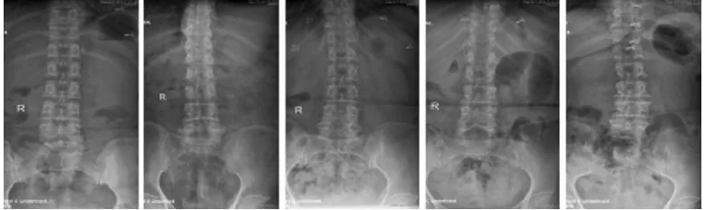
of 78 X-Ray scan images collected from various subjects. XSITRAY consist of 52 female and 26 male subjects. Each subject includes Spine, Femur, Clavicle, Extremity & Ankle, Extremity & Hand and Knee bones X-Ray scan images. The sample spine scan images of 5 subjects are shown in Figure.1. Similarly, the samples X-ray femur images of 5 subjects are shown in Figure.2. Table 1and Table 2 describes the details of the subjects. The sample X-Ray clavicle scan images of 5 subjects are shown in Figure.3. The XSITRAY
database is deliberated through subsequent stages:
1) Structure Details
2) Marking the XSITRAY images 3) Footnote
These are explained step by step.
Structure Details
The standard database is created from the
Indian X-ray images. In the 78 subjects, there are totally 9 Spine, 12 Femur, 28 Clavicle, 6 Extremity & Ankle, 12 Extremity & Hand and 11 Knee bones X-Ray scan images.
The images are harvested physically
and hoarded as discrete images in ‘png’ (portable network graphics) format. The detailed information about the X Ray scan images of all the subjects,
have also been delivered in the table format. The
entire dataset is grouped as six groups such as XSITRAY-SP, XSITRAY-FE, XSITRAY-CL, XSITRAY-EA, XSITRAY-EH and XSITRAY-KN. The XSITRAY-SP includes the X-Ray images of 9 spine bone images. Similarly XSITRAY-FE, XSITRAY-CL, XSITRAY-EA, XSITRAY-EH and XSITRAY-KN consists of 12 Femur, 28 Clavicle, 6 Extremity & Ankle, 12 Extremity & Hand and 11 Knee bone images. The labeling of the database,
encourage the investigators to understand and scrutinize the scores of the spine, femur, clavicle,
Extremity &Ankle, Extremity & Hand and Knee
bones separately.
Marking the XSITRAY Images
The proposed databases are labeled
Table1. Interpretation of Spine X-Ray images
Subject Id Gender Age Image
XRAY_SP_001 F 37 Spine XRAY _SP_002 F 70 Spine XRAY_SP_003 F 58 Spine XRAY_SP_004 F 65 Spine XRAY_SP_005 F 69 Spine
Table 2. Interpretation of femur X-Ray images
Subject Id Gender Age Image
XRAY _FE_001 F 37 Femur XRAY _FE_003 F 70 Femur XRAY _FE_006 F 58 Femur XRAY _FE_009 F 65 Femur XRAY _FE_010 F 69 Femur
Fig. 1. X-Ray scan images of spine of 5 selected subjects from the XSITRAY
Fig. 2. Sample X-Ray Femur images of 5 subjects
as: XRAY_SP_001.png. Here, in the initial, X-Ray refers to X-Ray scan image, SP interprets to the spine image of subject and 001 is the ID of the subject. Likewise XRAY_FE_002.png mentions
to X-Ray femur image of a subject with a subject ID of 002. Likewise XRAY_CL_002.png denotes to X-Ray clavicle image of a subject with a subject ID of 002, XRAY_EA_002.png states to X-Ray Extremity & Ankle image of a subject with a subject ID of 002, XRAY_EH_002.png denotes to X-Ray Extremity & Hand image of a subject with a subject ID of 002 and XRAY_KN_002 raises to X-Ray knee image of a subject with a subject ID of 002.
Footnote
XSITRAY affords a complete labelling through a careful investigation of all X-Ray scan images. All the images are annotated manually with
the following labels for each bone image.
• Specific ID • Gender • Age and • Type of Image
• View(Anterior/Posterior)
The database creation is through the motivation by the lot of problems associated to
Table 3. Interpretation of Clavicle X- Ray images
Subject Id Gender Age Image
XRAY_CL_001 F 29 Clavicle XRAY _CL_002 F 74 Clavicle XRAY _CL_003 F 60 Clavicle XRAY _CL_004 F 84 Clavicle XRAY _CL_005 F 52 Clavicle
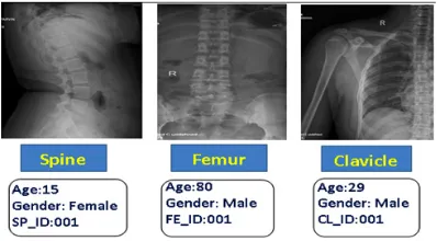
Fig. 3. Labelling of spine, femur and clavicle bone
images
Fig. 4. Sample images from XSITRAY database: From top to bottom (a) hand (b) Hand and extremity (c) Knee and extremity (d) Clavicle and (e) Ankle and extremity
values for the spine, Ankle, clavicle, femur and Knee bone images are presented which might be
convenient in the development of bone research.
Fig. 3 displays the sample labelling of a subject
from the dataset.
Clinical Data Analysis
Osteoporosis, a disease usually connected with humans, is categorized by diminish in mass of the bone and micro architectural integrity5. One critical problem in the growth of osteoporosis
is the achievement of apposite peak mass in
childhood and later stages6. A disappointment in
the attainment of youth peak bone mass may be
related with premature osteoporosis and augmented
fracture risk7. World Health Organization (WHO)
defines T-Score values for human beings in BMD plot as -1 SD for normal, -1 and -2.5for Osteopenia, below -2.5 SD for Osteoporosis
Table 1, Table 2 and Table 3 show the
medical report of the same persons.
Based on the BMD levels, T-score and Z-score, mild to destructive therapies are needed
in the form of Hormone replacement therapy
(HRT), Bisphosphonates, Calcitonin and SERMs
as suggested by the orthopedician. Moreover, all
dietary calcium (1200 mg/d) and vitamin D (400-800 IU daily). By exact study of the X-Ray bone
images and their reports, people can be prevented from the osteoporosis disease.
Based on the database medical reports
provided by the physician/experts, the biomedical
investigators can validate their accuracy of biomedical algorithms.
ConCluSIon
The projected paper presented a medical
image datasets called XSITRAY, a group of X-ray scan images for healthcare and orthopedic research. It is established with the meaning of
supplementing a standard for bone research and related development. The foremost features of this
XSITRAY database are:
a) spine X Ray images, femur, clavicle, Extremity and Ankle, Extremity and Hand and knee, X Ray images each. b) Labeling
the subject’s biological data. By creating and
making this database available to the research in
BME community, we optimizes to promote the investigation of many indeterminate problems.
XSITRAY database along with all medical
measures will be made accessible for investigation
purposes. The XSITRAY database can be observed
and downloaded at the institutional web address:
http://www.sethu.ac.in/ XSITRAY/.
ReFeRenCeS
1. Bonnick,S.L, “Bone Densitometry for
Technologists” Thesis Report: Springer, pp1-64, 2006.
2. S.M.Nazia Fathima, R.tamilselvi and M.Parisa Beham, “Role of Dual-Energy X-ray Absorptiometry in Assessment of Bone Mineral Density – A Review” Proceedings of International Conference on Informatics Computing in Engineering Systems ICICES,
(2018).
3. Rosa Lorente-Ramos Javier Azpeitia-Armán Araceli Muñoz-Hernández José Manuel arcía- Gómez Patricia Díez-Martínez Miguel rande- Bárez Dual-Energy X-Ray absorptiometry in the
Diagnosis of Osteoporosis: A Practical Guide, AJR ; 196: 897–904 0361–803X/11/1964–897, 2011.
4. M. K. Garg and Sandeep Kharb ,”Dual energy X-ray absorptiometry: Pitfalls in measurement
and interpretation of bone mineral density” Indian Journal of Endrocrinology and metabolism,
(2013).
5. Roth J, Palm C, Scheunemann I, Ranke MB, Schweizer R, Dannecker GE. “Musculoskeletal
abnormalities of the forearm in patients with juvenile idiopathic arthritis relate mainly to bone geometry”, Arthritis Rheum, 50: 12, pp.77–85 (2004).
6. Rabinovich CE. “Bone mineral status in juvenile
rheumatoid arthritis”, J Rheumatol Suppl, 58:
34–7 (2000).
7. S C Lacassagne, P N. Tyrrell, E Atenafu, A S. Doria, D Stephens, D Gilday, and E D. Silverman “Prevalence and Etiology of Low Bone Mineral Density in Juvenile Systemic Lupus Erythematosus”, Arthritis & Rheumatism,
56(6): pp 1966–1973 (2007).
8. R.M. Lorente Ramos, J. Azpeitia Armán, N. Arévalo Galeano, A. Mu˜noz Hernández, J.M. García Gómez, J. Gredilla Molinero “Dual energy X-ray absorptimetry: Fundamentals,