SHORT COMMUNICATION
Effects of two anthelmintics on gastrointestinal infestation by parasitic worms in
camels
Seddik Mabrouk Mouldi*, Ben Taleb Olfa, Zemezmi Najet, Salhi Imed and Khorchani Touhami
Laboratoire d’Elevage et de la Faune Sauvage, Arid LandsInstitute 4119 Medenine, University of Gabes, Tunisia
Abstract
The aim of this work was to follow the kinetics of parasitic eggs elimination after two anthelmintic treatments in dromedary camels. This study was performed on three groups of four female camels bred in semi-extensive system. The first group was the untreated control, while the second one received sub-cutaneous injection of Ivermectine (Ivermectyl 1% ® medivet) and the third received Albendazole (Dalben 1.9 ® ceva) by oral route. Microscopic examinations of 180 fecal samples showed predominance of Trichostrongylus (94%), then Trichuris (9%), Nematodirus (4%) and finally fasciola (0.5%). The statistical results of the fecal egg count showed a slow decrease of eggs elimination in the group treated by injection reaching the mean reducing rate of 55% at the end of the experimental period, whereas in the group drenched with Albendazole, the parasitic eggs elimination decreased rapidly with a mean reducing rate exceeding 95% beyond the third day of experimental period. It can be concluded that oral treatment by Albendazole has a very potent effect for deworming camels and reducing the fecal elimination of helminth eggs on pastures and consequently for improving camel productivity.
Key words: Camel, Digestive worms, Antihementic Treatment
Introduction
The dromedary (Camelus dromedarius)
occupies a central place in arid and desert regions thanks to its special feeding behaviour and its exceptional digestive physiology. In general, the feeding behaviour of camels on pastures is different from other animal species. Indeed, thanks to the liberty and the importance of grazed area which are available to him, the animal has the possibility to take some fragments of vegetation and can move on great distances. Despite its arid and dry environment, the dromedary food can accommodate some parasites that affect essentially the digestive tract. Helminthosis constitutes one part of camel pathology that affects productivity in quantitatively and qualitatively as it was stated for long time (Richard, 1989; Graber, 1967). The effect of the gastrointestinal helminths is characterized mainly by massive infestation that reduces the absorption of nutrients and increases tissue damage
of the digestive tract and represents one of the
major causes of camels’ mortality (Faye, 1997; Rewatkar, 2009). Therefore, it is necessary to choose the most effective treatment against the gastro-intestinal parasites. This work aims to study the effect of the two most used anthelmintics for deworming dromedary camels in southern Tunisia.
Material and Methods Animals
This work was carried out in a camel herd raised at the Institute of Arid Regions Medenine (IRA) during late winter to early spring 2013. A total number of 12 female camels were randomly divided into three groups of four she-camels with a means age of 13 years (range from 8 to 17 years) for the first group; 12 years (range from 8 to 15 years) for the second group and 14 years (range from 9 to 21 years) for the third group. The average of the body weight of the tree groups was about 350 kg. All the animals were raised freely with the rest of the herd in a semi extensive system. This herd spent 7 to 8 hours per day on the rangeland characterized by the presence of halophyte plants consisting of Artistida pungens, Astragalus
armatus, Tamarix galleca, Limomiastrum
Received 29 March 2014; Revised 13 July 2014; Accepted 10 December 2014; Published Online 01 April 2015
with hay and a mixture of concentrate and olive cake.
Treatments
The animals of the experiment were not treated against gastrointestinal parasites during the six previous months. To assess the effect of treatment by injection or by drenchers, it was proceeding as follows:
The first group was used as untreated control, the two other groups receiving (1) at day 0 one subcutaneous injection 0.2mg/kg of ivermectine (Ivermectyl 1% ® medivet) for group 2, and (2) drenched with Albendazole (Dalben 1.9 ® ceva) for group 3.
Specimen collection
Fecal egg counts were estimated at day 0 (just before treatment), then three times per week for one month. Immediately after the collection of 100 to 150 g of fecal material directly from the rectum, analysis was based on the flotation method. Fecal egg counts were estimated by dissolving 20g of faeces in 100ml saturated sodium chloride solution (d = 1.19). Eggs count and examination was performed under microscope at x40 and x100 magnification.
Statistical analysis
The statistical data were analysed by ANOVA test at the significance level of 0.05 with SPSS 17.0 package.
Results and Discussion Parasites identified
A total number of four genera were identified in this study, namely: Trichostrongylus,
Nematodirus, Trichuris and Fasciola.
Trichostrongylus genus was the most frequent: it
was identified in 170 collected samples (94%). The three other genera were rare and were identified in 17 (9%), 7 (4%) and 1 (0.5%) collected samples respectively. The high prevalence of
Trichostrongylus genus could be explained by
environmental conditions and the semi extensive breeding system which is favourable for the parasitic life cycle. Since nematode species
couldn’t be identified only by microscopic fecal
examination (Lichtenfels et al 1997), the most frequent Trichostrongylus species reported in camel was T. colubriformis, T. probolurus and T. vitrinus (Dakkak et al., 1987; Singh et al., 1993; Borji et al., 2010). T. colubriformis colonizes essentially the
duodenum but also the abomasum. The other two species are encountered only in the small intestines. In addition, haemonchosis due to Haemonchus
longistipes and H. contortus can occur throughout
the year but with a high prevalence during the rainy season. Disorders are however observed when the degree of infestation is high (more than 150 worms according to Graber et al. (1967). Clinical manifestations of gastrointestinal helminthic infections in camels have been described in numerous studies (Graber et al., 1967; Blaizot, 1975; Richard, 1986; Richard et al., 1989; El Bihari, 1985). Two forms of clinical chnages were reported; the low and the massive infestation. The lower infestations were manifested by signs of decreasing productivity: growth retardation, defect of fattening and decreasing milk production. In case of massive infestation, other signs can be observed: decreased and capricious appetite, progressive emaciation, especially with melting hump, alternation of constipation and diarrhoea, abdominal pain. The movement becomes difficult and the animal cannot feed adequately. The death may occur within a few weeks or months. This parasitism is often detected post-mortem (Faye, 1997).
Kinetics of infestation
1-State of infestation before the beginning of the experiment
By microscopic analysis of samples taken before the treatment (day 0), it was noticed the presence of a large number of parasitic eggs in all camels with a mean fecal egg count of 336 (±210), 405 (±179) and 340 (±218) per sample for the first, second and third group respectively.
2-Kinetic of infestation in each group a- Control Group (group 1)
Figure 1. Change in the number of helminth eggs during follow-up in the untreated group.
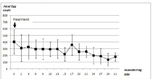
Figure 2. Helminth eggs elimination in the group treated with Ivermectin injection.
In group 2, the oldest she-camel was the most infested with a number of eggs detected by sample close to 600. On the other hand, the youngest she-camel was the least infested with a number of eggs at the beginning of the follow-up about 200 eggs per sample. Examination of the average number of eggs eliminated by this group throughout the follow-up period showed a slow decrease of excreted eggs (Figure 2). Indeed, the average number of eggs detected before treatment was 405 (± 179) eggs per sample which decreased after the treatment in a gradual way to reach at the end of the monitoring period an average of 180 (± 61) eggs per sample. By multiple comparisons, it appeared that only the number of eggs at the final state of monitoring was significantly different than initial state.
c- Group treated by Drenchers (group 3) In this group treated by oral route, the number of parasite eggs was very high 340 (± 218) for the first three days. From the third day until the end of the follow-up period, a rapid decline in the eliminated eggs number was observed. Elsewhere, all the examined samples had less than 25 eggs per sample. It was noticed also the absence of eggs in nine samples. Thus, the third day after treatment, a spectacular decrease in the number of eggs was observed and removed at very low levels (Figure 3). The low egg elimination rate was persistent throughout the rest of follow-up period.
Figure 3. Helminth eggs elimination in camels treated by oral administration of Albendazole.
Figure 4. Variation of the average helminth eggs elimination over time in all three groups.
Just before treatment performed on day 0, the three groups were highly infected and there was no significant difference between them. The average eggs elimination by animals was about 360 (± 186) eggs per sample. This state of infestation was favourable for our experimental protocol. However, except the first examination, done at 24 hours after treatment and which showed no significant difference between the three groups, all other observations done since the third day until the end of the trial period showed significant changes. Thus, the statistical comparison between the first group and the second group (Figure 4) showed no significant variation of the parasitic eggs elimination throughout the monitoring period, in spite of a gradual decrease in helminth eggs counts in the group treated with ivermectin injection. In addition, the comparison between the first and the third groups showed that there was a highly significant difference for the entire period after the third day of monitoring. All the examined samples for the group treated orally by albendazole have lower values, less than 50 eggs per sample, in most
marked from the third day of the monitoring by rapid decrease in the eggs elimination in the third group with an average reduction rate of 95% of eggs detected, while in the second group treated with ivermectin the decrease is lower with an average reduction rate of 18% in the same day. At the end of the monitoring period the rate reached 99% in group 3 vs 55% reduction rate of eggs elimination in group 2.
anthelmintic efficacy studies (Mukhwana, 1997) showed that albendazole (10mg/kg per os) and thiophanate (60mg/kg per os) were more effective in camels. However, Sharma (1991) reported that ivermectin (0.2 mg / kg subcutaneously) had a fast and maximum efficiency of 100% at 7th day after treatment which is not consistent with our results. Our present observations could imply the resistance of camel worms to ivermectin which was suggested in cattle by some authors (Gasbarre et al., 2009). The effect of the treatment with albendazole was fast, durable and easy to administer (non-invasive). However, this method requires a good containment of the animals, so there is the risk of losing part of the drug in case of insufficient immobilisation of
the animal’s head.On the other hand, the use of this method to control digestive parasites in animals reared in extensive system seems more convenient if we consider the difficulties for accessing to technical services and the plight of this farming system Thus, the camel breeders could choose every day the number of animals for whom the drug the drug is administered orally by respecting the required dose.
Conclusion
Camel is highly infested by gastro-intestinal parasitic worms with predominance of
Trichostrongylus species. The evaluation of the
effect of ivermectin (by injection) and albendazole (orally) treatments by coprological survey of two respectively treated groups compared to a none-treated one, showed the advantage of the oral treatment for the decrease of eggs elimination. The effect of Albendazole on reducing the elimination of helminth eggs in the digestive tract of camels was very efficient since it leads to an average reduction rate of eggs over 95% during all the period which follows the third day of treatment. Our study was performed on female adult camels bred in semi extensive system and during relatively dry year. Such experiment could be performed in other breeding conditions such as intensive or extensive system and during a rainy year in order to confirm the present observations
Acknowledgements
This study has been produced with the financial assistance of the European Union:
ENPI-CBC-MED “PROCAENPI-CBC-MED” Project: Promotion des
systèmes camelins innovants et des filières locales pour une gestion durable des territoires saharienne;
Author contributions
S. M. M. managed the study and wrote the paper, B. T. A., Z. N. and S. I. contributed to the experiment and to the data collection; T. K. supervised the study.
References
Armour, J. and J. Bogan. 1982. Anthelmintics for ruminants. British Vet. J. 138(5):371-382.
Blaizot, C. 1975. Etude des parasitoses gastro-intestinales du dromadaire dans la région de Dire Dowa (sud-est éthiopier), est essais therapeutiques. Doc Vet N°9 Alfort 62p.
Borji, H., G. H. Razmi, A. R. Movassaghi, A. G. Naghibi and M. Maleki. 2010. A study on gastrointestinal helminths of camels in Mashhad abattoir. Iran. Iranian J. Vet. Res. 11(2):174-179.
Dakkak, A. and H. Ouhelli. 1987. Helminthoses du dromadaire. Revue bibliographique. Rev. Sci. Off. Int. Epiz. 423-445.
El bihari, S. 1985. Helminthes of the camel: A rev. Br. Vet. 141(3):315-326.
Faye, B. 1997. Guide d’élevage du dromadaire.
Edition CIRAD-EMVT. Montpellier France. p. 126.
Gasbarre, L. C., L. L. Smith, J. R. Lichtenfels and P. A. Pilitt. 2009. The identification of cattle nematode parasites resistant to multiple classes of anthelmintics in a commercial cattle population in the US. Vet. Parasitol. 166(3-4):281-285.
Graber, M., R. Tabo and J. Service. 1967. Enquête sur les helminthes du dromadaire tchadien. Etudes des strongyloses gastro-intestinales et
de l’haemonchose à Haemonchus longistipes.
Rev. Elev. Méd. Vet. Pays Trop. 20(2):227-254.
Lichtenfels, J. R., E. P. Hoberg and S. Zarlengad. 1997. The Systematics of gastrointestinal nematodes domestic ruminants: advances between 1992 and 1995 proposals for future research. Vet. Parasitol.72:225-245.
Bhangale. 2009. Gastrointestinal helminths in migratory Camel. Vet World 2(7):258-258.
Richard, D. 1986. Manuel des maladies du dromadaire. IEMVT, Alfort.
Richard, D. 1989. L’haemonchose du dromadaire.
Revue Elev. Méd. Vét. Pays Trop. 42(1):45-53.
Sharma, I. K. 1991. Efficacy of some anthelmintics against gastrointestinal nematodes in camel (Camelus dromedarius). Indian Vet. J. 68(11):1069-1072.
Swai, E. S., W. Moshy, D. Mshanga, J. Lutatina and S. Bwanga. 2011. Intestinal parasitic infections of camels in the agro and pastoral areas of Northern Tanzania. Vet. Res. 4(2):34-38.
Singh, V., S. Kumar and C. L. Yadav. 1993. Establishment of camel isolates of
Haemonchus longistipes and Trichostrongylus colubriformis in goats. Vet. Parasitol.