Themed Section : Engineering and Technology © 2018 IJSRSET | Volume 5 | Issue 3 | Print ISSN: 2395-1990 | Online ISSN : 2394-4099
An Overview of Thermal Image Processing and Its Various
Applications
P.Kanimozhi1, Dr. S. Sathya2
1Assistant Professor, Aries Arts and Science College, Karunkuzhi, Tamil Nadu, India
2Assistant Professor, Department of Computer Science and Engineering, FEAT, Annamalai University, Chidambaram,
Tamil Nadu, India
ABSTRACT
Recent development in technology is focusing on modern and simple methods for diagnosing of fatal and incurable diseases using thermal image processing. Thermal Imaging is a type of imaging that determines an image based on the absolute temperature of the subject. The image is formed based on the heat signature of objects. Thermal imaging method is non-invasive and non-destructive method used even when the equipment is running, in production and on load. The image produces fast, accurate and immediate temperature measurement and helps in fault detection. The technique uses infrared radiations and includes most of the thermal radiation emitted by objects near room temperature. Using these images deadly diseases like tuberculosis, cancer, inflammatory diseases, complex pain syndromes can be identified. Diseases diagnosed early can be treated well and cured as quickly as possible.
Keywords: Thermal Images, Invasive, Inflammatory, Syndromes
I.
INTRODUCTIONThe expansion of software that could identify objects of interest sensed with thermal imaging video equipment, which the previous research specified with the use of infrared video cameras .Unlike traditional methods , video recording provides a real-time archive of what was observed and could be conducted at remote locations.[1] Thermal image processing concerns with infrared radiation. Infrared (IR) light is electromagnetic radiation with a wavelength longer than that of visible light, measured from the nominal edge of visible red light at 0.7 -300 micrometres. Thermal images use the heat emitted by any object captured by the infrared cameras which produces images usually grey scale in nature. Hot objects are indicated by white colour while cold objects with black. Different temperatures are identified by various colours which is seen later in this paper.
Figure 1. Ordinary Thermal Image
II.
DEFINITION FOR THERMAL IMAGEA thermal image (thermo gram) is a digital
representation of a scene and a measure of the
thermal radiation emitted by the pictured objects.
Thermal images are captured via thermo graphic
cameras, which are devices capable of sensing this
radiation in the form of infrared light. A thermal
image remotely senses the temperature of an object
or at least accurately tell its temperature relative to
its environment. The objects can be seen in the dark
as well as perceive the temperatures of many
objects remotely.
Thermal Imaging is the conversion of radiated or
reflected heat into real-time pictures or images. A
thermal image is an analogue visual representation
of temperature differences. All objects above
absolute zero (-273 degrees) emit radiation waves
that are infrared. Depending on temperature and
emissivity, most objects in the world can be
thermally imaged. Human beings emit the heat
pattern captured by the thermal IR sensors and
images are created
.
According to their temperature
and characteristics different objects emit different
range of Infra-red energy. The temperature of the
human body varies between 35.5°C to 37.5°C [2].
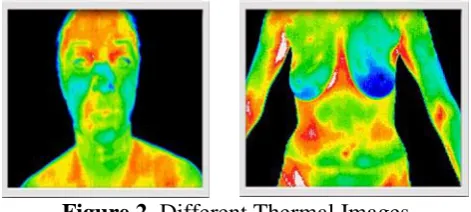
Figure 2.
Different Thermal Images
The
problems
related
with
difference
in
temperature can be easily detected using thermal
imaging. A variety of heat detection tasks uses
thermal imaging for checking any leakages , any
abnormal body temperatures when naked vision is
not possible. Thermal infrared images have
detectors and lens combination that provides a
visual representation of objects.
The most common tool used for thermal imaging
today is the thermal infrared camera. Thermal
infrared cameras can be used to detect and display
the presence of anything radiating heat above
absolute zero. They supply visual representation of
the infrared energy emitted by all objects.
The advantages of thermal imaging are
Non-invasive and non-destructive so used for any applications
Fast ,accurate, helps in fault detection
Easy installation of cameras at convenient times
Real time applications and low light conditions
Passive usage of cameras independent of light
III.
MECHANISM BEHIND THERMAL IMAGEAs thermal images allow to "see" in the dark, images are received clearly even in poor light conditions. The technique used is based on ‘blackbody radiation ’where most objects emit electromagnetic radiation as a function of its temperature. In room temperature this radiation is at infrared wavelengths. As temperature grows light emitted is in visible spectrum releasing energy in ultraviolet wavelengths. Infrared light consists of long wavelengths in the visible spectrum is absorbed by the objects soon increasing its kinetic energy thereby creating thermal image.
Thermal cameras are used to capture the images depicting thermal scenes by detecting IR radiation from an object, is a part of the thermal radiation which is the function of the objects temperature. An array of micro bolometers act as simple sensor in the cameras to detect long IR wavelengths of objects temperature by absorbing thermal radiation and changes resistance as result. This can be electrically measured and determine the images thermally.
A few thermal properties which are used in accurate producing of images are
Absorption—understanding absorption is more important because various materials absorb and emit certain wavelengths of light at varying levels affecting thermal energy.
Transmission—materials not only transmit light wavelengths of the objects alone but also some EM radiation is transmitted into the camera which affects the image
different temperature to sense the object emissivity is important.
Reflectivity—the radiation can be reflected based on the surface conditions of an object bounce and strike the camera sensors, similar to directly emitted radiation.
IV.
APPLICATIONS OF THERMAL IMAGESA thermal imaging camera can scan entire areas and objects all at once, never missing any overheating hazards. This enables the viewer for instant diagnostic insights showing the full extent of the problem. This property is used for various applications in certain areas like building Surveyors to detect moisture, identify energy loss & poor insulation. Electrical/Mechanical Engineers use thermal image to find electrical faults and use as fire prevention. Industrial/Civil Engineers use to identify water ingress in aeroplanes. Environmental Engineers use to detect anomalies in solar panels.
The study of temperature has widespread applications across science and medicine. [3]The human body is homoeothermic, i.e. self-generating and regulating the essential levels of temperature for survival. The association between human body temperature and disease is almost as old as medicine itself. So thermal images are used to study a number of diseases where skin temperature can reflect the presence of inflammation in underlying tissues, or where blood flow is increased or decreased due to a clinical abnormality. In principle, thermal imaging can be applied in medicine either as a diagnostic test or as outcome measure for clinical trials.
A. Diagnosis of Diseases
Most of the diseases could be cured if diagnosed in the early stage. This reduces the death rate in many dangerous and malignant diseases like cancer and contagious diseases like tuberculosis.
B. Breast Cancer Screening
Early-stage cancer detection could reduce breast cancer death rates significantly in the long-term. The most critical point for best prognosis is to identify early-stage cancer cells. Investigators have studied many breast diagnostic approaches, including mammography, magnetic resonance imaging, ultrasound, computerized tomography, positron emission tomography and biopsy.[4] However, these techniques have some limitations such as being expensive, time consuming and not suitable for young women. Moreover involves high radiation levels for some method and low specificity leading to over diagnosis.
Improving screening practice in order to reduce breast cancer mortality has been developed. Many established risk factors for breast cancer, including being a woman, aging, breast density, family history, genetics, or a prior breast cancer diagnosis, cannot be modified. Breast cancer screening, referred to as secondary prevention, is the routine testing of individuals without symptoms that aims to detect breast cancer at its earliest stages so effective treatment can be offered[5] .Thermal imaging enhances screening of breast cancer.
In this method there is no invasive problem, no radiation effect. This method is also painless and for this test there is no contact with the body.
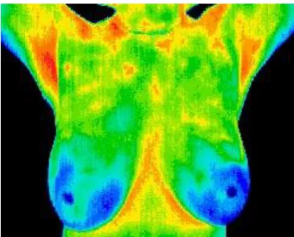
Figure 4. Thermal Image of Cancer Affected Patient
Fibrocystic Significant vascular activity in the left breast. Women at any age can begin utilizing thermography as a safe means of screening for breast disease. Thermography is not dependent on the density of a women’s breast or do implants or surgical procedures affect the process of detecting thermal pattern. Thermography is especially appropriate for younger women between 30 & 50 whose denser breast tissue makes it more difficult for mammography to pick up suspicious lesions. This test can provide a ‘clinical marker’ to the doctor or mammographer that a specific area of the breast needs particular close examination
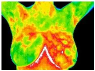
C. Tuberculosis Identifying Using Thermal Image Even if a person has symptoms of TB, it is often difficult to diagnose TB, and is particularly difficult to diagnose rapidly. Though there are various methods to diagnosis tuberculosis, there are certain disadvantages. Chest x-ray, sputum, skin test and culture methods are used in the hospitals to check if the patients are affected by mycobacterium tuberculosis. The problems faced are need of sophisticated infrastructure and trained professional. Time delay and negative results lead to fatal effects. Thermal images provide thermo gram, a representation in colour of radiation differences in affected areas. Thermal cameras uses infrared radiation related to temperature to realize the differences in heat emitted by the affected portion. The software of the thermal cameras emits a visible
alarm to the PC when a higher level of temperature is detected. High accuracy is assured in this method.
Figure 5. Thermal Images
Affected areas are shown by orange colour; other parts are shown by blue colour to indicate normal status.
V.
CONCLUSIONThermal imaging has developed for multiple applications because of practical technology, more reliable and portable mode. Standard software and devices when used in this technology is used for many applications in image processing. Thermal imaging applications are widely spread because of the advantages of thermal cameras. This technique is used for diagnosing dangerous diseases. There is a scope for working on development of innovative, efficient & fast interpreting algorithms which will help doctors in detecting the deadly disease
VI.
REFERENCES[1] CA Duberstein DJ Virden S Matzner J Meyer VI Cullinan AR Maxwel-Automated Thermal Image Processing for Detection and Classification of Birds and Bats- FY2012 Annual Report Offshore Wind Technology Assessment [2] VS. Elanthendral1 , R.K.Rekha2 , M.
Rameshkumar3- Thermal Imaging for Facial Expression – Fatigue Detection- CSE Department, Veltech Multitech Dr.Rangarajan Dr.Sakunthala Engineering College Chennai, Tamil Nadu
Rehabilitation, Hanuschkrankenhaus, Vienna, Austria
[4] Lulu Wang-Early Diagnosis of Breast Cancer- 1,2 1 School of Instrument Science and Opto-Electronics Engineering, Hefei University of Technology, Hefei 230009, China; [email protected] 2 Institute of Biomedical Technologies, Auckland University of Technology, Auckland 1142, New Zealand; [email protected]
[5] Earlier Detection and Diagnosis of Breast Cancer: A Report from It’s About Time! A Consensus Conference
[6] Amalric R, Altschuler C, Giraud D and Spitalier J M- 1984 Value of infrared thermography in the assessment of malignant melanoma of the skin Recent Advances in Medical Thermology ed E F J Ring and B Phillips (New York: Plenum) pp 623–9
[7] Amarja Adgaonkar , Aditi Atreya , Akshay D. Mulgund , Juhi R. Nath -Identification of Tuberculosis bacilli using Image Processing K.C. College Of Engineering And Management Studies And Research
[8] HarshalTalathi, Jinal Patel, KakoliLaha -Mrs.J.M.Kundargi -Identification of Tuberculosis Bacilli using image Processing , Department Of Electronics and Telecommunication ,K.J.Somaiya College of Engineering, Mumbai, India.
[9] Mohebian, M.R.; Marateb, H.R.; Mansourian, M.; Mañanas, M.A.; Mokarian, F. A hybrid computer-aided-diagnosis system for prediction of breast cancer recurrence (HPBCR) using optimized ensemble learning. Comput. Struct. Biotechnol. J. 2017, 15, 75–85. [CrossRef] [PubMed]
[10] U.S. Breast Cancer Statistics 2017. Available online:
http://www.breastcancer.org/symptoms/underst and_ bc/statistics (accessed on 2 July 2017). [11] Mark D. Perkins, MD; Marcus B. Conde, MD;
Marneili Martins, MD; Afranio L. Kritski, MD,
PhD-Serologic Diagnosis of Tuberculosis Using a Simple Commercial Multiantigen Assay*- [12] Grange JM, Lazlo A. -Serodiagnostic tests for