ISSN-1996-918X
Pak. J. Anal. Environ. Chem. Vol. 8, No. 1 & 2, (2007)
Nickel Adsorption by Wild type and Nickel Resistant
Isolate of Chlorella sp.
Pawan Raj Shakya
Department of Chemistry, Padma Kanya Multiple Campus, Tribhuvan University, Bagbazar, Kathmandu, Nepal.
---
Abstract
Use of unicellular green microalgae has several advantages over conventional methods for removing heavy metals from contaminated sites. Here, a comparative study was made to investigate nickel detoxification mechanisms between a wildtype (WT) Chlorella sp. and a nickel resistant, EMS-5 cell line isolated from the same species by EMS (Ethyl Methane Sulphonate) mutagenesis. Results showed that the growth rate of the tested algal cells was inhibited with increasing nickel concentrations in the liquid growth medium. Higher ID50 value of EMS-5
compared to the WT revealed some degree of resistance to nickel. Removal and adsorption of Ni2+ were found rapid during the first few hours in both the algal cultures when exposed to 50 M Ni2+. However, kinetic experiments showed significantly higher removal and adsorption of Ni2+ by EMS-5 compared to the WT throughout the treatment hours. Besides, the total nickel accumulation, surface bound and intracellular nickel in EMS-5 was significantly higher to that of the WT. Hence, the EMS-5 appeared more resistant to nickel.
Keywords: Wildtype, Nickel resistant isolate, adsorption, Chlorella sp., EMS mutagenesis
---
Introduction
Nickel is a widely used heavy metal in industries such as mining, electroplating and alloy production. It is one of the stable and persistent environmental contaminants since it cannot be biologically or chemically degraded or destroyed unlike many other organic toxic pollutants. Therefore, the metal has become a serious worldwide environmental problem. Many lakes elsewhere exposed to industrial pollution are highly contaminated with nickel (upto 100 M) and other metals [1]. The metal contamination in water bodies has posed an acute threatening to an ever-increasing portion of global population with respect to their general health consideration. The Environment Protection Agency (EPA) has, therefore established a water quality criteria limit of 13.4 ppb of nickel and considers it as one of 13 metals on the list of 129 priority pollutants [2]. Although nickel is a trace element required for living organisms, it is toxic when ingested in large amounts [3]. It’s toxic effects to human have also been well established. Nickel and nickel compounds are also well
recognized as carcinogens [4].
Among the various approaches proposed to remove the metal contamination from the environment, the use of eukaryotic algae, especially Chlorella species, is particularly appropriate because of their heavy metal-accumulating potential. They also possess large surface area and chelating potential that maximize binding of toxic metal ions. The metal chelation to cell walls of the biomass has the ability to reduce metal concentration in aqueous to 1 ppm or less [5]. In addition, Chlorella sp. is very common in wastewater and other surface water bodies. It is often used as a model organism to study metabolic processes in photosynthetic eukaryotic higher plants because of its similarity. Because the cell cultures are inexpensive, rapidly grow and easy to maintain in a simple mineral medium, the use of algae has been proposed and developed for numerous applications in different fields. The application includes water treatment, wastewater treatment, biological detoxification and heavy metal
controls in natural and/or industrial waste streams [6]. Therefore, the Chlorella sp. was a proposed organism to use in the present work to study mechanisms confirming resistance to nickel toxicity.
Although many studies have been conducted on the detoxification of metal ions by various micro-algae, little information is available concerning the nickel-resistance mechanisms of Chlorella sp. Hence, the present study was designed with an attempt to understand the effect of nickel on the growth of the WT (wild type) culture and nickel-resistant Chlorella cell line and to characterize mechanism(s)-confirming resistance to nickel toxicity in them.
Experimental
Growth conditions
The living organism used for the present study was Chlorella sp. Cultures of the WT alga were maintained in modified BG-11 mineral medium. They were incubated in a gyratory shaker (180 rev./min) at 270C continuously illuminated with a light intensity of 20 - 50 mol by cool white fluorescent lamps.
EMS (Ethyl Methane Sulphonate) mutagenesis
Isolation of nickel resistant cell lines of
Chlorella sp. was carried out following the Herskowitz
Lab Protocol for EMS mutagenesis (modified) [7]. Of the isolated nickel resistant cell lines, one of the cell lines named EMS-5 was used for further investigation. Cultures of the isolated cell lines were also maintained in BG-11 mineral medium and incubated in the gyratory shaker as described above.
Experiments involving effects of nickel on growth rate
To study the effect of nickel on growth rate of the WT and EMS-5, the cultures with initial cell densities of 5.0 - 5.5 x 10⁵ cells per milliliter of the medium were treated with 0, 1, 10, 50 and 100 M of the stocked nickel solution respectively. The algal growth was monitored by measuring the change in the absorbance of the algal culture at 540 nm. The measurement was taken at the time of inoculation and each day thereafter until it reached the stationary phase. The growth rate of the algal culture was determined between the 2nd and the 6th days by the following equation [8]:
= (ln X6 - ln X2 ) / (T6 - T2)
Where is the specific growth rate of the algal culture, X6 is the A 540 nm of the algal culture at time T6, and X2
is the A 540 nm of the algal culture at time T2. All the
experiments were carried out in triplicates in 250-ml Erlenmeyer flasks containing a volume of 100 ml of the liquid growth medium.
Experiments involving mechanisms confirming resistance to nickel toxicity (adsorption and absorption process)
To study the adsorption kinetics at different time intervals, Erlenmeyers flasks containing cell densities of 109 per milliliter were added with 50 M of the nickel solution and samples were drawn at different time intervals such as zero, half, one, two, four, eight, twelve, twenty-four and forty eight hours respectively. The samples taken at each time interval were spun down in a bench centrifuge (3500 rpm, 10 min) and the supernatants collected were analyzed for the residual nickel concentration. The cell pellets were washed with 5-ml of EDTA (10 g/lit.) three times. Each time, the cells were spun down (3500 rpm, 10 min) and the supernatants containing EDTA were collected for metal analysis. This analysis revealed the concentration of Ni2+ adsorbed at the cell wall at varying time intervals. Finally, the remaining cell pellets were subjected to acid digestion (Conc. HNO3, 230ºC, 3h) followed by metal
analysis in AAS. This result revealed the total nickel accumulated inside the cells at different time intervals. The experiment was carried out in triplicates.
Results and Discussion
Effects of nickel on growth rate
The effects of various Ni2+ concentrations on growth rate of WT Chlorella sp. and the selected cell line, EMS-5 are shown in Table 1. It was observed that the growth of WT and EMS-5 cell lines was reduced with the increasing of nickel concentrations in the liquid growth medium. The presence of 1 M Ni2+ had a negligible inhibitory effect on the growth compared to the control. The inhibitory effect was more pronounced in the presence of 10 and then 50 M Ni2+, and the growth was completely arrested upon exposure to 100
M Ni2+. In general, increasing of metal concentrations in growth medium is causing inhibition of growth [9, 10]. This is also revealed in the present study. However, the effect of nickel was more pronounced on the growth rate of WT than that of EMS-5 cell line. In other words, the percentage growth rate of EMS-5 was found comparatively higher to that of the WT even at higher nickel concentration. This was supported by the calculation of inhibition of 50% in growth rate (ID50) of
0 0.5 1 1.5 2 2.5 3 3.5 4
0 10 20 30 40 50
Ni residual Ni adsorption
[N
i
2
+ ]
u
m
o
l
/1
0
9
ce
ll
s
A
Wild Type Culture
0.25 0.5 1 2 4 8 12 24 48
P
er
ce
n
ta
g
e
o
f
n
ic
k
el
r
em
o
v
al
Time (hours)
B
for the WT and EMS-5 were 30 and 45 M Ni2+ respectively. The EMS-5 cell line with slightly higher ID50 value compared to the WT, might be expected to
have some degree of resistance to nickel (Table 1). Hence, the cell line exhibited more resistance to nickel toxicity comparatively higher to that shown by the WT.
Table 1. Percentage growth rate of the WT and EMS-5 in various nickel concentrations
Ni-concentration (µM) Cell lines
0 (Control) 1 10 50 100
WT 100 95 65 35 05
EMS-5 100 98 80 60 25
ID50 Value: EMS-5 (45 µM) > WT (30 µM)
Mechanisms confirming resistance to nickel toxicity (adsorption and absorption process)
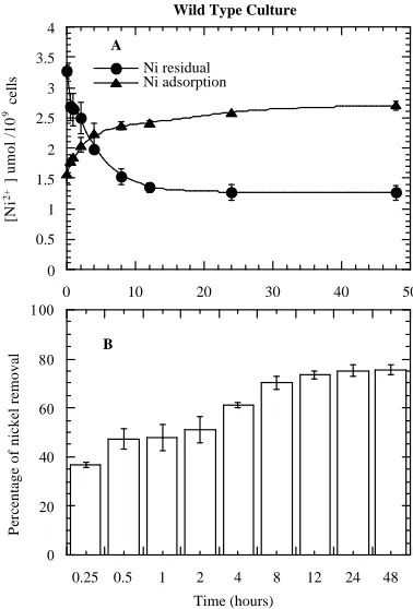
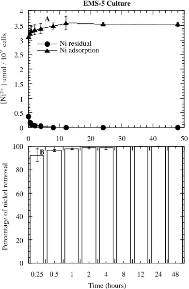
The mechanism for removing metal ions from aqueous solutions by algae is based on biosorption, which is a two-stage process, consisting of a rapid passive surface adsorption followed by a slow active metabolic uptake of metal ions into the cells. Studies have also shown that higher metal binding to the cell surface i.e., extracellular adsorption, occurs within few minutes and the binding reaches a steady state within few hours [11, 12]. Results of the present study are also in agreement with the above studies. On exposure of the WT to 50 M Ni2+, a gradual removal of Ni2+ from the medium was observed (Figure 1B). The removal of Ni2+ from the medium was indicated by a gradual decreasing residual Ni2+ concentration in the medium (Figure 1A). The Ni2+ concentration dropped rapidly during the first hour, decreased gradually until 12 hours and no further decline in Ni2+ concentration beyond this time point in the medium was detected. In parallel, the removal of Ni2+ from the medium corresponded to the increase in Ni2+ adsorption in the WT i.e., extracellular adsorption (Figure 1A). It appears that the extracellular adsorption of Ni2+ was rapid within the first hour of treatment (Figure 1A). The reason may probably be due to the availability of binding sites for the metal ions on the cell surface. After this rapid adsorption of Ni2+, further adsorption by the algal cells occurred slowly and reached equilibrium. No obvious increase in Ni2+ adsorption was observed thereafter. This also suggests that the adsorption capacity will level off when all the specific binding sites of the cells are saturated or occupied. The EMS-5 cell line, however demonstrated a totally different behavior in terms of the kinetics of Ni2+ removal and extracellular adsorption (Figures 2A & 2B). On exposure to 50 M Ni2+, a very rapid removal
Figure 1. Extracellular adsorption by WT and residual concentrations of Ni2+ in the growth medium (A); and percentage
removal of Ni2+ from the medium (B) with treatment hours. Each value in the curves and bars is the mean SD of three experiments.
of Ni2+ was observed from the medium and the removal was completed (100%) within few hours unlike that of the WT. In other words, residual Ni2+ concentration in the medium was almost negligible within the first few hours (Figure 2A). Of the total Ni2+ supplemented in the medium, this cell line showed the highest removal of Ni2+ (92%) within 15 minutes. Similarly, a gradual increase of the extracellular Ni2+ adsorption occurred simultaneously and reached a point of saturation within few hours (Figure 2A). Unlike WT, almost all Ni2+ were adsorbed with the cell surfaces during the first few hours. This unique behavior of EMS-5 might be due to some genetic modification in the cell line due to the mutagenic activity that increased the metal tolerance capacity.
0 0.5 1 1.5 2 2.5 3 3.5 4
0 10 20 30 40 50
Ni residual Ni adsorption
[N
i
2
+ ]
u
m
o
l
/
1
0
9
c
e
ll
s
A
EMS-5 Culture
0 20 40 60 80 100
0.25 0.5 1 2 4 8 12 24 48
P
e
rc
e
n
ta
g
e
o
f
n
ic
k
e
l
re
m
o
v
a
l
Time (hours)
B
the process occurred simultaneously. However, the intracellular Ni2+ uptake by the algal cells was found relatively slow and therefore the metal ions accumulated inside the cells were significantly low compared to the metal associated with external cell fractions (Table 2).
Figure 2. Extracellular adsorption by EMS-5 and residual concentrations of Ni2+ in the growth medium (A); and percentage removal of Ni2+ from the medium (B) with treatment hours. Each value in the curves and bars is the mean SD of three experiments.
It is possible that when the binding sites of the algal cells became exhausted or nearly saturated, the cells would start taking up Ni2+ by active physiological mechanisms. On the other side, the EMS-5 cell line demonstrated comparatively higher accumulation of the total Ni2+ per 109 cells (Table 3). This cell line confirmed the total Ni2+ accumulation about 2 fold higher than that of the WT. The algal cells accumulated almost all of the metal supplemented within few hours of exposure to Ni2 + suggesting that a stabilization point approached at the early hours of treatment. Like the WT, Ni2+ concentration in external cell fractions also increased with duration of exposure to the metal. However, Ni2+ distribution inside the cells appeared to have saturated during the first few hours. Compared to the WT, amount of Ni2+ distributed between the external
and internal cell fractions were higher. Of the total Ni2+ accumulated by the cell line at 48 hours, the cell density had 70% externally bound Ni2+ and 30% inside the cells (Table 3).
Table 2. Surface-bound and intracellular Ni2+ for WT. Each value
in the Table is the mean SD of three experiments.
Nickel Association (mol /109 cells)
Culture Time interval
(hr.) External Internal Total Ni
2+
accumulated
WT 0.25 1.56 0.13 0.27 0.25 1.89 0.91
0.5 1.78 0.04 0.62 0.26 2.40 0.82
1.0 1.84 0.04 0.60 0.31 2.44 0.88
2.0 2.05 0.12 0.55 0.38 2.60 1.06
4.0 2.23 0.18 0.89 0.24 3.12 0.95
8.0 2.37 0.07 1.19 0.21 3.56 0.83
12.0 2.42 0.04 1.32 0.12 3.74 0.78
24.0 2.59 0.00 1.23 0.13 3.82 0.96
48.0 2.70 0.08 1.13 0.19 3.83 1.11
Table 3. Surface-bound and intracellular Ni2+ for EMS-5. Each value in the Table is the mean SD of three experiments.
Nickel Association (mol /109 cells)
Culture Time interval
(hr.) External Internal Total Ni
2+
accumulated
EMS-5 0.25 3.09 0.23 1.54 0.05 4.63 1.10
0.5 3.23 0.18 1.62 0.13 4.85 1.14
1.0 3.31 0.15 1.61 0.12 4.92 1.20
2.0 3.34 0.17 1.61 0.12 4.95 1.22
4.0 3.39 0.17 1.57 0.10 4.96 1.29
8.0 3.47 0.11 1.55 0.11 5.02 1.36
12.0 3.58 0.24 1.43 0.24 5.01 1.52
24.0 3.54 0.06 1.48 0.07 5.02 1.46
48.0 3.53 0.07 1.48 0.07 5.01 1.45
start taking up Ni2+ by active physiological mechanisms. On the other side, the EMS-5 cell line demonstrated comparatively higher accumulation of the total Ni2+ per 109 cells (Table 3). This cell line confirmed the total Ni2+ accumulation about 2 fold higher than that of the WT. The algal cells accumulated almost all of the metal supplemented within few hours of exposure to Ni2 + suggesting that a stabilization point approached at the early hours of treatment. Like the WT, Ni2+ concentration in external cell fractions also increased with duration of exposure to the metal. However, Ni2+ distribution inside the cells appeared to have saturated during the first few hours. Compared to the WT, amount of Ni2+ distributed between the external and internal cell fractions were higher. Of the total Ni2+ accumulated by the cell line at 48 hours, the cell density had 70% externally bound Ni2+ and 30% inside the cells (Table 3).
Apparently, the intracellular Ni2+ uptake by the tested cell lines had significantly less contribution towards reducing the metal toxicity as evident from the results of the present study. The fate of Ni2+ inside the algal cells reveals intracellular detoxification mechanisms in response to the metal toxicity. Phytochelatins (PCs) were reported extensively as heavy metal detoxifiers in plants [13] and are induced when plants are exposed to heavy metals [14]. The synthesis of such metal binding peptides or other organic materials may be possible once the metal ions are inside the algal cells. The present study could not, however, cover the fate of the metal ions inside the cell. On the basis of the experiments, EMS-5 was found as the resistant line that has higher rate capacity to remove, adsorption and uptake of Ni2+ compared to that of the WT. However, more detailed studies on cell wall composition and the availability of these functional groups on the tested cell lines will be essential to further understanding the mechanism of metal ion adsorption. Furthermore, comprehensive understandings of physiological, biochemical and molecular mechanisms conferring Ni2+ resistance in Chlorella sp. are of utmost importance. This will enable the engineering of metal accumulating organisms so that they could serve as a tool in the remediation of metal contaminated sites.
Acknowledgements
I am very much grateful to Department of Environmental Resources, Technical University of Denmark for providing me the opportunity to work on the related area.
References
1. P. M. Stoke, In: Effects of Nickel in Canadian
Environment, National Research Council of
Canada, Environmental Secretarial Pub. No. NRCC 18568 (1981) 77.
2. EPA (Environment Protection Agency), Ambient
Water Quality Criteria for Nickel, United States
EPA Document EPA, (1980) 440/5-80-060. 3. P. Kotrba and T. Ruml, Czech. Chem. Commun.,
65 (2000) 1205.
4. ATSDR (Agency for Toxic Substance and Diseases Registry) Toxicological Profile for
Nickel, ATSDR/U.S. Public Health Service
(1988) ATSDR/TP - 88/19.
5. D. Roy, P. N. Greenlaw and B.S. Shane, J.
Environ. Sci. Health 28 (1993) 37.
6. J. P. Gaur and L. C. Rai, In: Algal adaptation to
environmental stresses: Physiological,
Biochemical and Molecular Mechanisms. (L. C.
Rai and J. P. Gaur, Eds), Springer Verlag Pub., Heidelberg (2001) 363.
7. A. Sil and J. Chenevert, EMS Mutagenesis, Hersckowitz Lab Protocol, Dept. of Biochemistry and Biophysics, University of California, San Francisco, (1998) 7.
8. M. Ajmal and A. U. Khan, Environ. Res., 38 (1985) 248.
9. X. Jin, C. Nalewajko and D. J. Kushner, Microb.
Ecol. 31 (1996) 103.
10. S. M. Macfie and P. M. Welbourn, Arch.
Environ. Contam. Toxicol. 39 (2000) 413.
11. J. P. K. Wong, Y. S. Wong and N. F. Y. Tam,
Biores. Tech., 73 (2000)133.
12. P. Kaewsarn and Q. Yu, Environ. Pollut. 112 (2001) 209.
13. E. Grill, S. Loffler, E. L. Winnacker and M. H. Zenk, Proc. Nat. Acad. Sci. USA, (1989) 6838. 14. M. H. Zenk, Gene 179 (1996) 21.
Pawan R. Shakya is presently the Executive Member of Environment Management Committee, Padma Kanya Multiple Campus, Tribhuvan University, Kathmandu, Nepal. His activities are focused on interactions between living organisms and toxic heavy metals and environmental related issues.