1
Seven challenges in the multiscale modelling of multicellular tissues
Alexander G. Fletcher
School of Mathematics and Statistics, University of Sheffield, Sheffield S3 7RH, UK Bateson Centre, University of Sheffield, Sheffield S10 2TN, UK
E-mail address: [email protected] ORCID iD: https://orcid.org/0000-0003-0525-4336
James M. Osborne
School of Mathematics and Statistics, University of Melbourne, Parkville 3010, Victoria, Australia
ABSTRACT
INTRODUCTION
How multicellular tissues self-organise and remodel is a fundamental question in developmental biology with profound implications for tissue engineering and regeneration (Sasai, 2013). Recent advances in molecular biology, live-imaging and ex vivo techniques have resulted in a wealth of detailed, but isolated, descriptions of these processes (St Johnston, 2015). To truly comprehend such complexity, we need computational models that can link observations in a quantitative, predictive, and reproducible way (Sharpe, 2017). However, before these goals can be achieved there are substantial methodological challenges to overcome. This review aims to highlight these challenges and how they might be overcome.
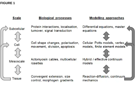
The growth and dynamics of multicellular tissues are multiscale in nature, involving tightly regulated and coordinated morphogenetic cell behaviours, such as shape changes, movement, and division, which are governed by subcellular machinery and involve coupling through short- and long-range signals (Figure 1). A key challenge is to understand how relationships between scales produce emergent tissue-scale self-organisation.
State-of-the-art experimental approaches are revealing emergent behaviour at different scales: super-resolution microscopy allows quantification of complex, dynamic feedback interactions and subcellular localisation (Liu & Keller, 2016) underlying cell-level patterning; live-imaging and force-inference techniques uncover ‘mesoscale’ structures, comprising groups of cells, driving tissue morphogenesis (Blanchard et al., 2019); while ex vivo organoid cultures offer a window into the contribution and timing of different signals to tissue self-organisation (Simunovic & Brivanlou, 2017). To fully leverage these techniques and obtain a more comprehensive understanding of the causal relationships underlying normal and abnormal tissue dynamics, computational modelling approaches are being developed that span multiple spatial and temporal scales, which couple cell shape, growth, mechanics and signalling.
facilitated the accelerated development of discrete agent-based and multiscale modelling approaches, which more naturally allow investigation of the influence of intracellular and cellular level behaviours and heterogeneity in tissue-scale phenomena. Recent work on such models has facilitated successful interdisciplinary studies of emergent behaviours in developing tissues (Nissen et al., 2017); for a detailed review, see (Glen et al., 2019).
Despite their utility, agent-based and multiscale models of multicellular tissue remain technically challenging: modelling at each scale requires different areas of technical skills, while integration across scales necessitates the solution to novel mathematical and computational problems (Figure 1). In this review, we set out seven ongoing challenges in multicellular modelling, ordered roughly in the order in which they may be experienced by someone developing a new model of this form: from model construction and calibration; to numerical and software/hardware implementation; to analysis and validation; to issues associated with model re-use and comparing modelling assumptions and paradigms. For each challenge, we summarise recent progress in specific applications as well as efforts to tackle the general case, highlighting one key open research question that we believe requires attention in future computational research.
When commenting on applications of agent-based and multiscale modelling in the following, we focus primarily on animal developmental biology. This is a natural area in which to apply these modelling techniques, because of the availability of data across multiple spatial and temporal scales - from gene regulatory networks and signalling pathways to cellular interactions and larger-scale morphogen transport processes - and the feedbacks between scales that underlies many emergent processes (Sharpe, 2017). We refer the interested reader to several recent reviews on the use of these modelling approaches in plant biology (Bucksch et al., 2017), cancer biology (Metzcar et al., 2019), synthetic biology (Gorochowski et al., 2020), and tissue engineering (Montes-Olivas et al., 2019).
CHALLENGE 1: MODEL CONSTRUCTION
In mathematical biology, modelling assumptions must be carefully tailored to the biological system of interest, the available data, and the scientific hypotheses. In the case of multiscale modelling, one must choose the level of complexity with which one describes subcellular processes, cellular interactions, and larger-scale processes such as morphogen transport. This leads to our first ongoing challenge: how to choose an appropriate model for a given problem.
A variety of multiscale modelling approaches have been developed for multicellular tissues (Fletcher et al., 2017). These approaches generally either discretise space into a regular (Lehotzky & Zupanc, 2019) or irregular (Scianna & Preziosi, 2016) lattice, or allow cells to move continuously in space (Drasdo & Höhme, 2005). Such approaches also vary in their geometric description: cells may be treated as point-like particles or prescribed shapes such as ellipsoids (Xu et al., 2008); or cell shape may be more complex and dynamic, evolving according to a balance of forces (Farhadifar et al., 2007) or stochastic fluctuations (Graner & Glazier, 1992). There can also be increased geometric complexity moving from two- and three-dimensional representations, such as a larger number of possible cell rearrangements (Okuda et al., 2015). Another distinction can be made based on the level of biophysical complexity used: considering cell surface mechanics only (Magno et al., 2015), or including subcellular heterogeneity such as the presence and mechanical role of the nucleus and other organelles (Jamali et al., 2010).
CHALLENGE 2: MODEL CALIBRATION
Having arrived at a set of modelling assumptions, one is faced with the issue of how to choose appropriate parameter values and initial conditions. This leads to our second ongoing challenge: how to calibrate a multiscale model against data. Here we refer both to estimation of model parameters and their associated uncertainty, as well as calibration of initial and boundary conditions (for example, initial cell and tissue geometry). In an ideal world, we would parameterise any model and initialise our simulations using appropriate data at each level of the model. In practice, however, we must resort to using the understanding and data that are available to us (Pargett & Umulis, 2013).
Initial conditions are specified based on our biological understanding. In some cases, this can be straightforward, for example when modelling tissue development from a single stem cell (Atwell et al., 2015). This ad hoc approach works well if there are initially few cells, or if the initial state is highly conserved. Another common approach is to evolve the system to a dynamic equilibrium that is not sensitive to the choice of initial condition, as in the case of many homeostatic tissues such as the skin (Li et al., 2013) or gut epithelium (van Leeuwen et al., 2009). For developing tissues undergoing morphogenesis, an alternative approach is to derive initial conditions such as cell packing directly based on microscopy images (Iber et al., 2016) or via extracted summary statistics (Vicente-Munuera et al., 2019). Here, one should bear in mind variability in structures and it may be more important to capture essential patterns and conserved properties of tissue organization rather than use a specific example.
Local and global sensitivity analysis methods have also been developed to assess to which parameters a model is most sensitive, and to identify the effects of uncertainties in parameter values on its output (Boas et al., 2015).
Several challenges remain when specifying initial and boundary conditions and estimating parameters of multiscale models. In particular, more tailored and efficient parameter inference techniques are needed that rely less heavily on repeated simulations of computationally expensive models, for example by exploring parameter space more intelligently and by exploiting ‘low-fidelity' or surrogate modelling (Prescott & Baker, 2020). Where simulation outputs are intended to inform decision-making, greater use of uncertainty quantification techniques is needed, for which software tools are increasingly available (Richardson et al., 2020).
CHALLENGE 3: NUMERICAL SOLUTION
Due to their complexity, multiscale models of multicellular tissues are typically analytically intractable and must be solved numerically. This leads to our third challenge: how to solve multiscale models efficiently, stably and accurately. Their numerical solution remains challenging since the coupling of processes or sub-models across scales can make it harder to avoid numerical instabilities, and many such models include parameters of numerical approximation; for example, thresholds for changes in cell topology (Fletcher et al., 2013). Thus, we need to be aware of any impacts that numerical implementation choices may have on model predictions (Kursawe et al., 2017).
Typically, subcellular processes such as protein production, degradation and complex formation evolve on a faster timescale than cellular level processes such as division and movement, which in turn play out on a faster timescale than tissue-scale processes such as paracrine signalling. Therefore, based on quasi-steady state approximations or similar, multiscale models may be decomposed into subsystems that are solved numerically over discrete time steps during which other subsystems are held constant.
complicated due to processes such as cell death and division changing the size of the dynamical system over each time step (Atwell, 2016). For some cell-based models, numerical methods must be carefully chosen to respect physical conservation laws (Cooper et al., 2017). Efficient numerical techniques have also been developed to simulate stochastic reaction-diffusion processes in spatially complex or evolving domains (Drawert et al., 2012). At the cellular level, algorithms have been developed to simulate the evolution of stochastic models such as the cellular Potts model efficiently while forbidding physically unrealistic phenomena such as cell fragmentation (Durand & Guesnet, 2016). Several efforts have also been made to develop efficient solvers that couple cell-based models and tissue-scale processes such as nutrient transport (Ghaffarizadeh et al., 2016), morphogen signalling (Smith et al., 2012) and fluid flow (Grogan et al., 2017; Osborne & Bernabeu, 2018), based on finite element, finite volume, finite difference, and lattice Boltzmann methods.
Looking ahead, ongoing challenges include the application of adaptive methods, which allow spatial or temporal discretizations to vary according, for example, to the amount of cell movement of proliferation in a domain. This also leads to the prospect of models that allow one to change the level of spatial complexity when and where needed, for example by using a cell-based description in regions where the bulk of growth occurs, and a continuum description elsewhere (Kim et al., 2007).
CHALLENGE 4: SOFTWARE AND HARDWARE IMPLEMENTATION
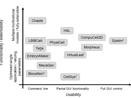
If every researcher implemented their own model from scratch we would have thousands of implementations of each model at a massive overall time cost. The solution to this is to develop and use computational tools to build and interrogate multicellular models, which leads to our fourth ongoing challenge: how to fully exploit modern software and hardware architectures. Multiple numerical libraries and software tools have been developed for building, running and interrogating multicellular models. These range from user-friendly GUIs with limited functionality intended for use by biologists to more extensible software libraries, which require significant programming skills to utilise (Figure 2).
approaches, are: to run a large number of simulations, each at reasonable computational cost (for example, when performing parameter sweeps); and to run a small number of simulations, each at significant computational cost (for example, when simulating the collective dynamics of tens or hundreds of thousands of cells).
The first use-case is ‘trivially’ parallelizable, in that multiple instances may be run independently on multiple processors in parallel. In contrast, the second use-case requires one to partition a simulated model into smaller components, for example through spatial decomposition, that are simulated in each processor. Parallel interfaces such as MPI (for distributed memory devices) and OpenMP (for shared memory devices) are then used to communicate transfer information between processors. Such algorithms have been developed, for example, for the cellular Potts model (Chen et al., 2007) and for off-lattice cell-centre models (Harvey et al., 2015). Further speedups may be obtained through the use of GPGUs, for example through using CUDA or OpenCL; again, published examples include implementations of the cellular Potts model (Tapia & D’Souza, 2011) and off-lattice cell-centre models (Germann et al., 2019).
Several software and hardware challenges remain. When developing and refining software tools for multiscale modelling, one must carefully balance functionality and usability; such tools are unlikely to be taken up by non-specialists unless barriers are further lowered, for example through the greater use of GUIs and APIs. Modern computing resources, such as cloud computing, should also be more fully exploited; a laudable example of work towards this is xml2jupyter (Heiland et al., 2019). Finally, there remains remarkably little in the way of cross-validation of different model implementations; we return to this point when discussing Challenges 6 and 7.
CHALLENGE 5: MODEL VALIDATION
against data? This strongly relates to the issue of parameter inference and model selection discussed in Challenge 2, but is typically encountered later in the modelling process. In each case, we typically have access only to partial information about the system at each spatial or temporal scale, and so we may need to resort to validating separate model components individually (Viceconti et al., 2020).
Testing model predictions, like estimating model parameters (see Challenge 2), often involves using various different types and sources of experimental data. Subcellular processes, such as protein dynamics, may be compared - at least semi-quantitatively - against experiments on isolated cells or on in vitro cell cultures (Tan et al., 2012), with due care for possible discrepancies with the in vivo setting. At the cellular and tissue level, rheology experiments, laser ablations, and non-invasive force inference techniques can all be used to interrogate how tissues respond to applied stresses (Sugimura et al., 2016) and compare these measurements with model predictions. Cell tracking can also be used to match cell velocities and other summary statistics. In this context, recent work demonstrating the pitfalls of inferring cell-level heterogeneity from such data (Schumacher et al., 2017) has implications for inferring parameters from the ‘wrong’ model; we return to the issue of comparing models and modelling assumptions when discussing Challenge 7.
A complementary approach to validating multiscale models is to apply mathematical or computational techniques to perform system model reduction, for example through the use of asymptotic or equation-free methods. Here, the intention is to arrive at a coarse-grained model that may be more amenable to efficient simulation or qualitative mathematical analysis; such approaches have been applied, to lattice-based models (Johnston et al., 2012) and cellular Potts models (Alber et al., 2007), as well as off-lattice cell-centre models (Murray et al., 2012) and vertex models (Fozard et al., 2010). Further work is needed in this area, as well as advances in our statistical understanding of how best to use information at different scales, the extent to which we can fully validate a given model, and which experiments will yield the greatest information for model calibration and validation.
There are myriad simulation tools, as mentioned above. This is unlike fields like molecular dynamics where one or two libraries are used by everyone or ‘subcellular’ systems biology where the modelling formalism is shared across the field. This leads to our sixth ongoing challenge: how to assess robustness of simulation results to software and hardware implementation.
There are ongoing discussions in the wider field of computational biology regarding the need for model replicability and reproducibility (Grüning et al., 2018; Lewis et al., 2016). In the case of multiscale modelling of multicellular tissues, model construction, exchange and re-use remain hampered by the lack of easily shareable model definitions, for example in the form of a fully declarative model definition language. SBML and CellML have largely solved this problem in the context of reaction kinetics, and have associated APIs and model repositories, but have limited functionality for spatial models; some initiatives are ongoing in this area, such as the MultiCellML project. Many multicellular tools have support for SBML specification of subcellular processes, for example CompuCell3D (Swat et al., 2012), Chaste (Romijn et al., 2020) and Morpheus (Starruß et al., 2014). Another aspect of this is separating out the definition of the biological process from the definition of any in silico experiments or perturbations, allowing easier comparison of model behaviour with experimental data. Relatively little work has been done on this (J. Cooper & Osborne, 2013) compared to more mature areas of systems biology such as cardiac electrophysiology (Cooper et al., 2016).
Several challenges remain in this area. In particular, we must work out how to improve standards, reproducibility and interoperability among new and existing software tools (Macklin, 2019). Here, there may be lessons to be learned from other domains such as astrophysics, weather/climate modelling, and molecular dynamics.
CHALLENGE 7: COMPARING MODELLING ASSUMPTIONS AND APPROACHES
It can be difficult to disentangle differences in model behaviour arising from algorithmic, software, and hardware implementation. However, some progress has been made on aspects of this issue. Simulation studies have been used to compare different mechanical assumptions and constitutive equations for particular classes of cell-based models, such as cell-centre models (Mathias et al., 2020; Pathmanathan et al., 2009) and vertex models (Fletcher et al., 2013). Efforts have also been made to compare and contrast five competing cell-based model paradigms, including lattice-based and off-lattice models, in a consistent computational framework (Osborne et al., 2017), to help elucidate where one may expect to see qualitative differences between model behaviours. Four exemplar simulations were chosen to compare the models, which represent facets of development: adhesion; proliferation, death and differentiation; short-range signalling; and long-range signalling. At a tissue level, each of the modelling paradigms investigated was able to implement these processes and, outside a few edge cases where artifacts were introduced, the tissue level behaviour was qualitatively the same. A natural extension of this work is to compare models across different software implementations, such as those discussed in Challenge 6. This is feasible in principle, though one needs to ensure the same model definition and numerical method of solution (to within some tolerance), which is difficult in the absence of model standards discussed above.
Recent work has begun comparing the effect of both cellular and subcellular models especially in the field of intestinal crypt development (Dunn et al., 2013) compare how multiple cell level models of the crypt behave under artificial radiation and compare this to decades-old experimental results, adding new insight into the mechanism for cell migration in the absence of mitosis; while elsewhere a framework to couple SBML subcellular models with multicellular crypt simulations has been used to compare different approaches to modelling the Wnt pathway (Romijn et al., 2020).
CONCLUSION
In this paper we have presented seven open challenges associated with multiscale modelling of multicellular tissue dynamics. These challenges range from the mathematical (how to couple processes across multiple scales) and statistical (how to infer multiscale model parameters from partial information on processes at each scale) to the computational (how to unambiguously define such models in a shareable format) and even societal (how to exploit synergies among competing software tools). In our view, overcoming these challenges will require a significant community-driven effort in order to move such modelling approaches to a point where they can be a standard tool for stem cell and developmental biologists and tissue engineers alongside experimental approaches. Addressing these challenges, particularly those associated with quantifying uncertainty in model predictions, is even more important if such approaches are to become a greater part of clinical research and decision-making, as is starting to be seen for example in mathematical oncology (Rockne et al., 2019).
FUNDING INFORMATION
AGF acknowledges the support of the UK Biotechnology and Biological Sciences Research Council (BBSRC, grant BB/R016925/1) and the University of Sheffield (Vice-Chancellor's Fellowship).
ACKNOWLEDGMENTS
REFERENCES
Atwell, K. (2016). Investigating the interplay between cellular mechanics and decision-making in the C. elegans germ line [PhD Thesis]. University of Oxford.
Atwell, K., Qin, Z., Gavaghan, D., Kugler, H., Hubbard, E. J. A., & Osborne, J. M. (2015). Mechano-logical model of C. elegans germ line suggests feedback on the cell cycle. Development, 142(22), 3902–3911. https://doi.org/10.1242/dev.126359
Blanchard, G. B., Fletcher, A. G., & Schumacher, L. J. (2019). The devil is in the mesoscale: Mechanical and behavioural heterogeneity in collective cell movement. Seminars in Cell & Developmental Biology, 93, 46–54. https://doi.org/10.1016/j.semcdb.2018.06.003
Boas, S. E. M., Navarro Jimenez, M. I., Merks, R. M. H., & Blom, J. G. (2015). A global sensitivity analysis approach for morphogenesis models. BMC Systems Biology, 9(1), 85. https://doi.org/10.1186/s12918-015-0222-7
Bravo, R. R., Baratchart, E., West, J., Schenck, R. O., Miller, A. K., Gallaher, J., Gatenbee, C. D., Basanta, D., Robertson-Tessi, M., & Anderson, A. R. A. (2020). Hybrid Automata Library: A flexible platform for hybrid modeling with real-time visualization. PLOS Computational Biology, 16(3), e1007635. https://doi.org/10.1371/journal.pcbi.1007635
Bressloff, P. C., & Kim, H. (2019). Search-and-capture model of cytoneme-mediated morphogen gradient formation. Physical Review E, 99(5), 052401.
https://doi.org/10.1103/PhysRevE.99.052401
Cooper, F., Baker, R., Bernabeu, M., Bordas, R., Bowler, L., Bueno-Orovio, A., Byrne, H., Carapella, V., Cardone-Noott, L., Cooper, J., Dutta, S., Evans, B., Fletcher, A., Grogan, J., Guo, W., Harvey, D., Hendrix, M., Kay, D., Kursawe, J., … Gavaghan, D. (2020). Chaste: Cancer, Heart and Soft Tissue Environment. Journal of Open Source Software, 5(47), 1848. https://doi.org/10.21105/joss.01848
Cooper, F. R., Baker, R. E., & Fletcher, A. G. (2017). Numerical analysis of the immersed boundary method for cell-based simulation. SIAM Journal on Scientific Computing, 39(5), B943–B967. https://doi.org/10.1137/16M1092246
Cooper, J., Scharm, M., & Mirams, G. R. (2016). The Cardiac Electrophysiology Web Lab. Biophysical Journal, 110(2), 292–300. https://doi.org/10.1016/j.bpj.2015.12.012
Dasbiswas, K., Hannezo, E., & Gov, N. S. (2018). Theory of epithelial cell shape transitions induced by mechanoactive chemical gradients. Biophysical Journal, 114(4), 968–977. https://doi.org/10.1016/j.bpj.2017.12.022
Delile, J., Herrmann, M., Peyriéras, N., & Doursat, R. (2017). A cell-based computational model of early embryogenesis coupling mechanical behaviour and gene regulation. Nature Communications, 8(1), 13929. https://doi.org/10.1038/ncomms13929
Drasdo, D., & Höhme, S. (2005). A single-cell-based model of tumor growth in vitro: Monolayers and spheroids. Physical Biology, 2(3), 133–147.
https://doi.org/10.1088/1478-3975/2/3/001
Drawert, B., Engblom, S., & Hellander, A. (2012). URDME: A modular framework for stochastic simulation of reaction-transport processes in complex geometries. BMC Systems Biology, 6(1), 76. https://doi.org/10.1186/1752-0509-6-76
Dunn, S.-J., Näthke, I. S., & Osborne, J. M. (2013). Computational models reveal a passive mechanism for cell migration in the crypt. PLoS ONE, 8(11), e80516.
https://doi.org/10.1371/journal.pone.0080516
fragmentation. Computer Physics Communications, 208, 54–63. https://doi.org/10.1016/j.cpc.2016.07.030
Farhadifar, R., Röper, J.-C., Aigouy, B., Eaton, S., & Jülicher, F. (2007). The influence of cell mechanics, cell-cell interactions, and proliferation on epithelial packing. Current Biology, 17(24), 2095–2104. https://doi.org/10.1016/j.cub.2007.11.049
Fletcher, A. G., Cooper, F., & Baker, R. E. (2017). Mechanocellular models of epithelial
morphogenesis. Philosophical Transactions of the Royal Society B: Biological Sciences, 372(1720), 20150519. https://doi.org/10.1098/rstb.2015.0519
Fletcher, A. G., Osborne, J. M., Maini, P. K., & Gavaghan, D. J. (2013). Implementing vertex dynamics models of cell populations in biology within a consistent computational framework. Progress in Biophysics and Molecular Biology, 113(2), 299–326. https://doi.org/10.1016/j.pbiomolbio.2013.09.003
Fozard, J. A., Byrne, H. M., Jensen, O. E., & King, J. R. (2010). Continuum approximations of individual-based models for epithelial monolayers. Mathematical Medicine and Biology, 27(1), 39–74. https://doi.org/10.1093/imammb/dqp015
Germann, P., Marin-Riera, M., & Sharpe, J. (2019). ya||a: GPU-powered spheroid models for mesenchyme and epithelium. Cell Systems, 8(3), 261-266.e3.
https://doi.org/10.1016/j.cels.2019.02.007
Ghaffarizadeh, A., Friedman, S. H., & Macklin, P. (2016). BioFVM: An efficient, parallelized diffusive transport solver for 3-D biological simulations. Bioinformatics, 32(8), 1256– 1258. https://doi.org/10.1093/bioinformatics/btv730
Ghaffarizadeh, A., Heiland, R., Friedman, S. H., Mumenthaler, S. M., & Macklin, P. (2018). PhysiCell: An open source physics-based cell simulator for 3-D multicellular systems. PLOS Computational Biology, 14(2), e1005991.
https://doi.org/10.1371/journal.pcbi.1005991
systems: Advantages and challenges. PLOS Computational Biology, 15(3), e1006577. https://doi.org/10.1371/journal.pcbi.1006577
Gorochowski, T. E., Hauert, S., Kreft, J., Marucci, L., Stillman, N. R., Tang, T. D., Bandiera, L., Bartoli, V., Dixon, D. O. R., Fedorec, A. J. H., Fellermann, H., Fletcher, A. G., Foster, T., Giuggiolo, L., Matyjazkiewicz, A., McCormick, S., Montes Olivas, S., Naylor, J., Rubio Deniss, A., & Ward, D. (2020). Towards engineering biosystems with emergent collective functions. In press.
Graner, F., & Glazier, J. A. (1992). Simulation of biological cell sorting using a two-dimensional extended Potts model. Physical Review Letters, 69(13), 2013–2016.
https://doi.org/10.1103/PhysRevLett.69.2013
Grogan, J. A., Connor, A. J., Markelc, B., Muschel, R. J., Maini, P. K., Byrne, H. M., & Pitt-Francis, J. M. (2017). Microvessel Chaste: An open library for spatial modeling of vascularized tissues. Biophysical Journal, 112(9), 1767–1772.
https://doi.org/10.1016/j.bpj.2017.03.036
Grüning, B., Chilton, J., Köster, J., Dale, R., Soranzo, N., van den Beek, M., Goecks, J.,
Backofen, R., Nekrutenko, A., & Taylor, J. (2018). Practical computational reproducibility in the life sciences. Cell Systems, 6(6), 631–635.
https://doi.org/10.1016/j.cels.2018.03.014
Heiland, R., Mishler, D., Zhang, T., Bower, E., & Macklin, P. (2019). xml2jupyter: Mapping parameters between XML and Jupyter widgets. Journal of Open Source Software, 4(39), 1408. https://doi.org/10.21105/joss.01408
Hoehme, S., & Drasdo, D. (2010). A cell-based simulation software for multi-cellular systems. Bioinformatics, 26(20), 2641–2642. https://doi.org/10.1093/bioinformatics/btq437
Iber, D., Karimaddini, Z., & Ünal, E. (2016). Image-based modelling of organogenesis. Briefings in Bioinformatics, 17(4), 616–627. https://doi.org/10.1093/bib/bbv093
population mechanics. PLoS ONE, 5(8), e12097. https://doi.org/10.1371/journal.pone.0012097
Johnston, S. T., Simpson, M. J., & Baker, R. E. (2012). Mean-field descriptions of collective migration with strong adhesion. Physical Review E, 85(5), 051922.
https://doi.org/10.1103/PhysRevE.85.051922
Kang, S., Kahan, S., McDermott, J., Flann, N., & Shmulevich, I. (2014). Biocellion: Accelerating computer simulation of multicellular biological system models. Bioinformatics, 30(21), 3101–3108. https://doi.org/10.1093/bioinformatics/btu498
Kaucka, M., Zikmund, T., Tesarova, M., Gyllborg, D., Hellander, A., Jaros, J., Kaiser, J.,
Petersen, J., Szarowska, B., Newton, P. T., Dyachuk, V., Li, L., Qian, H., Johansson, A.-S., Mishina, Y., Currie, J. D., Tanaka, E. M., Erickson, A., Dudley, A., … Adameyko, I. (2017). Oriented clonal cell dynamics enables accurate growth and shaping of vertebrate cartilage. ELife, 6, e25902. https://doi.org/10.7554/eLife.25902
Klinger, E., Rickert, D., & Hasenauer, J. (2018). pyABC: Distributed, likelihood-free inference. Bioinformatics, 34(20), 3591–3593. https://doi.org/10.1093/bioinformatics/bty361
Kursawe, J., Baker, R. E., & Fletcher, A. G. (2017). Impact of implementation choices on quantitative predictions of cell-based computational models. Journal of Computational Physics, 345, 752–767. https://doi.org/10.1016/j.jcp.2017.05.048
Kursawe, J., Baker, R. E., & Fletcher, A. G. (2018). Approximate Bayesian computation reveals the importance of repeated measurements for parameterising cell-based models of growing tissues. Journal of Theoretical Biology, 443, 66–81.
https://doi.org/10.1016/j.jtbi.2018.01.020
Lambert, B., MacLean, A. L., Fletcher, A. G., Combes, A. N., Little, M. H., & Byrne, H. M. (2018). Bayesian inference of agent-based models: A tool for studying kidney branching morphogenesis. Journal of Mathematical Biology, 76(7), 1673–1697.
Lan, H., Wang, Q., Fernandez-Gonzalez, R., & Feng, J. J. (2015). A biomechanical model for cell polarization and intercalation during Drosophila germband extension. Physical Biology, 12(5), 056011. https://doi.org/10.1088/1478-3975/12/5/056011
Lehotzky, D., & Zupanc, G. K. H. (2019). Cellular automata modeling of stem‐cell‐driven
development of tissue in the nervous system. Developmental Neurobiology, dneu.22686. https://doi.org/10.1002/dneu.22686
Lewis, J., Breeze, C. E., Charlesworth, J., Maclaren, O. J., & Cooper, J. (2016). Where next for the reproducibility agenda in computational biology? BMC Systems Biology, 10(1), 52. https://doi.org/10.1186/s12918-016-0288-x
Li, X., Upadhyay, A. K., Bullock, A. J., Dicolandrea, T., Xu, J., Binder, R. L., Robinson, M. K., Finlay, D. R., Mills, K. J., Bascom, C. C., Kelling, C. K., Isfort, R. J., Haycock, J. W., MacNeil, S., & Smallwood, R. H. (2013). Skin stem cell hypotheses and long term clone survival—Explored using agent-based modelling. Scientific Reports, 3(1), 1904.
https://doi.org/10.1038/srep01904
Liu, Z., & Keller, P. J. (2016). Emerging Imaging and Genomic Tools for Developmental Systems Biology. Developmental Cell, 36(6), 597–610.
https://doi.org/10.1016/j.devcel.2016.02.016
Macklin, P. (2019). Key challenges facing data-driven multicellular systems biology. GigaScience, 8(10), giz127. https://doi.org/10.1093/gigascience/giz127
Magno, R., Grieneisen, V. A., & Marée, A. F. (2015). The biophysical nature of cells: Potential cell behaviours revealed by analytical and computational studies of cell surface
mechanics. BMC Biophysics, 8(1), 8. https://doi.org/10.1186/s13628-015-0022-x Marin-Riera, M., Brun-Usan, M., Zimm, R., Välikangas, T., & Salazar-Ciudad, I. (2015).
Computational modeling of development by epithelia, mesenchyme and their interactions: A unified model. Bioinformatics, 32(2), 219–225.
Mathias, S., Coulier, A., Bouchnita, A., & Hellander, A. (2020). Impact of force function
formulations on the numerical simulation of centre-based models [Preprint]. Biophysics.
https://doi.org/10.1101/2020.03.16.993246
Metzcar, J., Wang, Y., Heiland, R., & Macklin, P. (2019). A review of cell-based computational modeling in cancer biology. JCO Clinical Cancer Informatics, 3, 1–13.
https://doi.org/10.1200/CCI.18.00069
Montes-Olivas, S., Marucci, L., & Homer, M. (2019). Mathematical models of organoid cultures. Frontiers in Genetics, 10, 873. https://doi.org/10.3389/fgene.2019.00873
Murray, P. J., Edwards, C. M., Tindall, M. J., & Maini, P. K. (2012). Classifying general nonlinear force laws in cell-based models via the continuum limit. Physical Review E, 85(2),
021921. https://doi.org/10.1103/PhysRevE.85.021921
Nissen, S. B., Perera, M., Gonzalez, J. M., Morgani, S. M., Jensen, M. H., Sneppen, K., Brickman, J. M., & Trusina, A. (2017). Four simple rules that are sufficient to generate the mammalian blastocyst. PLOS Biology, 15(7), e2000737.
https://doi.org/10.1371/journal.pbio.2000737
Osborne, J. M., & Bernabeu, M. O. (2018). A fully discrete open source framework for the simulation of vascular remodelling. 2018 40th Annual International Conference of the IEEE Engineering in Medicine and Biology Society (EMBC), 4552–4555.
https://doi.org/10.1109/EMBC.2018.8513223
Osborne, J. M., Fletcher, A. G., Pitt-Francis, J. M., Maini, P. K., & Gavaghan, D. J. (2017). Comparing individual-based approaches to modelling the self-organization of multicellular tissues. PLOS Computational Biology, 13(2), e1005387.
https://doi.org/10.1371/journal.pcbi.1005387
Pargett, M., & Umulis, D. M. (2013). Quantitative model analysis with diverse biological data: Applications in developmental pattern formation. Methods, 62(1), 56–67.
https://doi.org/10.1016/j.ymeth.2013.03.024
Pathmanathan, P., Cooper, J., Fletcher, A., Mirams, G., Murray, P., Osborne, J., Pitt-Francis, J., Walter, A., & Chapman, S. J. (2009). A computational study of discrete mechanical tissue models. Physical Biology, 6(3), 036001.
https://doi.org/10.1088/1478-3975/6/3/036001
Prescott, T. P., & Baker, R. E. (2020). Multifidelity approximate Bayesian computation. SIAM/ASA Journal on Uncertainty Quantification, 8(1), 114–138.
https://doi.org/10.1137/18M1229742
Richardson, R. A., Wright, D. W., Edeling, W., Jancauskas, V., Lakhlili, J., & Coveney, P. V. (2020). EasyVVUQ: A library for verification, validation and uncertainty quantification in high performance computing. Journal of Open Research Software, 8, 11.
https://doi.org/10.5334/jors.303
Rockne, R. C., Hawkins-Daarud, A., Swanson, K. R., Sluka, J. P., Glazier, J. A., Macklin, P., Hormuth, D. A., Jarrett, A. M., Lima, E. A. B. F., Tinsley Oden, J., Biros, G., Yankeelov, T. E., Curtius, K., Al Bakir, I., Wodarz, D., Komarova, N., Aparicio, L., Bordyuh, M., Rabadan, R., … Scott, J. G. (2019). The 2019 mathematical oncology roadmap. Physical Biology, 16(4), 041005. https://doi.org/10.1088/1478-3975/ab1a09
Romijn, L. B., Almet, A. A., Tan, C. W., & Osborne, J. M. (2020). Modelling the effect of subcellular mutations on the migration of cells in the colorectal crypt. BMC Bioinformatics, 21(1), 95. https://doi.org/10.1186/s12859-020-3391-3
Sasai, Y. (2013). Cytosystems dynamics in self-organization of tissue architecture. Nature, 493(7432), 318–326. https://doi.org/10.1038/nature11859
Schumacher, L. J., Maini, P. K., & Baker, R. E. (2017). Semblance of Heterogeneity in Collective Cell Migration. Cell Systems, 5(2), 119-127.e1.
https://doi.org/10.1016/j.cels.2017.06.006
Scianna, M., & Preziosi, L. (2016). A node-based version of the cellular Potts model. Computers in Biology and Medicine, 76, 94–112. https://doi.org/10.1016/j.compbiomed.2016.06.027
Sharpe, J. (2017). Computer modeling in developmental biology: Growing today, essential tomorrow. Development, 144(23), 4214–4225. https://doi.org/10.1242/dev.151274 Simunovic, M., & Brivanlou, A. H. (2017). Embryoids, organoids and gastruloids: New
approaches to understanding embryogenesis. Development, 144(6), 976–985. https://doi.org/10.1242/dev.143529
Smith, A. M., Baker, R. E., Kay, D., & Maini, P. K. (2012). Incorporating chemical signalling factors into cell-based models of growing epithelial tissues. Journal of Mathematical Biology, 65(3), 441–463. https://doi.org/10.1007/s00285-011-0464-y
St Johnston, D. (2015). The renaissance of developmental biology. PLOS Biology, 13(5), e1002149. https://doi.org/10.1371/journal.pbio.1002149
Starruß, J., de Back, W., Brusch, L., & Deutsch, A. (2014). Morpheus: A user-friendly modeling environment for multiscale and multicellular systems biology. Bioinformatics, 30(9), 1331–1332. https://doi.org/10.1093/bioinformatics/btt772
Strutt, H., Warrington, S. J., & Strutt, D. (2011). Dynamics of core planar polarity protein
turnover and stable assembly into discrete membrane subdomains. Developmental Cell, 20(4), 511–525. https://doi.org/10.1016/j.devcel.2011.03.018
Sugimura, K., Lenne, P.-F., & Graner, F. (2016). Measuring forces and stresses in situ in living tissues. Development, 143(2), 186–196. https://doi.org/10.1242/dev.119776
Semantic integration of quantitative SBML in graphical multi-cellular models and simulations with EPISIM and COPASI. Bioinformatics, 29(2), 223–229.
https://doi.org/10.1093/bioinformatics/bts659
Swat, M. H., Thomas, G. L., Belmonte, J. M., Shirinifard, A., Hmeljak, D., & Glazier, J. A. (2012). Multi-scale modeling of tissues using CompuCell3D. In Methods in Cell Biology (Vol. 110, pp. 325–366). Elsevier. https://doi.org/10.1016/B978-0-12-388403-9.00013-8 Tan, C. W., Gardiner, B. S., Hirokawa, Y., Layton, M. J., Smith, D. W., & Burgess, A. W. (2012).
Wnt signalling pathway parameters for mammalian cells. PLoS ONE, 7(2), e31882. https://doi.org/10.1371/journal.pone.0031882
Tanaka, S., Sichau, D., & Iber, D. (2015). LBIBCell: A cell-based simulation environment for morphogenetic problems. Bioinformatics, 31(14), 2340–2347.
https://doi.org/10.1093/bioinformatics/btv147
Tetley, R. J., Blanchard, G. B., Fletcher, A. G., Adams, R. J., & Sanson, B. (2016). Unipolar distributions of junctional Myosin II identify cell stripe boundaries that drive cell intercalation throughout Drosophila axis extension. ELife, 5.
https://doi.org/10.7554/eLife.12094
Trichas, G., Smith, A. M., White, N., Wilkins, V., Watanabe, T., Moore, A., Joyce, B., Sugnaseelan, J., Rodriguez, T. A., Kay, D., Baker, R. E., Maini, P. K., & Srinivas, S. (2012). Multi-cellular rosettes in the mouse visceral endoderm facilitate the ordered migration of anterior visceral endoderm cells. PLoS Biology, 10(2), e1001256. https://doi.org/10.1371/journal.pbio.1001256
https://doi.org/10.1111/j.1365-2184.2009.00627.x
Viceconti, M., Juarez, M. A., Curreli, C., Pennisi, M., Russo, G., & Pappalardo, F. (2020). Credibility of in silico trial technologies—A theoretical framing. IEEE Journal of Biomedical and Health Informatics, 24(1), 4–13.
https://doi.org/10.1109/JBHI.2019.2949888
Vicente-Munuera, P., Gómez-Gálvez, P., Tetley, R. J., Forja, C., Tagua, A., Letrán, M.,
Tozluoglu, M., Mao, Y., & Escudero, L. M. (2019). EpiGraph: An open-source platform to quantify epithelial organization. Bioinformatics, btz683.
https://doi.org/10.1093/bioinformatics/btz683
Wolff, H. B., Davidson, L. A., & Merks, R. M. H. (2019). Adapting a plant tissue model to animal development: Introducing cell sliding into VirtualLeaf. Bulletin of Mathematical Biology, 81(8), 3322–3341. https://doi.org/10.1007/s11538-019-00599-9
FIGURE 1
FIGURE 2