Title. Molecular modelling and docking experiments examining the interaction between SARS-CoV-2 spike glycoprotein and neuronal nicotinic acetylcholine receptors
Konstantinos Farsalinos1#, Elias Eliopoulos2#, Demetres D. Leonidas3#, Georgios E. Papadopoulos3, Socrates Tzartos1, Konstantinos Poulas1*
1Laboratory of Molecular Biology and Immunology, Department of Pharmacy, University of
Patras, Panepistimiopolis, 26500, Rio-Patras, Greece.
2Department of Biotechnology, Laboratory of Genetics, Agricultural University of Athens,
Iera Odos 75, 11855 Athens, Greece.
3 Department of Biochemistry and Biotechnology, University of Thessaly, Biopolis, 41500
Larissa, Greece.
# These authors equally contributed to the study and the manuscript.
*Author to whom correspondence should be addressed: Konstantinos Poulas, email:
[email protected], Tel. +30 2610 962353
Abstract
While SARS-CoV-2 uses angiotensin converting enzyme 2 (ACE2) as the receptor for cell entry, it is important to examine for other potential interactions between the virus and other cell receptors. Based on the clinical observation of low smoking prevalence among
hospitalized COVID-19 patients, we recently identified a “toxin-like” amino acid (aa) sequence on the receptor binding domain of the spike glycoprotein of SARS-CoV-2 (aa 375-390) with homology to a sequence of a snake venom toxin, which could interact with
nicotinic acetylcholine receptors (nAChRs). We now present computational molecular modelling and docking experiments using 3D structures of the SARS-CoV-2 spike glycoprotein and the extracellular domain of the nAChR alpha9 subunit. We identified an
interaction between the aa381-386 of the SARS-CoV-2 spike glycoprotein and the aa189-192
of the extracellular domain of the nAChR alpha9 subunit, a region which forms the core of the “toxin-binding site” of the nAChRs. The mode of interaction is very similar to the
interaction between the alpha9 nAChR and alpha-bungarotoxin. A similar interaction was observed between the pentameric alpha7 AChR and the SARS-CoV-2 spike glycoprotein. Our findings support the hypothesis that severe COVID-19 may be associated with disruption of the nicotinic cholinergic system which could be caused by an interaction between SARS-CoV-2 and nAChRs.
Introduction
Corona Virus Disease 2019 (COVID-19), a disease caused by a novel coronavirus (SARS-CoV-2), was recently declared a pandemic and has caused substantial health, economic and social disruption globally. It has been recognized that the virus uses the angiotensin
converting enzyme 2 (ACE2) as a receptor for cell entry.1 The 3-D structure and function of SARS-CoV-2 spike glycoprotein has been published2 and the structure of the complex of SARS-CoV-2 spike glycoprotein with the ACE-2 has been solved by cryo-EM experiments.3 This interaction between SARS-CoV-2 and ACE2 has been the focus of the global research community, examining factors and disease conditions that affect ACE2 expression.4,5 One of the risk factors that have been examined is smoking, considering that it is associated with respiratory infection susceptibility and severity.6 While it was previously known that
smoking was associated with down-regulation of ACE2,7 recent studies suggest that it causes ACE-2 up-regulation and this may propagate viral spread and disease severity.8,9 However, there are consistent clinical observations from retrospective cases series of COVID-19 patients showing a low smoking prevalence among hospitalized COVID-19 patients.10 In early April, we hypothesized that the nicotinic cholinergic system (NCS) may be implicated in the pathophysiology of severe COVID-19 and we recently expanded our research on this hypothesis.10,11 Immune dysregulation and cytokine storm are important features in severe COVID-19,12 and the cholinergic anti-inflammatory pathway is an important regulator of the inflammatory response.13 Since other clinical manifestations of COVID-19 could also be explained by dysfunction of the NCS, we hypothesized that there may be a direct interaction between SARS-CoV-2 and the NCS.11
Considering the above, we focused on the potential interaction between SARS-CoV-2 and the NCS, particularly alpha7 nicotinic acetylcholine receptors (nAChRs).11 We previously
375-390 - STFKCYGVSPTKLNDL) which is homologous with a peptide from the Neurotoxin homolog NL1, part of its “three-finger” interacting motif. This peptide fragment of the
SARS-COV-2 spike glycoprotein is part of the Receptor Binding Domain (aa 319-541) and is located close to the ACE2 Receptor Binding Motif (aa 437-508).14 In order to further
examine this hypothesis, molecular modelling and docking experiments were performed to identify potential binding interactions between SARS-CoV-2 and AChRs.
Materials and Methods
Sequence retrieval
The protein sequences of the three finger toxin and the SARS-CoV-2 spike glycoprotein were retrieved from the National Center for Biotechnology Information (NCBI, Bethesda, MD, USA) database with details (designation and accession numbers) listed in Figure 1. BLAST searches were performed using Mega BLAST15 at the UNIPROT protein database (and PDB and SwissProt) using Blastp (BLASTP) (protein–protein BLAST) with default parameters.
Sequence alignment
ClustalOmega, the multiple sequence alignment program (Clustal-O),16,17 was used to
perform all alignments of the SARS-CoV-2 spike glycoprotein sequence. Default parameters
were used for the alignment. UGENE18 was used to depict the sequence alignments and to
incorporate the available conservation or diversity information.
Structure retrieval, alignment and modelling
The three dimensional structures of the SARS-CoV-2 spike glycoprotein (S1) in complex
determined complex of spike glycoprotein S ̶ ACE2 ̶ B0AT1 neutral amino acid transporter
(PDB id: 6M18), the structure of a neutralizing to SARS-CoV monoclonal antibody that also
cross reacts with the S protein of SARS-CoV-2 when the latter is in complex with the ACE2
receptor (PDB id: 6NB7) and the extracellular domain of the nAChR alpha9 subunit in
complex with alpha-bungarotoxin (PDB id: 3U8M) were downloaded from the Protein Data
Bank.19 They were used to analyze the structural and functional consequences of the
interaction between S1 and nAChR. Since the structure of the human alpha7 nAChR is not solved yet, we used the crystal structure of the extracellular domain of the homologous alpha9 nAChR bound to alpha-bungarotoxin.20 The crystal structures of the ligand binding domain of a pentameric alpha7 nicotinic receptor chimera (PDB id: 3SQ9) was used to model the alpha7 AChR structure.21 All figures depicting 3D models were created using the
molecular graphics program PyMOL V.2.2.22 Molecular modelling and molecular docking studies were performed using the software packages MOE and PyMOl.22,23
Results – Discussion
The identified “toxin-like” sequence, located in the Receptor Binding Domain of the
SARS-CoV-2 spike glycoprotein, which was hypothesized to interact with the nAChRs is presented in Figure 1. The proposed interface region derived from the molecular docking experiments, displayed in Figure 2, is formed between the aa381-386 of the SARS-CoV-2 spike
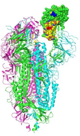
Figure 1. Structural location of the toxin-like sequence (aa 375-390) within the SARS-CoV-2
spike glycoprotein. The Receptor Binding Domain is displayed in a space-filling model, with
Figure 2. (A). Molecular docking of the S1 subunit of the SARS-CoV-2 spike glycoprotein
(purple) with alpha9 AChR extracellular subunit (cyan). The interaction between the two
proteins is caused by hydrogen bonds and shape complementarity. The S1 subunit of the
spike glycoprotein interacts with the toxin-like sequence (aa375-390) (pink color). (B). The
interaction between alpha9 nAChR subunit (aa189-195 are forming the “toxin binding
domain”) and the binding site in more details. C. Surface models.
Α
B
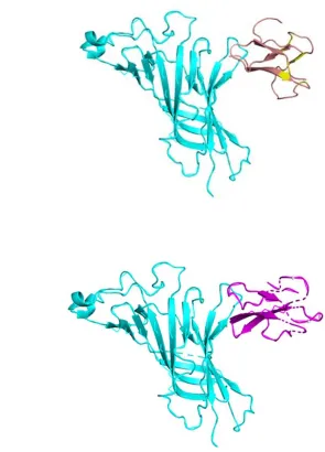
The mode of interaction is similar to the interaction between alpha9 nAChR and alpha-bungarotoxin. (Figure 3). Figure 4 displays the interaction between two alpha9 nAChR subunits and both alpha-bungarotoxin and SARS-CoV-2 spike glycoprotein.
Figure 3. (A). Interaction between alpha9 AChR (cyan - on the left) and alpha-bungarotoxin
(salmon - on the right). (B). Interaction between alpha9 AChR (cyan – on the left) and
neurotoxin homolog NL1 (purple – on the right). The interaction with one alpha9 AChR
subunit is displayed.
Α
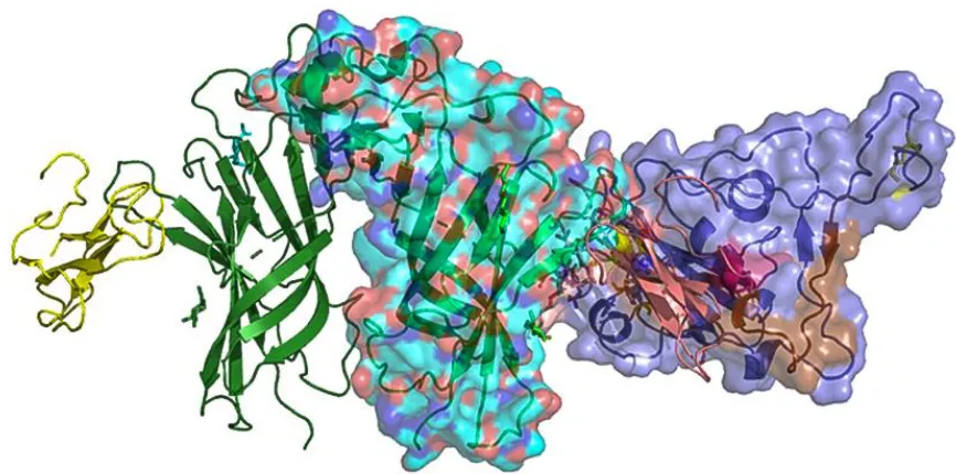
Figure 4. Interaction between two (of the five) alpha9 nAChR subunits (cyan – in the
middle), alpha-bungarotoxin (yellow - on the left) and SARS-CoV-2 spike glycoprotein
(purple – on the right). Alpha-bungarotoxin is shown interacting with the toxin-binding
domain of one alpha9 subunit and the S1 subunit of the SARS-CoV-2 is shown interacting
with the other alpha9 subunit.
Figure 5. (A). Interaction between the ligand binding domain of a pentameric alpha7
nicotinic receptor chimera (left) and the S1 subunit of the SARS-CoV-2 spike glycoprotein
(purple – on the right). (B) Alpha-bungarotoxin and Neurotoxin homolog NL1 bound to the
pentameric nicotinic receptor. (C). S1-SARS-CoV-2 spike glycoprotein (purple – on the right)
bound to the pentameric nicotinic receptor (top view) and side view. (D). The beginning of
the toxin-like sequence (S375) and the top of the finger-like fragment (F392) are shown. E.
Close top view of the interacting surfaces. (F). Neurotoxin homolog NL1 bound to the
pentameric nicotinic receptor in close from top view, emphasizing the interaction between the
toxin and the nicotinic receptor.
Α
C
D
E
Τhe sequence aa185-200 represents an important binding site in the extracellular domain of
AChRs where several neurotoxins, such as alpha-bungarotoxin, bind and exert their receptor antagonist activity.24,25 The molecular modelling and docking experiments suggest an
interaction between alpha9 nAChRs and the SARS-CoV-2 spike glycoprotein, with the sequence aa189-195 of the receptor being at the core of this interaction. Endogenous (acetylcholine) or exogenous (e.g. nicotine) agonists to any nAChR open the ion channel in the receptor, allowing the flow of cations and inducing a wide variety of biological responses. nAChR antagonists, such as alpha-neurotoxins, compete with typical agonists for binding, and their binding is restricted to nAChR alpha-subunits.25
According to our hypothesis, the SARS-CoV-2 spike glycoprotein has a “toxin-like”
sequence on its receptor binding domain which could bind to the toxin binding domain of the alpha subunit of the nAChRs. This binding could produce a number of adverse effects upon viral infection by dysregulating the NCS, in which alpha7 nicotinic receptors are mainly involved. One of the effects could be the dysfunction of the cholinergic anti-inflammatory pathway, resulting in cytokine storm and failure for the immune response to return to homeostasis. Several other clinical manifestations of COVID19 could be explained by
cholinergic dysfunction.11 Nicotine may partially reverse this binding while other compounds may also compete for binding with the SARS-CoV-2 spike glycoprotein; this needs to be studied further and eventually verified in in vitro and in vivo studies.
References
1. Hoffmann M, Kleine-Weber H, Schroeder S, Krüger N, Herrler T, Erichsen S, Schiergens TS, Herrler G, Wu NH, Nitsche A, Müller MA, Drosten C, Pöhlmann S.Leung JM, Yang CX, Tam A, Shaipanich T, Hackett TL, Singhera GK, Dorscheid DR, Sin DD. ACE-2 Expression in the Small Airway Epithelia of Smokers and COPD Patients: Implications for COVID-19. Eur Respir J. 2020 Apr 8. pii: 2000688. doi: 10.1183/13993003.00688-2020. SARS-CoV-2 Cell Entry Depends on ACE2 and TMPRSS2 and Is Blocked by a Clinically Proven Protease Inhibitor. Cell. 2020 Apr 16;181(2):271-280.e8. doi: 10.1016/j.cell.2020.02.052.
2. Wang Q, Zhang Y, Wu L, Niu S, Song C, Zhang Z, Lu G, Qiao C, Hu Y, Yuen KY, Wang Q, Zhou H, Yan J, Qi J. Structural and Functional Basis of SARS-CoV-2 Entry by Using Human ACE2. Cell. 2020 Apr 7:S0092-8674(20)30338-X. doi:
10.1016/j.cell.2020.03.045. Online ahead of print.PMID: 32275855.
3. Walls AC, Park YJ, Tortorici MA, Wall A, McGuire AT, Veesler D. Structure, Function, and Antigenicity of the SARS-CoV-2 Spike Glycoprotein. Cell. 2020 Apr 16;181(2):281-292.e6. doi: 10.1016/j.cell.2020.02.058. Epub 2020 Mar
9.PMID: 32155444.
4. Vaduganathan M, Vardeny O, Michel T, McMurray JJV, Pfeffer MA, Solomon SD. Renin-Angiotensin-Aldosterone System Inhibitors in Patients with Covid-19. N Engl J Med. 2020. doi: 10.1056/NEJMsr2005760.
5. Verdecchia P, Cavallini C, Spanevello A, Angeli F. The pivotal link between ACE2 deficiency and SARS-CoV-2 infection. Eur J Intern Med. 2020 Apr 20. pii: S0953-6205(20)30151-5. doi: 10.1016/j.ejim.2020.04.037.
7. Oakes JM, Fuchs RM, Gardner JD, Lazartigues E, Yue X. Nicotine and the renin-angiotensin system. Am J Physiol Regul Integr Comp Physiol. 2018;315(5):R895-R906. doi: 10.1152/ajpregu.00099.2018.
8. Leung JM, Yang CX, Tam A, Shaipanich T, Hackett TL, Singhera GK, Dorscheid DR, Sin DD. ACE-2 Expression in the Small Airway Epithelia of Smokers and COPD Patients: Implications for COVID-19. Eur Respir J. 2020 Apr 8. pii: 2000688. doi: 10.1183/13993003.00688-2020.
9. Brake SJ, Barnsley K, Lu W, McAlinden KD, Eapen MS, Sohal SS. Smoking
Upregulates Angiotensin-Converting Enzyme-2 Receptor: A Potential Adhesion Site for Novel Coronavirus SARS-CoV-2 (Covid-19). J Clin Med. 2020 Mar 20;9(3). pii: E841. doi: 10.3390/jcm9030841.
10. Farsalinos K, Barbouni A, Niaura R. Systematic review of the prevalence of current smoking among hospitalized COVID-19 patients in China: could nicotine be a therapeutic option? Intern Emerg Med. 2020 May 9. doi: 10.1007/s11739-020-02355-7.
11. Farsalinos K, Niaura R, Le Houezec J, Barbouni A, Tsatsakis A, Kouretas D,
Vantarakis A, Poulas K. Editorial: Nicotine and SARS-CoV-2: COVID-19 may be a disease of the nicotinic cholinergic system. Toxicol Rep. 2020 Apr 30. doi:
10.1016/j.toxrep.2020.04.012.
12. Mehta P, McAuley DF, Brown M, Sanchez E, Tattersall RS, Manson JJ. HLH. Across Speciality Collaboration, UK. COVID-19: consider cytokine storm syndromes and immunosuppression. Lancet. 2020 Mar 28;395(10229):1033-1034. doi:
10.1016/S0140-6736(20)30628-0.
14. Farsalinos K, Eliopoulos E, Tzartos S, Poulas K. Nicotinic Cholinergic System and COVID-19: Identification of a Potentially Crucial Snake Toxin-Like Sequence in the SARS-CoV-2 Spike Glycoprotein. Preprints 2020, 2020050301 (doi:
10.20944/preprints202005.0301.v1).
15. Basic Local Alignment Search Tool. Available online:
http://www.ncbi.nlm.nih.gov/BLAST/
16. Sievers F, Wilm A, Dineen D, Gibson TJ, Karplus K, Li W, Lopez R, McWilliam H, Remmert M, Söding J, Thompson JD, Higgins DG. Fast, scalable generation of high-quality protein multiple sequence alignments using Clustal Omega. Mol Syst Biol. 2011;7:539.
17. Clustal Omega. Available online: https://www.ebi.ac.uk/Tools/msa/clustalo/
18. Okonechnikov K, Golosova O, Fursov M, the UGENE team. Unipro UGENE: a unified bioinformatics toolkit. Bioinformatics. 2012;28:1166-1167. doi.
10.1093/bioinformatics/bts091.
19. Berman HM, Westbrook J, Feng Z, Gilliland G, Bhat TN, Weissig H, Shindyalov LN, Bourne PE. The Protein Data Bank. Nucleic Acids Res. 2000;28:235–242.
20. Zouridakis M, Giastas P, Zarkadas E, Chroni-Tzartou D, Bregestovski P, Tzartos SJ. Crystal structures of free and antagonist-bound states of human α9 nicotinic receptor extracellular domain. Nat Struct Mol Biol. 2014;21(11):976‐980.
doi:10.1038/nsmb.2900.
21. Li SX, Huang S, Bren N, Noridomi K, Dellisanti CD, Sine SM, Chen L. Ligand-binding domain of an alpha 7-nicotinic receptor chimera and its complex with agonist. (2011) Nat Neurosci 14: 1253-1259.
23. MOE (Molecular Operating Environment) available from the Chemical Computing Group (CCG; www.chemcomp.com)
24. Zouridakis M, Zisimopoulou P, Poulas K, Tzartos SJ. Recent Advances in Understanding the Structure of Nicotinic Acetylcholine Receptors. IUBMB Life 2009;61(4):407–423.
25. Marinou M and Tzartos SJ. Identification of regions involved in the binding of