http://www.pharmacophorejournal.com 701
Pharmacophore 2014, Vol. 5 (5), 701-710 USA CODEN: PHARM7 ISSN 2229-5402
Pharmacophore
(An International Research Journal)
Available online at http://www.pharmacophorejournal.com/
Original Research Paper
IN VITRO
FREE RADICAL SCAVENGING ACTIVITY OF AQUEOUS FRUIT
EXTRACT OF
COCCINA INDICA
Swathi Putta
* and Eswar Kumar kilari
CSIR-SRF-Extended, Department of Pharmacology, A.U. College of Pharmaceutical
Sciences, Andhra University,Visakhapatnam, India
ABSTRACT
In this study, we evaluated the antioxidant potential of aqueous fruit extract of Coccinia indica (AFCI) using different in vitro assays including the scavenging activities of super oxide radical, hydroxyl radical, hydrogen peroxide, DPPH radical, Nitric oxide radical, Fe+3 radical scavenging activity. The extract was evaluated for antioxidant potential by the phosphomolybdenum method, Fe+2 chelating activity, ferric reducing ability power (FRAP) and ABTS+ radical scavenging assay. The activity of AFCI was compared with standard antioxidant ascorbic acid. The results indicated that the AFCI possesses the highest antioxidant activity. The antioxidant activity of AFCI is due to its rich source of β- carotene, lycopene, taraxerone, taraxerol and β-sitosterol. The results obtained in the present study indicate that the fruit of
Coccinia indica are a potential source of antioxidants.
Keywords:
Coccinia indica,Antioxidant, Free radical.INTRODUCTION
Oxygen consumption inherent in cell growth leads to the generation of a series of reactive oxygen species (ROS) (Barros L et al., 2006). They are continuously produced by the body’s normal use of oxygen such as respiration and some cell mediated immune functions. ROS include free radicals such as superoxide anion radicals (O2•−), hydroxyl radicals (OH•) and non-free radical species such as hydrogen peroxide (H2O2) and singlet oxygen (1O2) (Gu lc I, 2006). Antioxidants may be defined as compounds that inhibit or delay the oxidation of other molecules by inhibiting the initiation or propagation of oxidizing chain reactions (Velioglu et al., 1998). Antioxidants can protect the human body from free radicals and ROS effects. They retard the progress of many chronic diseases as well as lipid peroxidation (Lai LS et al., 2001). Hence, a need for identifying alternative natural and safe sources of food antioxidants has been created, and the
search for natural antioxidants, especially of plant origin, has notably increased in recent years. Natural antioxidants are known to exhibit a wide range of biological effects including antibacterial,
antiviral, antiinflammatory, antiallergic,
antithrombotic and vasodilatory activities. In fact, a fundamental property important for life is the antioxidant activity and this property may give rise to anticarcinogenicity, antimutagenicity and antiaging activity, among others (Cook and Samman, 1996). The ethnobotanical information reports that about 800 plants may possess an antidiabetic potential including Coccinia indica
(synonym Coccinia cordifolia, ivy gourd)
(Alarcon-Aguilara et al., 1998). The active constituents present in the fruit are taraxerone, taraxerol, and (24R)-24- ethylcholest- 5- en- 3β-
ol glucoside. β- carotene, lycopene,
Swathi Putta et al. / Pharmacophore 2014, Vol. 5 (5), 701-710
K et al., 1972; Bhakuni DS et al., 1962). C. indica is used in Ayurveda and Unani systems of medicine for the treatment of diabetes, skin eruptions, tongue sores, earaches, etc. in India (Chopra et al., 1956; Chopra et al., 1958). The root is cooling, aphrodisiac, stops vomiting urinary losses, burning of hands and feet, given for uterine discharges (Chandra S et al., 2001). The ethanolic extracts of leaves, stems and fruits showed significant anti-inflammatory activity (Juneia D et al., 2007; Bambal VC et al., 2010). The methanolic extract of leaves and fruits of C. indica possesses significant antimicrobial activity with different potency of selected micro organisms (Dewanjee S et al., 2007; Saheen SZ et al., 2009). The present study was designed to evaluate the In Vitro antioxidant potential of aqueous fruit extract of Coccinia indica by using different in vitro models of radical scavenging activities.
MATERIALS AND METHODS
Materials
Nitroblue tetrazolium (NBT), were obtained from Merck. 1,1-diphenyl-2-picrylhydrazyl (DPPH),
2,4 dinitrophenylhydrazine (DNPH),
trichloroacetic acid (TCA) were obtained from Sigma (St. Louis, MO, USA). 2.4.6-Tri- (2ʹ-pyridyl)-1, 3, 5-triazine (TPTZ), 2, 2ʹ- Azinobis- (3-ethylbenzothiazoline- 6-sulfonic acid) (ABTS) and Trolox were obtained from Sigma Aldrich Chemical Co., Ltd. (England). All other reagents were of analytical reagent (AR) grade.
Plant Materials and Preparation
The aqueous fruit extract of Coccinia indica was obtained from laila Implex, vijiyawada.
Super Oxide Radical Scavenging Activity
The assay was based on the capacity of the aqueous extract to inhibit formazan formation by scavenging the superoxide radicals generated in riboflavin-light- NBT system (Beauchamp & Fridovich, 1971). The reaction mixture contained 58 mM phosphate buffer, pH 7.6, 20 µM riboflavin, 6 mM EDTA, and 50 µM NBT, final volume made up to 3 ml, added in that sequence. Initiated with the reaction (Kumaran R et al., 2006) the reaction mixture with the different concentrations was exposed to 40 volts under
fluorescence lamp for 15 min to initiate the reaction. Immediately after illumination, the absorbance was measured at 560 nm. The entire reaction assembly was enclosed in a box lined with aluminium foil. Identical tubes, with reaction mixture, above were kept in the dark and served as blanks. The percentage inhibition of superoxide anion generation was calculated using the following formula:
% Inhibition = AO – A1 100 AO
Where A0 was the absorbance of the control, and
A1 was the absorbance of the aqueous
extract/standard. All experiments were performed in triplicate.
Hydroxyl Radical Scavenging Activity
Scavenging activity of hydroxyl radical was measured by the method of Halliwell et al., 1985. Hydroxyl radicals were generated by a Fenton reaction (Fe3+-ascorbate-EDTA-H2O2 system), and the scavenging capacity of the extract and standard towards the hydroxyl radicals was measured by using deoxyribose method. The reaction mixture contained 2-deoxy-2-ribose (2.8 mM), phosphate buffer (0.1 mM, pH 7.4), ferric chloride (20 μM), EDTA (100 μM), hydrogen peroxide (500 μM), ascorbic acid (100 μM) and various concentrations (10-1000 μg/ml) of the test sample in a final volume of 1 ml. The mixture was incubated for 1 h at 37 °C. After the incubation an aliquot of the reaction mixture (0.8 ml) was added to 2.8% TCA solution (1.5 ml), followed by TBA solution (1% in 50 mM sodium hydroxide, 1 ml) and sodium dodecyl sulphate (0.2ml). The mixture was then heated (20 min at 90 °C) to develop the colour. After cooling, the absorbance was measured at 532 nm against an appropriate blank solution. All experiments were performed in triplicates. The percentage of inhibition was expressed, according to the following equation:
% Inhibition = AO – A1 100 AO
Swathi Putta et al. / Pharmacophore 2014, Vol. 5 (5), 701-710
http://www.pharmacophorejournal.com 703
Hydrogen Peroxide Radical Scavenging Activity
The hydrogen peroxide scavenging assay was carried out following the procedure of Ruch et al., 1989. The principle of this method is that there is a decrease in absorbance of H2O2 upon oxidation of H2O2. A solution of 43 mM H2O2 was prepared in 0.1M phosphate buffer (pH 7.4). The AFCI of different concentrations were prepared in 3.4 mL phosphate buffer were added to 0.6 mL of H2O2 solution (43 mM) and absorbance of the reaction mixture was recorded at 230 nm. All experiments were performed in triplicates. The percentage of inhibition was expressed, according to the following equation:
% Inhibition = AO – A1 100 AO
Where A0 was the absorbance of the control without a sample, A1 is the absorbance in the presence of the sample.
DPPH Radical Scavenging Activity
The potential of extract and AA was determined on the basis of the scavenging activity of the stable 1, 1-diphenyl-2-picrylhydrazyl (DPPH) free radical. Aliquots of 1ml of a methanolic solution containing each concentration of extract were added to 3 ml of 0.004% MeOH solution of DPPH. Absorbance at 517 nm, against a blank of methanol without DPPH, was determined after 30
min (UV, Perkin-Elmer-Lambda 11
spectrophotometer) and the percent inhibition activity was calculated (Braca et al., 2001). The percentage of inhibition was expressed, according to the following equation:
% Inhibition = AO – A1 100 AO
Where A0 was the absorbance of the control without a sample, A1 is the absorbance in the presence of the sample. All tests were run in triplicate and averaged.
Nitric Oxide Radical Scavenging Activity
Nitric oxide generated from sodium nitroprusside in aqueous solution at physiological pH interacts
with oxygen to produce nitrite ions, which were measured by the Griess reaction (Ebrahimzadeh MA et al., 2010). The reaction mixture (3 ml) containing sodium nitroprusside (10 mM) in phosphate buffered saline (PBS) and AFCI and the AA in different concentrations were incubated at 25°C for 150 min. After incubation 1.5 ml of the Griess reagent (1% sulphanilamide and 0.1% naphthyl ethylene diamine dihydrochloride in 2% H3PO4) was added. The absorbance of the chromophore formed was measured at 546nm. The percentage of inhibition was expressed, according to the following equation:
% Inhibition = AO – A1 100 AO
Where A0 was the absorbance of the control without a sample, A1 is the absorbance in the presence of the sample.
Reducing Power
The reducing power of the extract was determined according to the method of Oyaizu et al., 1986. Various concentrations of the extracts (mg/ml) in distilled water were mixed with phosphate buffer (2.5 ml, 0.2 M, pH 6.6) and 1% of potassium ferricyanide water solution (2.5 ml, K3 [Fe (CN)6 ]). The mixture was incubated at 50 oC for 20 min. Aliquots of trichloracetic acid (2.5
ml, 10% aqueous solution) were added to the mixture which was then centrifuged at 3000 rpm for 10 min. The supernatant (2.5 ml) was mixed with distilled water (2.5 ml) and a freshly prepared FeCl3 solution (0.5 ml, 0.1%). The absorbance was measured at 700 nm. The percentage of inhibition was expressed, according to the following equation:
% Inhibition = AO – A1 100 AO
Where A0 was the absorbance of the control without a sample, A1 is the absorbance in the presence of the sample.
Phosphomolybdinum Method
Swathi Putta et al. / Pharmacophore 2014, Vol. 5 (5), 701-710
Prieto et al., 1999. An aliquot of 0.1 ml of sample solution (equivalent to 100 lg) was combined with 1 ml of reagent solution (0.6M sulfuric acid, 28 mM sodium phosphate, and 4 mM ammonium molybdate). In the case of the blank, 0.1 ml of methanol was used in place of sample. The tubes were capped and incubated in water bath at 950C for 90 min. After the samples were cooled to RT, the absorbance of the aqueous solution of each was measured at 695 nm.
The percentage of inhibition was expressed, according to the following equation:
% Inhibition = AO – A1 100 AO
Where A0 was the absorbance of the control without a sample, A1 is the absorbance in the presence of the sample.
Fe+2Chelating Activity
The chelating activity of the extracts for ferrous ions (Fe2+)was measured according to the method of Dinis et al., 1994. To 0.5 ml of extract, 1.6 ml of deionized water and 0.05 ml of FeCl2 (2 mM) was added. After 30 sec, 0.1 ml ferrozine (5 mM) was added. Ferrozine reacted with the divalent iron to form stable magenta complex species that were very soluble in water. After 10 min at room temperature, the absorbance of the Fe2+/Ferrozine complex was measured at 562 nm. The percentage of inhibition was expressed, according to the following equation:
% Inhibition = AO – A1 100 AO
Where A0 was the absorbance of the control without a sample, A1 is the absorbance in the presence of the sample.
Ferric Reducing Ability Power
The FRAP method measures the absorption change that appears when the TPTZ (2,4,6 -tri pyridyl-s-triazine)-Fe3+ complex is reduced to the TPTZ-Fe2+ form in the presence of antioxidants (Benzie IF, 1996). An intense blue colour develops with absorption maximum at 595 nm. The FRAP reagent contained 2.5 ml of 10 mM tripyridyltriazine (TPTZ) solution in 40 mM HCl plus 2.5 ml of 20 mM FeCl3 and 25 ml of 0.3 M
acetate buffer, pH 3.6, was freshly prepared. The extracts were dissolved in ethanol at a concentration of 1 mg/ml. An aliquot of 0.2 ml of solution was mixed with 1.8 ml of FRAP reagent and the absorption of the reaction mixture was measured at 595 nm. Ethanolic solutions of known Fe (II) concentration, in the range of 50-1000 μM (FeSO4), were used for obtaining the calibration curve (Figure 2). The FRAP value represents the ratio between the slope of the linear plot for reducing Fe3+-TPTZ reagent by plant extract compared to the slope of the plot for FeSO4.
ABTS+ Assay
The ABTS assay was based on the method of Re
et al., 1999 with slight modifications. ABTS radical cation (ABTS+) was produced by reacting 7 mM ABTS solution with 2.45 mM potassium persulphate and allowing the mixture to stand in the dark at room temperature for 12-16 h before use. The ABTS+ solution was adjusted to an absorbance 0.70±0.02 by diluting with ethanol at 734 nm. The 25µl of sample or standard Trolox was added to 2 ml of diluted ABTS+ solution, and the absorbance was measured after 6 min. The decrease in absorption with the addition of different concentrations of extract was used for calculating TEAC values. A standard curve was prepared by measuring the reduction in absorbance of ABTS+ solution at different concentrations of Trolox (Figure 4). Appropriate blank measurements were carried out and the values recorded. The reduction in the absorbance of different concentrations of extract was measured from the trolox standard graph a TEAC values. Results were expressed as Trolox equivalent antioxidant capacity (TEAC).
Animals
Swathi Putta et al. / Pharmacophore 2014, Vol. 5 (5), 701-710
http://www.pharmacophorejournal.com 705 (Hindustan liver Bangalore). After randomization
before the experiment, the rats were acclimatized for a period of two weeks. The animal housing and handling were in accordance with CPSCEA guidelines. Our college was approved by CPCSEA for conducting animal experiments with the registration No. 516/01/A/CPCSEA. The prior permission for the study was obtained from our Institutional Animal Ethics Committee (IAEC).
RESULTS AND DISCUSSION
Several phytochemicals possessing polyphenolic structures are advocated as neutraceuticals to supplement food for better health care during recent years. Most of them are claimed to possess antioxidant activity. Ayurveda and naturopathy the medical systems indigenous to India advocate the use of plant extracts/ mixtures of extracts for treating various disorders apart from others from times immemorial in humans without preclinical evidence, which is required to make the systems popular and scientific. The claimed usefulness of herbs in several disorders might be due to their antioxidant activity. To support the use of the selected plant extracts in herbal mixture and in Ayurveda and naturopathy, the antioxidant potential of the aqueous extracts of Coccinia indica of Indian origin were investigated in comparison with the known antioxidant ascorbic acid (AA) following in vitro studies. Herbal drugs containing antiradical constituents are gaining importance in prevention and treatment of stress related disorders. The free radical scavengers like polyphenolics are well known for their therapeutic activity in disorders such as cancer, diabetes and skin diseases (Jose JK, 1995). Earlier reports on the above plants indicated the presence of compounds representing polyphenols, flavonoids, terpenoids, tannins, alkaloids etc (Rasstogi, 1991; Monika B, 2005).
The superoxide radical (O2-) scavenging activity of the AFCI, as measured by the riboflavin- NBT-light system in vitro. Superoxide radical is known to be very harmful to cellular components as a precursor of more reactive oxygen species (Halliwell B et al., 1985). During the electron transport chain in mitochondria one of the carrier electrons was replaced by oxygen, so produce
superoxide radical. To prevent the superoxide radical mediated injury, cells contain superoxide dismutase (SOD) as a cellular antioxidant enzyme, which removes this ubiquitous superoxide metabolic product by converting it into hydrogen peroxide and oxygen and this hydrogen peroxide radical readily decomposed into hydroxyl radical in the presence of catalase in biological systems (Chan PC et al., 1995; Lehmann et al., 2000; Giorgio et al., 2007). The AFCI were shown similar activity like AA in scavenging the superoxide and hydroxyl radicals. Hydrogen peroxide itself is not very reactive, but reacts with transitional metal ion dependent OH radical mediated oxidative damage to the DNA. The AFCI were shown better activity than AA in scavenging the hydrogen peroxide radical. Hydroxyl radicals can occasionally produced as a byproduct of immune action by macrophages. They can damage virtually all types of macromolecules like carbohydrates, nucleic acids, lipids and amino acids. The hydroxyl radicals cannot be eliminated by enzymatic reactions, only endogenous antioxidants can scavenge these radicals (Sies et al., 1993; Reiter RJ et al., 1995). The DPPH known to abstract labile hydrogen and the ability to scavenge the DPPH radical is related to the inhibition of lipid peroxidation. The AA was shown better activity than AFCI in scavenging the DPPH radical (table 1). The antioxidant products are capable of scavenging free radicals at pH 7.4 via electron or hydrogen donating mechanisms and thus should be able to prevent the initiation of deleterious free radical mediated chain reactions. Nitric oxide is a free radical in terms its unpaired electron. It reacts with O2- in termination reactions in the mitochondrial matrix, yielding peroxynitrite (ONOO-). These oxyradical and peroxynitrite induce oxidative damage to mitochondrial DNA damage and protein inactivation and ATP synthesis (Wink DA et al., 1998). The antioxidant capability of AFCI was found to have better than ascorbic acid because of presence of active
principles like tannins, phenols and
Swathi Putta et al. / Pharmacophore 2014, Vol. 5 (5), 701-710
Iron is essential for life due to its unusual flexibility to serve as both an electron donor and acceptor. Among the transition metals, iron is known as the most important lipid oxidation. In nature it can be found as either ferrous (Fe2+) or ferric (Fe3+). If iron is free within the cell it can catalyze the conversion of hydrogen peroxide into free radicals (Andrew NC et al., 1992; Conred ME et al., 2000). Minimizing these ions may afford protection against oxidative damage by inhibiting production of ROS. Reducing power is often used as an indicator of electron donating activity, which is an important mechanism of phenolic antioxidant action, can be strongly correlated with other antioxidant properties. The presence of reductants such as antioxidant substances in the AFCI causes the reduction of the Fe3+/ferricyanide complex to the ferrous form (Fe+2). In the phosphomolybdinum method is based on the reduction of Mo (VI) to Mo (V) by the antioxidant compounds and the formation of green Mo (V) complexes (Halliwell et al., 1985). In the metal chelating assay, ferrozine can quantitatively form complexes with Fe2+. In the presence of other chelating agents, the complex formation is disrupted with consequent decrease in the intensity of the red color of the complex. Our results indicated that the AFCI showed a dose dependent effect with an increase in the concentration (Graph 1). FRAP assay measures the reducing ability of antioxidant that react with ferric tripyridyltriazine (Fe3+-TPTZ) complex and produce a coloured ferrous tripyridyltriazine (Fe2+- TPTZ) (Benzie IF.,1996). Using this assay, the FRAP value of AFCI shown to have dose
dependent effect with an increase in the concentration (Graph 2).
The ABTS+ radical assay can be used to measure the substances, i.e., both aqueous phase radicals and lipid peroxyl radicals. The scavenging activity of the extract on the radical ABTS, generated by potassium persulfate was compared with a standard amount of trolox. In general, medicinal plants may play an important role in chemical protection from oxidative damage by possessing endogenous antioxidants such as phenolic compounds. Hagerman et al., 1998 have reported that the high molecular weight phenolics (tannins) have more ability to quench free radicals (ABTS+) (Hagerman AE et al., 1998). The TEAC value of AFCI was found be better than AA.
CONCLUSIONS
It is concluded that, using different in vitro
models for estimation the antioxidant potential of
Coccinia indica Sshowed better activity in scavenging the different free radicals. In addition to its antioxidant activity it also showed better activity against the formation of DPPH and ABTS radicals. All the activities might be due to high levels of β- carotene, lycopene, taraxerone, taraxerol and β – sitosterol present in fruit extract of Coccinia indica.
ACKNOWLEDGMENTS
The author acknowledges the financial support for completion of research work from Council for Scientific Industrial research, New Delhi (REF: 111177/2K11/1).
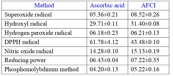
Table 1: Effect of Ascorbic acid and APPG on IC50 values of different in vitro models
Method Ascorbic acid AFCI
Superoxide radical 05.36±0.21 08.52±0.26
Hydroxyl radical 29.71±0.11 31.40±0.08
Hydrogen peroxide radical 06.18±0.23 06.21±0.13
DPPH radical 61.78±4.12 43.48±0.10
Nitric oxide radical 14.28±0.10 15.13±0.19
Reducing power 06.43±0.04 07.22±0.35
Swathi Putta et al. / Pharmacophore 2014, Vol. 5 (5), 701-710
http://www.pharmacophorejournal.com 707
Graph 1: Effect of AA and AFCI on Fe+ chelating activity
Graph 2: Effect of AA and AFCI on ferric reducing ability power (FRAP) assay
Graph 3: Effect of AA and AFCI in ABTS radical scavenging activity
REFERENCES
1. Alarcon-Aguilara, FJ, Roman-Romos, R;
Perez-Gutierrez, S; Aguilar-Contreras, A; Contreras-Weber, CC and Flores-Saenz, JL (1998), “Study of the anti-hyperglycemic effect of plants used as antidiabetics”,
Journal of Ethnopharmacology, 61, 101-110.
2. Andrews, NC (1992), “Disorders of iron metabolism”, N Engl J Med, 341(26), 1986-95.
3. Bambal, VC; Wani, NS; Chaudhari, SP;
Anti-Swathi Putta et al. / Pharmacophore 2014, Vol. 5 (5), 701-710
inflammatory Activity of Coccinia indica Wight and Arn. (Cucurbitaceae) fruits”, Latin
American Journal of Pharmacy, 29 (5),
820-24.
4. Barros, L;Ferreira, M and Queiros, B (2006), “Total phenols, ascorbic acid, β-carotene and
lycopene in Portuguese wild edible
mushrooms and their antioxidant activities”,
Food Chem, 103, 413-419.
5. Basu, K and Ghosh, BK (1972), “Chemical
investigation of Coccinia indica”,
Transactions of the Bose Research Institute
(Calcutta), 35(2), 43-4.
6. Beauchamp, C and Fridovich, I (1971),
“Superoxide dismutase: improved assays and an assay applicable to acrylamide gels”,
Analytical Biochemistry,44, 276-277.
7. Benzie, IF and Strains, JJ (1996), “The ferric reducing ability of plasma (FRAP) as a measure of "antioxidant power" The FRAP assay”, Anal Biochem,239, 70-6.
8. Bhakuni, DS; Srivastava, SN; Sharma, VN and Kaul, KN (1962), “Chemical examination of the fruits of Coccinia indica”, Journal of Scientific and Industrial Research, Section
B: Physical Sciences, 21, 237-8.
9. Braca, A; Tommasi, N; Bari, LD; Pizza, C; Politi, M and Morelli, I (2001), “Antioxidant principles from Bauhinia terapotensis”,
Journal of Natural Products, 64, 892-895.
10. Chan, PC; Sills, RC; Braun, AG; Haseman, JK and Bucher, JR (1995), “Toxicity and
carcinogenicity of delta
9-tetrahydrocannabinol in Fischer rats and
B6C3F1 mice”, Fundam Appl Toxicol, 30,
109-117.
11. Chandra, S (2001), “The Ayurvedic Pharmacopoeia of India. Government of India, Ministry of Health and Family Welfare, Department of Indian System of Medicine and
Homeopathy, Part 1”, 1st Ed., Vol.III,
National Institute of Science
Communication, New Delhi, 32-34.
12. Chopra, RN; Chopra, IL; Handa, KL and Kapur, LD (1958), “Indigenous drugs of
India”, 2nd Ed. Calcutta, UN Dhar and Sons
Pvt.Ltd. India, 314-316.
13. Chopra, RN; Nayar, SL and Chopra, IC (1956), “Glossary of Indian Medicinal plants”, CSIR, New Delhi.
14. Conrad, ME and Umbreit, JN (2000), “Disorders of iron metabolism”, N Engl J Med, 342(17), 1293-4.
15. Cook, NC and Samman, S (1996),
“Flavonoids: chemistry, metabolism,
cardioprotective effects and dietary sources”,
Journal of Nutritional Biochemistry, 7, 66.
16. Dewanjee, S; Kundu, M; Maiti, A; Majumdar,
R; Majumdar, A and Mandal, SC (2007), “In
vitro evaluation of anti-micribial activity of
crude extract from plants Diospyros
peregrine, Coccinia indica and Swietenia microphylla”
, Trp J Pharm Res, 6(3), 773-78.
17. Dinis, CP; Madeira, VMC and Almeida, LM (1994), “Action of phenolic derivatives
(acetaminophen, salicylate, and
5-aminosalicylate) as inhibitors of membrane lipid peroxidation and as peroxyl radical
scavengers”, Arch Biochem Biophy, 315,
161-9.
18. Ebrahimzadeh, MA and Nabavi, SF (2010), “In Vitro antioxidant and free radical scavenging activity of Leonurus cardiaca
subsp. Persicus, Grammosciadium
platycarpum and Onosma demawendicum”,
Afr J of Biotech, 9(51), 8865-8871.
19. Giorgio, M; Trinei, M; Migliaccio, E and Pelicci, PG (2007), “Hydrogen peroxide a metabolic byproduct or a common mediator of ageing signals”, Nat Rev Mol Cell Biol, 8(9), 722-8.
20. Gu, lc I (2006), “Antioxidant and antiradical activities of l-carnitine”, Life Sci, 78, 803-811.
21. Hagerman, AE; Riedl, KM; Jones, GA; Sovik, KN; Ritchard, NT and Hartzfeld, PW (1998),
“Higher molecular weight oplant
polyphenolics(Tannins) as biological
antioxidant”, J Agric Food Chem, 46, 1887-1892.
Swathi Putta et al. / Pharmacophore 2014, Vol. 5 (5), 701-710
http://www.pharmacophorejournal.com 709 radicals in biology and medicine”, Oxford
Clarendron Press, 279-315.
23. Jose, JK and Kuttan, R (1995), “Antioxidant activity of Emblica officinalis”, J Clin
Biochem Nutr, 19, 63 -70.
24. Juneja, D; Shrivastava, PN; Guna, MK and
Saxena, RC (2007), “Preliminary
Phytochemical Screening of some folklore medicinal plants for their anti-inflammatory activity”, Phcog Mag, 11, 201-03.
25. Kumaran, A and Karunakaran, RJ (2006), “In vitro antioxidant activities of methanol extracts of five Phyllanthus species from India”, LWT, 40, 344-352.
26. Kumaran, A and Karunakaran, RJ (2007), “In vitro antioxidant activities of methanol extracts of five Phyllanthus species from India”, LWT, 40, 344-352.
27. Kundu, Sujata and Ray, AB (1987), “Chemical examination of Coccinia indica fruits”, Journal of the Indian Chemical
Society, 64(12), 776-7.
28. Lai, LS; Chou, ST and Chao, WW (2001), “Studies on the antioxidative activities of Hsian-tsao (Mesona procumbens Hemsl) leaf gum”, J Agric Food Chem, 49, 963-968. 29. Lehmann, R and Schleicher, ED (2000),
“Molecular mechanism of diabetic
nephropathy”, Clin Chim Acta,297, 135-44. 30. Monika, B; Anurag, P; Tiwari, SK and
Prakash, D (2005), “Phenolic contents and antioxidant activity of some food and medicinal plants”, International Journal of
Food and Nutrition, 56(4), 287-291.
31. Oyaizu, M (1986), “Studies on product of browning reaction prepared from glucose amine”, Japanese Journal of Nutrition, 44, 307-315.
32. Prieto, P; Pineda, M and Aguilar, MM (1999),
“Spectrophotometric quantitation of
antioxidant capacity through the formation of a phoshomolybdenum complex: specific application to the determination of vitamin
E”, Anal Biochem,269, 337-341.
33. Rastogi, RP and Mahrotra, BN (1991), “Compendium of Indian Medicinal Plants”,
Publication and Information Directorate,
New Delhi, India, 2
34. Re, R; Pellegrini, N; Proteggente, A; Pannala, A; Yang, M and Rice-Evans, C (1999), “Antioxidant activity applying an improved ABTS radical cation decolorization assay”,
Free Radic Biol Med , 26(9-10), 1231-7.
35. Reiter, RJ; Melchiorri, D and Sewerynek, E (1995), “A review of the evidence supporting melatonin’s role as an antioxidant”, J Pineal Res, 18(1), 1-11.
36. Ruch, RJ; Cheng, SJ and Klaunig, JE (1989), “Prevention of cytotoxicity and inhibition of intracellular communication by antioxidant catechins isolated from Chinese green tea”,
Carcinogenesis, 10, 1003-1008.
37. Shaheen, SZ; Bolla, K; Vasu, K and Singara, charya MA (2009), “Anti-microbial activity of the fruit extracts of Coccinia indica”,
African Journal of Biotechnology, 8(24),
7073-76.
38. Sies, Helmut (1993), “Strategies of
antioxidant defense”, Euro Jour Bio Chem, 215(2), 213-219.
39. Velioglu, YS; Mazza, G; Gao, L and Oomah, BD (1998), “Antioxidant activity and total phenolics in selected fruits, vegetables and grain products”, Journal of Agricultural and
Food Chemistry, 46, 4113.
40. Wink, DA and Mitchell, JB (1998), “Chemical biology of nitric oxide; Insights into regulatory, cytitoxic, cytoprotective mechanisms of nitric oxide”, J Biol Chem, 273, 11044-11048.
Correspondence Author: Swathi Putta
Swathi Putta et al. / Pharmacophore 2014, Vol. 5 (5), 701-710