R E S E A R C H
Open Access
Posttranslationally modified progesterone
receptors direct ligand-specific expression
of breast cancer stem cell-associated gene
programs
Todd P. Knutson
1, Thu H. Truong
1, Shihong Ma
2, Nicholas J. Brady
3, Megan E. Sullivan
4, Ganesh Raj
2,
Kathryn L. Schwertfeger
5and Carol A. Lange
1*Abstract
Background:Estrogen and progesterone are potent breast mitogens. In addition to steroid hormones, multiple signaling pathways input to estrogen receptor (ER) and progesterone receptor (PR) actions via posttranslational events. Protein kinases commonly activated in breast cancers phosphorylate steroid hormone receptors (SRs) and profoundly impact their activities.
Methods:To better understand the role of modified PRs in breast cancer, we measured total and phospho-Ser294 PRs in 209 human breast tumors represented on 2754 individual tissue spots within a tissue microarray and assayed the regulation of this site in human tumor explants cultured ex vivo. To complement this analysis, we assayed PR target gene regulation in T47D luminal breast cancer models following treatment with progestin (promegestone; R5020) and antiprogestins (mifepristone, onapristone, or aglepristone) in conditions under which the receptor is regulated by Lys388 SUMOylation (K388 intact) or is SUMO-deficient (via K388R mutation to mimic persistent Ser294 phosphorylation). Selected phospho-PR-driven target genes were validated by qRT-PCR and following RUNX2 shRNA knockdown in breast cancer cell lines. Primary and secondary mammosphere assays were performed to implicate phospho-Ser294 PRs, epidermal growth factor signaling, and RUNX2 in breast cancer stem cell biology.
Results:Phospho-Ser294 PR species were abundant in a majority (54%) of luminal breast tumors, and PR promoter selectivity was exquisitely sensitive to posttranslational modifications. Phospho-PR expression and target gene programs were significantly associated with invasive lobular carcinoma (ILC). Consistent with our finding that activated phospho-PRs undergo rapid ligand-dependent turnover, unique phospho-PR gene signatures were most prevalent in breast tumors clinically designated as PR-low to PR-null (luminal B) and included gene sets associated with cancer stem cell biology (HER2,PAX2, AHR,AR,RUNX). Validation studies demonstrated a requirement for RUNX2 in the regulation of selected phospho-PR target genes (SLC37A2). In vitro mammosphere formation assays support a role for phospho-Ser294-PRs via growth factor (EGF) signaling as well as RUNX2 as potent drivers of breast cancer stem cell fate.
(Continued on next page)
* Correspondence:[email protected] 1
Departments of Medicine (Division of Hematology, Oncology, and Transplantation) and Pharmacology, Masonic Cancer Center, University of Minnesota, Delivery Code 2812, Cancer and Cardiovascular Research Building, 2231 6th St SE, Minneapolis, MN 55455, USA
Full list of author information is available at the end of the article
(Continued from previous page)
Conclusions:We conclude that PR Ser294 phosphorylation is a common event in breast cancer progression that is required to maintain breast cancer stem cell fate, in part via cooperation with growth factor-initiated signaling
pathways and key phospho-PR target genes includingSLC37A2andRUNX2. Clinical measurement of phosphorylated
PRs should be considered a useful marker of breast tumor stem cell potential. Alternatively, unique phospho-PR target gene sets may provide useful tools with which to identify patients likely to respond to selective PR
modulators that block PR Ser294 phosphorylation as part of rational combination (i.e., with antiestrogens) endocrine therapies designed to durably block breast cancer recurrence.
Keywords:Progesterone receptor (PR), Phosphorylation, ERK/MAP kinase (MAPK), SUMOylation, Antiprogestin, Onapristone, Estrogen receptor (ER), Breast cancer, RUNX2, Cancer stem cells
Background
Breast cancer is the most commonly diagnosed cancer in women with over 246,000 new cases estimated for 2016 and the second leading cause of cancer-related death in women [1]. Recent publications by The Cancer Genome Atlas (TCGA) Network and others have revealed funda-mental molecular characteristics of breast cancer [2–5]. Most notably, the four major breast cancer subtypes (luminal A, luminal B, HER2-enriched, and basal-like) were identified and comprehensively analyzed across data-sets that included mRNA expression, protein expression/ activation, microRNA expression, DNA copy number variation, DNA methylation, and exome sequencing [5]. These results revealed that breast cancers display a wide range of tumor heterogeneity caused by alterations in multiple factors, including somatically mutated driver genes (e.g., TP53, PIK3CA, AKT1, CBFB, GATA3, and
MAP3K1), gene amplifications (ERBB2), and hormonally regulated gene programs (driven primarily by estrogen, progesterone, and androgen steroids) [2, 3, 6]. Further genomic analyses reveal additional stratification of breast cancer subtypes. For example, recent TCGA analysis com-pared ductal and lobular histological subtypes, revealing new molecular factors strongly associated with lobular subtypes, including mutations that lead to activation of AKT and increased FOXA1 expression and activity (i.e., key inputs to amplified estrogen receptor (ER) signaling) [7]. Despite these important findings, targeting the domin-ant molecular pathways has not been completely success-ful and many women experience tumor relapse after treatment with targeted therapies (i.e., ~40% of women re-ceiving tamoxifen suffer relapse [8–14]). Thus, a deeper mechanistic understanding of the complex molecular pathways that drive breast cancer progression and how they may emerge accompanied by more precise bio-markers is urgently needed to successfully impede tumor initiation, optimize and customize treatment strategies, as well as prevent disease progression while undergoing long-term (i.e., up to 10 years) endocrine therapy.
In breast cancer models, progesterone receptor (PR) target gene selectivity is profoundly affected by cellular
context. Like other steroid hormone receptor (SR) family members, PRs are heavily posttranslationally modified and thus act as molecular sensors for abnormally elevated or active signaling pathways. Little overlap exists between PR transcriptomes assayed in normal relative to neoplastic breast cells [15]. In cancer cells, posttranslational modifications (namely, phosphorylation and SUMOylation; see Fig. 1a) create unique PR species whose altered behavior as ligand-dependent transcrip-tion factors is predicted to impact tumor initiatranscrip-tion and progression [16–20]. As an estrogen-induced target gene and downstream effector of ER action, PR is a master transcription factor that, via both autocrine and para-crine mechanisms, regulates the expansion of luminal and basal mammary epithelial cells during breast development and pregnancy [21–23]. In breast cancer models, numerous ER target genes require PR expres-sion (i.e., via scaffolding actions in ER/PR complexes) for optimal estrogen responsiveness in the absence of ex-ogenously added progesterone [24], while PR can dom-inantly repress or modify ER actions as part of ER/PR complexes when both ligands (i.e., estrogen and proges-terone agonists or antagonists) are combined [25, 26]. These studies illustrate both the complexity and elo-quence of SR actions, alone or as frequent collaborators with other SRs and signaling pathways. Recently, PR in cooperation with HER2 signaling was directly implicated as a potent promotor of the “seeds” of early disseminat-ing tumors [27]. These and other studies [28–30] under-score the importance of fully understanding the context(s) for deleterious PR actions relevant to breast cancer biology and particularly to luminal breast tumor progression.
(i.e., cycling) breast cancer cells. Phosphorylation of PR Ser294 is associated with transcriptional hyperactivity at select phosphorylation-responsive PR target genes re-quired for increased cell proliferation and survival in vitro. Activated phospho-PRs are deSUMOylated and subject to more rapid ligand-dependent receptor down-regulation by the ubiquitin proteasome relative to de-phosphorylated and SUMOylated PR species [31, 32], explaining the inverse relationship between PR pro-tein levels and peak expression of PR target gene transcripts observed in in vitro models [31, 33–36]. In this heightened signaling context, we predict that a subset of ER+ luminal breast tumor cells that appear to be PR-low (or PR-null) by standard clinical protein
immunohistochemistry (IHC) assays may in fact ex-press short-lived phospho-PR species that are SUMO-deficient and thus transcriptionally hyperactive at phospho-PR target genes important for breast cancer cell proliferation and pro-survival.
Herein, we detected Ser294-phosphorylated PRs in 54% of luminal breast cancers (n= 209). Notably, phos-phorylated PR was significantly associated with invasive lobular carcinoma (ILC), an understudied breast cancer subtype that consists of 10–15% of all ER+ breast cancers. Additionally, consistent with our finding of rapid PR protein loss (i.e., by turnover) of activated (deSUMOylated and phosphorylated) receptors, we de-tected phospho-PR gene signatures in breast tumors
A
B
D
E
C
clinically designated as PR-low to PR-null (luminal B) and identified novel gene sets (HER2, PAX2, AHR, AR, and RUNX) uniquely regulated by modified PRs that are associated with cancer stem cell biology. In vitro mam-mosphere formation assays confirmed that phosphory-lated PRs and RUNX2 are potent drivers of breast cancer stem cell expansion. In sum, our data demon-strate that phospho-Ser294 PRs represent regulatory
“gatekeepers” for subsequent expression of key tran-scription factors implicated in tumor heterogeneity and stem cell biology. Unique phospho-PR target gene sets (including RUNX-regulated genes) may provide useful tools with which to identify patients likely to respond to selective PR antagonists that uniquely block PR Ser294 phosphorylation (i.e., onapristone) as part of novel com-bination endocrine therapies designed to block ER as well as the gene selective actions of phospho-Ser294 PRs.
Methods
Cell culture and reagents
T47D human breast cancer cell lines engineered to sta-bly express PR variants (null, WT, K388R, or S294A) were previously described [32]. T47D cells were main-tained in complete minimal essential medium (cMEM) supplemented with 5% fetal bovine serum (FBS), 1% non-essential amino acids (NEAA), 1% penicillin/ streptomycin, 6 ng/ml insulin (CellGro, Manassas, VA, USA, catalog #10-010-CV). The T47D cells described above were engineered to also stably express RUNX2 shRNAs via the pLKO.1 knockdown expression vector system, which required 25μg/ml puromycin for the vec-tor. In various experiments, cells or explants were treated with E2, R5020, mifepristone, aglepristone, or onapristone (kindly provided by Arno Therapeutics, Inc.).
Breast tumor explants
De-identified breast tumor samples were collected after surgery and immediately processed for tissue explant maintenance on sponges in cell culture medium, as pre-viously described [37]. The samples were derived from six patients pathologically diagnosed with invasive ductal carcinoma and scored positive for ER (94–100%) and PR (1–100%), and negative for HER2 expression. Tissue ex-plants were starved in media containing hormone-stripped FBS for 24 h, and then treated for 48 h with (1) vehicle, (2) 1 nM estradiol, (3) 10 nM estradiol, (4) 1 nM progesterone, and (5) 10 nM progesterone prior to pro-cessing for quantitation of Ki-67 levels by IHC. Across treatment conditions, we tested for statistical signifi-cance using one-way analysis of variance (ANOVA) followed by pairwise testing of all treatment groups using the TukeyHSD posttest with R statistical software
[38]. In additional experiments, six explants were simi-larly treated with estrogen or progesterone (2 h) but in combination with 1μM U0126 (to inhibit ERKs 1/2) and phospho-Ser294 PR levels were measured by IHC.
Immunohistochemistry, immunofluorescence, and immunoblotting
A custom phospho-specific antibody targeting PR Ser294 (clone 8508) was generated in rabbit against peptide se-quence: C-PMAPGR(pS)PLATTV-amide (Thermo Fisher Scientific). PR expression was measured by immunohisto-chemistry methods: 1 × 10⁷ improved MEM (IMEM)-starved T47D cell lines were treated, fixed in 10% neutral buffered formalin for 15 min, embedded in HistoGel (Richard-Allan Scientific), and embedded in paraffin blocks. The samples were sectioned, de-paraffinized, and microwaved for antigen retrieval in 10 mM sodium citrate and stained according to the Vectastain Elite ABC perox-idase (catalog # PK-6101) and ImmPACT DAB (catalog # SK-4105) kits. The slides were counterstained with hematoxylin before imaging.
Immunocytochemistry was performed on T47D cells expressing PR variants to measure total and phospho-Ser294 PR levels. Five hundred thousand cells were grown on coverslips in six-well plates, starved in IMEM plus 5% charcoal-stripped FBS, treated, and fixed with 4% paraformaldehyde for 20 min. The cells were perme-abilized with 0.3% Triton X-100 before incubating with total PR (Thermo Scientific, clone Ab8) or custom phospho-Ser294-PR (clone 8508) antibodies. The cells were incubated with fluorescent secondary antibodies (Alexa Fluor 488) and DAPI mounting medium (Life Technologies) before visualizing on a Zeiss microscope with A4 and L5 filter cubes. Immunoblotting was per-formed as previously described [31].
Tissue microarray
(percent of cells positive) and staining intensity (weak, moderate, strong). These two values were combined into a single histology score (H-score) that was used in sub-sequent analyses [39, 40]. H-scores represent a combin-ation of staining intensity and percent positive cells. H-scores range from 0 to 300, where the staining intensity score (negative (0), weak (1), moderate (2), or strong (3)) is multiplied by the percent positive cells. For example, an H-score of 20 could represent weak staining of 20% of the cells. For multiple regression analysis, H-scores were log2 transformed and standardized prior to model fitting and feature selection. The linear model was fit using the glm function (family = gaussian) in the R statistical soft-ware. After backward elimination of non-significant vari-ables, six variables remained significant, resulting in the following regression formula: H-scorePR-Ser294= 0.40256− 0.40579(ERPos) + 0.25459(PRPos)−0.21988(LNPos) + 0.88609 (TumorTypeILC)−0.02332(Grade1)−0.18223(Grade2)− 0.56517(TissueTumor). All variables were standardized prior to fitting the model, and the coefficients are plotted with their respective 95% CI.
Gene expression profiling
For genome-wide microarray expression analysis, T47D cells expressing pIRES-neo3 empty vector, WT PR, or K388R PR were serum starved in modified IMEM (Gibco) for 1 day before treatment. Eight groups were treated with vehicle control, progesterone (10−8 M), mifepristone (10−7 M), aglepristone (10−7 M), onapris-tone (10−7M), or combined treatment of progesterone + mifepristone, progesterone + aglepristone, or progester-one + onapristprogester-one for 6 h before RNA extraction using an RNeasy kit (QIAGEN). DNase I-treated (QIAGEN) RNA samples from triplicate experiments were prepared for expression analysis using the Illumina HT-12v4 beadchip platform according to standard protocols. Raw data from agonist-treated cells (progesterone or R5020) collected from two identically performed independent experiments (from this study and our previous study: GSE34148 [32]) was combined, normalized, and batch corrected to ensure that gene expression values were in-formative across samples from separate experiments. Data were analyzed within multiple common R [38] and Bioconductor [41] packages. Raw intensities were log2 transformed and quantile normalized using lumi [42] and batch corrected using sva [43], and multiple probes for a single gene were collapsed using genefilter. Differ-entially expressed genes (pairwise comparisons between all eight groups) were analyzed in limma, where empir-ical Bayes was used to better estimate the variance of the genes. Biological comparisons (for example, R5020/ vehicle) were presented as log2 fold change including the Benjamini and Hochberg (BH)-adjustedP value [44] to account for multiple hypothesis testing. Expression
data is available in the GEO database (accession: GSE94363).
For reverse transcription quantitative polymerase chain reaction (RT-qPCR) assays, 5 × 10⁵ cells/well were plated in six-well dishes, serum starved in modi-fied IMEM for 1 day before treatment. RNA was ex-tracted using TriPure reagent (Roche), and cDNA was created using the qScript cDNA SuperMix kit (Quanta Biosciences). Relative expression levels were determined by qPCR assays performed on a Roche LightCycler II using SYBR Green Master Mix (Roche). Target gene quan-tification levels were normalized to the expression of standard housekeeper genes: TBP, ACTB, 18S, and/or
GAPDH.
Non-negative matrix factorization and hierarchical clustering
Normalized gene expression data (see above) were fil-tered to isolate only high-variance genes using the Bioconductor package genefilter [45] using an interquar-tile range cutoff value of 0.85. Non-negative matrix factorization (NMF) [46] was performed within R using the NMF package version 0.20.5 [47], where matrix fac-tors were rank (2–10) and algorithm (brunet or snmf/r) optimized using the nmf function with parameters nrun = 30 and seed = 123456. Based on these results, we chose to fully process our gene expression matrix using the brunet algorithm, rank = 5, nrun = 150, seed = 123456. Hierarchical clustering was performed with normalized expression data (see above), filtered to isolate only high-variance genes (i.e., log2 fold change ≥2 with Benjamini and Hochberg-adjusted P value ≤0.01 in any pairwise comparison). Clustering and plots were per-formed in R (NMF package, a heat map function) using Euclidean distance and UPGMA (average) linkage.
T47D gene signature analysis within TCGA samples
upregulated by progestin in T47D cells expressing KR PR (Additional file 1).
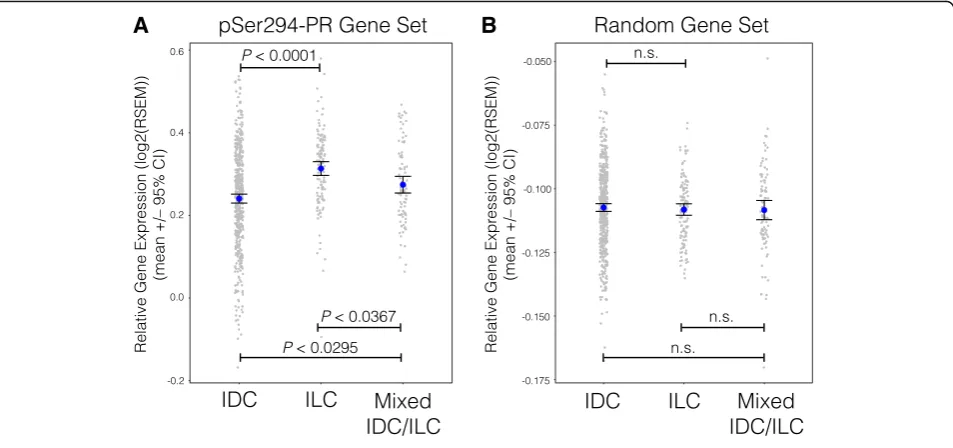
The ductal and lobular TCGA data were downloaded from the Sloan Kettering data freeze (freeze set 3/26/14): http://cbio.mskcc.org/cancergenomics/tcga/brca_tcga [7]. The RNA-seq gene expression values (RSEM) were merged from 705 ductal and lobular samples. Downloaded values were provided as centered z-scores and were log2 transformed across all genes before analysis. The mean expression values for genes within each gene set (PR or random) were plotted for each sample, according to their pathological characteristic (IDC, ILC, or mixed).
Gene set enrichment analysis
Gene set enrichment analysis (GSEA) software [49, 50] was used to identify gene sets from the Molecular Signatures Database (MSigDB) collections 1–7 that were significantly regulated in cells stably expressing SUMO-deficient PR (K388R) compared to WT PR. Our analysis compared two phenotype groups: KR +R5020/KR
−R5020 versus WT +R5020/WT−R5020. GSEA was ex-ecuted using the default settings, except the permutation type was set to Gene_set with 1000 permutations and the metric for ranking genes was set to Diff_of_Classes because our dataset contained log-scale data.
Mammosphere culture
Primary mammospheres
Adherent cells were washed with PBS and dissociated enzymatically in 0.25% trypsin-EDTA (Invitrogen). The cells were sieved through a 40-μm sieve (BD Fal-con) and analyzed microscopically for single cellular-ity. Single cells were plated in ultra-low attachment plates (Corning) and cultured in a humid incubator. The cells were grown in a serum-free mammary epithelial basal medium (MEBM; Lonza) containing 1% B27 Supplement (Invitrogen), 1% penicillin-streptomycin (Invitrogen), 5 μg/ml insulin (Invitro-gen), 20 ng/ml EGF (Sigma), 1 ng/ml hydrocortisone (Sigma), and 100 μM β-mercaptoethanol. Mammo-spheres were allowed to grow for approximately 14 days. Mammosphere forming efficiency (MFE, %) is calculated by the number of mammospheres per well/number of cells seeded per well.
Secondary mammospheres
Primary mammospheres were collected by centrifuga-tion (5 min, 1000 rpm) and dissociated enzymatically in 0.25% trypsin-EDTA. Cells were sieved through a 40-μm tip strainer (Bel-Art SP Scienceware) and ana-lyzed microscopically for single cellularity. Single cells were plated in ultra-low attachment plates and cul-tured in a humid incubator. The cells were grown in conditioned media for approximately 14 days. The
conditioned media consists of a 1:1 mixture of mam-mosphere media (described above) and media from cultured parental cells. MFE (%) is calculated by the number of mammospheres per well/number of cells seeded per well.
Results
A majority of breast tumors contain phospho-Ser294 PR
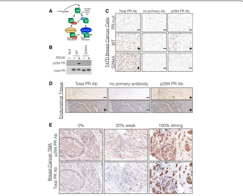
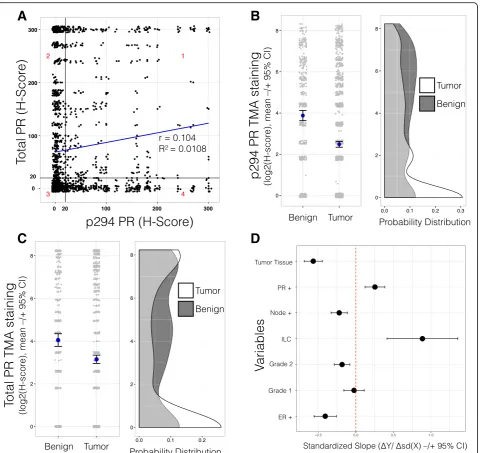
We previously demonstrated functional roles for phos-phorylation of PRs by mitogenic protein kinase pathways commonly elevated in breast cancers, including MAPKs, CDKs, and CK2 [33, 51–55]. These events are predicted to enable gene promoter selection by uniquely modified PR species according to cell context (Fig. 1a). To dem-onstrate the prevalence of PR Ser294 phosphorylation in human luminal breast tumors in vivo, we completed IHC staining of a tissue microarray (TMA) containing 209 patient breast tumors (split into 2754 tissue spots) for both total PR and phospho-Ser294 PR (Table 1). Note that phospho-Ser294 antibodies are unable to dis-tinguish between PR isoforms. However, PR-B but not PR-A is primarily phosphorylated on Ser294 in breast cancer models [33, 56]. We thus validated total and phospho-Ser294-specific PR antibodies by performing western blotting and IHC on PR-null and PR+ T47D cells containing either WT PR-B or S294A mutant PR-B (Fig. 1b) and further optimized PR staining protocols for IHC using human PR+ healthy uterine tissues (Fig. 1c). For the majority of tumor samples in our breast cancer TMA, four pathological regions of each tumor were identified by a clinical pathologist (designated as inva-sive, inflammatory, DCIS, or normal-like) and repre-sented as separate tissue spots. Following staining with either total PR or phospho-Ser294 PR antibodies, a histology score (H-score) was calculated for each tissue spot based on the percent of positively stained cells and their staining intensity (strong, moderate, or weak, Fig. 1d, e) [39, 40].
express relatively low levels of total PR (quadrant 4, 19%). Conversely, ~23% of tumors (quadrant 2) expressed high total PR and low phospho-Ser294 PR. Thirty-nine percent of tumors were negative for both total PR and phospho-Ser294 PR (quadrant 3) while 17% of tumors were positive for both total and Ser294-phosphorylated receptors (quadrant 1). Positive staining for phospho-Ser294 PR was greatest in tissue
spots pathologically classified as “Normal” (54%; normal-like tissue within tumor-containing tissue), followed by “DCIS” (47%), “Inflammatory” (43%), and
“Invasive” (38%) sections, having the lowest H-score levels for expression of phospho-Ser294 PR. Positive staining for total PR levels by tissue type were as fol-lows: Normal (72%), DCIS (55%), Inflammatory (53%), and Invasive (52%). These data indicate that total PR and phospho-Ser294 PR staining are not directly re-lated in this TMA as measured using distinct antisera and that PR levels are diminished in tissues with invasive characteristics relative to normal tissues or regions of DCIS.
We predicted that lowered PR expression in tumors relative to benign breast tissue (BBT) is indicative of heightened (i.e., activated) PR transcriptional activity that occurs during the process of tumor progression. We thus compared the levels of phospho-Ser294 PR or total PR expression between these two tissue classifications. IHC scoring was completed by an independent breast cancer pathologist, who also classified the tissue spot as BBT or tumor tissue (TT). As predicted, H-scores among the BBT samples were significantly greater than those among the TT samples (Ser294: P< 2.2e−16, Mann-Whitney test, Fig. 2b; total PR: P< 7.8e−06, Mann-Whitney test, Fig. 2c). These data suggest that while both total and phospho-PR expression is varied within regions of established tumors, greater levels of both total and phospho-PR are typically found in epithe-lial layers displaying early lesions or normal-like path-ology. Overall, the majority of tumors (TT) represented in this TMA contained less total PR/phospho-Ser294 PR relative to BBT. Interestingly, at least 21 tumor samples of invasive lobular carcinoma (ILC) were represented in our TMA as 468 spots. These tumors also expressed heterogeneous levels of total and phospho-Ser294 PRs. Phosphorylated PR levels were similar within each quad-rant: 1–4 (29, 20, 21, and 27%), again bearing no direct relationship to total PR levels (Additional file 2 and fur-ther discussed below).
We next probed the relationship between PR Ser294 phosphorylation and the available patient tumor charac-teristics (Table 1). We investigated whether any of the tumor characteristics (independent variables) could pre-dict PR Ser294 phosphorylation H-scores (dependent variable) using a multiple regression method. All inde-pendent variables were initially included in the model, and non-significant variables were removed stepwise by backward elimination until a core set of significant vari-ables remained. In this model, in addition to PR positiv-ity (at clinical diagnosis), only invasive lobular carcinoma (ILC tumor type) was a significant indicator of PR Ser294 phosphorylation. Multiple factors were negative predictors of PR Ser294 phosphorylation, Table 1Breast cancer tissue microarray patient characteristics
Number (n= 209) Percent
ER/PR status
ER-positive 163 78.0
ER-negative 40 19.1
PR-positive 120 57.4
PR-negative 83 39.7
ER-positive and PR-positive 117 56.0
ER-positive and PR-negative 46 22.0
ER-negative and PR-negative 37 17.7
Unknown 6 2.9
HER2 status
HER2-positive 59 28.2
HER2-negative 140 67.0
Intermediate 1 0.5
Unknown 9 4.3
Lymph node status
LN-positive 75 35.9
LN-negative 111 53.1
Unknown 23 11.0
Grade
1 34 16.3
2 96 45.9
3 66 31.6
Unknown 13 6.2
Tumor type
Invasive ductal carcinoma 168 80.4
Invasive lobular carcinoma 21 10.0
DCIS 2 1.0
Other 14 6.7
Unknown 4 1.9
Tumor volume
<10 cm3 64 30.6
≥10, <20 cm3 48 23.0
>20 cm3 68 32.5
Unknown 29 13.9
including tumor tissue pathology (vs. benign breast tis-sue), lymph node positivity (vs. node negativity), grade 3 status (vs. grade 2 or 1), and ER positive status (vs. nega-tive status at clinical diagnosis) (Fig. 2d). These findings suggest that PR Ser294 phosphorylation is a relatively common but early event in breast cancer development. The presence of phospho-PR species may indicate that
these early lesions contain sufficient levels of local pro-gesterone and/or express activated MAPK or CDK sig-naling (i.e., downstream of growth factor receptors, for example) relative to tissues that are strongly PR+ but lack appreciable levels of phospho-Ser294 PR (i.e., ex-pressing largely inactive/dephosphorylated and stable receptors) [19, 31].
A
B
C
D
Progesterone treatment of breast tumor explants cultured ex vivo drives proliferation and induces PR Ser294 phosphorylation
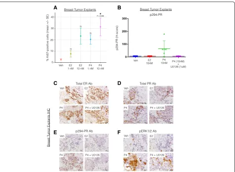
Because PR expression is primarily estrogen-induced in a majority of PR+ tissues and cancer models, isolating the unique contributions of progesterone/PR in breast cancer biology can be difficult to study in breast cancer models without the confounding (i.e., proliferative) ef-fects of estrogen/ER. We thus tested the proliferative re-sponse to progesterone treatment in ex vivo 3D cultures of human breast tumor tissue (i.e., tumor explants). Fresh tumor fragments from ER+/PR+ tumors were dis-sected into 1-mm3 sections and maintained on gelatin sponges submerged in cell culture medium as previously described [37, 57, 58]. Explants were treated with 1 or 10 nM estrogen or progesterone for 48 h before the tumor fragments were embedded in paraffin, sectioned, and analyzed by IHC for Ki-67 expression. ER+ tumor explants treated with progesterone (10 nM) but not es-trogen (1 and 10 nM) had a significantly higher percent-age of Ki67-positive cells (a marker of cell proliferation), compared to vehicle treatment (P= 0.006, ANOVA with TukeyHSD posttest;n= 6) (Fig. 3a). Note that 10 nM es-trogen alone induced a weak proliferative response in breast tumor tissue explants [25] that was similar to the proliferative trend observed herein (Fig. 3a). These data show that progesterone (P4) treatment alone signifi-cantly stimulates proliferation in ex vivo breast tumor tissue samples.
We previously demonstrated a proliferative and pro-survival role for MAPK-dependent phosphorylation of PR on Ser294 in breast cancer cells [32]. To assess whether PR Ser294 is a regulated phosphorylation site in human tumors ex vivo, we again employed the human tumor explant model as above (Fig. 3a). ER+ luminal tu-mors were maintained as explants as above and instead treated with either vehicle or progesterone (10 nM) for 2 h in the presence or absence of the MEK1/2 inhibitor U0126 (1μM) prior to IHC staining using specific anti-bodies for total and phospho-Ser294 PR as well as ER-alpha and phospho-ERK1/2 (n= 6; see “Methods”). As predicted, progesterone treatment induced robust PR Ser294 phosphorylation that was blocked by inclusion of the MEK inhibitor U0126 (Fig. 3b). IHC staining demon-strated that all explants were ER+ and PR+ (representa-tive examples are shown; Fig. 3c, d). However, only progesterone treatment (P4) induced robust phosphoryl-ation of PR Ser294 that was accompanied by activphosphoryl-ation of ERK1/2 (representative examples are shown; Fig. 3e, f ). Phospho-ERK1/2 staining confirmed that progester-one (but not estrogen) activated ERKs 1/2 and that U0126 also blocked the majority of hormone-induced ERK 1/2 activity in breast tumor tissues (Fig. 3f ). These data demonstrate that progesterone, at a physiologic
dose, is a potent mediator of breast tumor cell prolifera-tion (independent of estrogen) in a model system that maintains breast tumor 3D structure, microenvironment, and epithelial cell polarity (known factors required for PR expression and paracrine actions [59, 60]) and indi-cate that PR-dependent transcriptional programs (i.e., that drive proliferation) including those enacted by MAPK-dependent phosphorylation of PR on Ser294 are likely to be activated in human breast cancers cultured ex vivo.
Mifepristone and aglepristone, but not onapristone, induce PR Ser294 phosphorylation and act as partial agonists
apparent in the presence of progesterone and selected antiprogestins relative to liganded WT PR, consistent with increased turnover of deSUMOylated active re-ceptors relative to intact WT PRs [31]. Notably, only onapristone blocked Ser294 phosphorylation in the presence of progesterone. These data show that PR Ser294 phosphorylation is stimulated by multiple ligands including the common PR antagonists mife-pristone and aglemife-pristone. However, onamife-pristone treat-ment does not permit Ser294 phosphorylation, even in the presence of progesterone, predicting that cells treated with this ligand will exhibit distinct gene ex-pression profiles relative to other ligands (i.e., both agonists and antagonists) that stimulate PR Ser294 phosphorylation.
PR ligand-mediated promoter selectivity remains under-studied, especially in the context of antiprogestins and posttranslationally modified PR species. Breast tumors clearly express phosphorylated PR molecules (Fig. 1e and 2a) predicted to be deSUMOylated and transcrip-tionally hyperactive at a subset of SUMO-sensitive and phosphorylation-dependent gene promoters [32]. To further explore altered phospho-PR promoter se-lectivity (Fig. 1a), we conducted global gene expres-sion analyses in T47D breast cancer cells expressing either WT or K388R PR-B receptors treated as above (Fig. 4a). PR-null cells or cells expressing unmodified WT or SUMO-deficient KR PR species were serum starved (24 h) prior to ligand treatment (6 h). The cells were then treated with vehicle control (ethanol),
A B
C D
E F
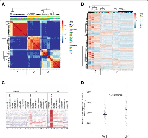
progesterone (P), mifepristone (M), aglepristone (A), onapristone (O), or combined (progesterone agonist plus each antagonist) treatments of P + M, P + A, or P + O. Total RNA was collected and subjected to microarray gene expression analysis using the Illumina HT-12v4 beadchip platform. Our normalized gene expression data-set included 84 different samples under the above treat-ment conditions, necessitating identification of commonly regulated sample clusters (i.e., groups of similarly regu-lated samples) in an unbiased manner. First, we isoregu-lated genes under high variance and performed NMF analysis (a clustering method that does not depend on prior know-ledge of the sample labels or molecular classifications of breast cancer; see “Methods”) [46, 63–65]. The resulting consensus matrix indicated five uniquely regulated sample clusters within the gene expression dataset (Fig. 5a). Annotating the consensus matrix with cell line and treat-ment labels revealed five sample clusters: (1) PR-null cells (all treatments), (2) WT cells (all antiprogestin and hicle treatments), (3) KR cells (onapristone and ve-hicle treatments), (4) WT cells (progestin treatments), and (5) KR cells (progestin, aglepristone, and mife-pristone treatments) (Fig. 5a). These data clearly show
that in cells expressing activated PR (i.e., KR or deSUMOylated PR), the antiprogestins mifepristone and aglepristone significantly regulated a similar gene expression program to that of progestin agonists (pro-gesterone or R5020). Further, depending on the phos-phorylation/SUMOylation status of PR, the receptor regulates completely different target genes when bound to different classes of antiprogestin (mifepris-tone/aglepristone vs. onapristone). The antiprogestins aglepristone and mifepristone substantially regulate multiple genes in cells expressing KR PR, but not WT PR, whereas in contrast, onapristone does not substantially regulate PR target genes in either cell line. These data suggest that the status of PR post-translational modifications can substantially impact PR target gene selectivity in a ligand-selective manner. Namely, deSUMOylated (KR) PRs recognize selected antagonists (mifepristone and aglepristone) as potent receptor agonists relative to onapristone.
In addition to unsupervised NMF clustering (above), we performed differential gene expression analysis be-tween various biologically interesting cell line/treat-ment comparisons and identified 251 genes that were
A
B
up- or downregulated greater than twofold in any comparison (Fig. 5b). Using this set of PR regulated genes, unsupervised hierarchal clustering revealed that progestin-treated samples clustered closely together in cells expressing either WT or KR. Hierarchical
clustering revealed two major branches of closely re-lated samples: (1) WT cells treated with progestin and KR cells treated with progestin, mifepristone, or agle-pristone and (2) WT cells treated with any antipro-gestin or vehicle, KR cells treated with onapristone or
A
B
C
D
Fig. 5Gene expression analysis in T47D cells treated with various ligand combinations demonstrates unique promoter selection.aGene expression arrays were used to measure global changes in gene expression levels in T47D cells treated with different PR ligands: vehicle, progestin (P), mifepristone (M), aglepristone (A), onapristone (O), P + M, P + A, or P + O. Genes under high variance across these samples were isolated, and expression values were used for NMF clustering. Presented is the consensus matrix that indicates five major clusters are present in the samples.
vehicle, and all PR-null cells regardless of treatment (Fig. 5b). This result demonstrates that various anti-progestins have different transcriptional effects de-pending on the dominant PR species. However, based on this clustering analysis, all samples (expressing WT or KR PR) treated with onapristone were closely related (and members of the second branch), suggest-ing that onapristone effectively inhibited PR (either WT or KR) transcriptional activity comparable to the level found in PR-null (control) cells (Fig. 5b, cluster 2, right). Furthermore, importantly, onapristone did not stimulate SUMO-deficient PR target gene expres-sion in KR-containing T47D cells, as did mifepristone and aglepristone (Fig. 5b, cluster 1).
PR-low (by IHC) breast tumors significantly express “activated PR”target gene signatures
We demonstrated that PR transcriptional activity is directly linked to rapid proteasome-mediated turnover of bound receptors [33, 51] and that ligand-dependent PR downregulation is greatly augmented by phosphorylation of PR Ser294 in response to acti-vated MAPK or CDK2 signaling pathways [33]. To address this context-dependent complexity, we identi-fied activated PR target genes that were specifically regulated in cells expressing SUMO-deficient PRs (as markers of phosphorylated or hyperactivated PR tran-scriptional activity) and examined their average ex-pression levels in the TCGA breast cancer patient cohort [5]. First, we isolated only luminal A, luminal B, and HER2-enriched tumors that were diagnosed as ER+ but PR-negative by clinical IHC, as we suspected some of these tumors would contain undetected but hyperactivated PRs. Next, in these tumors, we com-pared the expression of genes known to be primarily upregulated by deSUMOylated (i.e., phosphorylated) activated PRs relative to genes known to be regulated by SUMOylated PRs (Fig. 5c) [32]. As we predicted, tumors clinically classified as PR-negative were char-acterized by elevated expression of activated PR target genes (Fig. 5d). These exciting results suggest that a cohort of “PR-negative” breast tumors assigned using standard clinical IHC protocols in fact express signifi-cantly high levels of phospho-Ser294 PR target gene mRNA transcripts whose collective expression (i.e., the activated PR transcriptome) signifies the presence of activated phospho-Ser294 PRs. Our data suggest that modified PRs cannot be reliably detected in the clinical setting by measurement of PR protein expres-sion (as determined by IHC) as the sole marker of PR activity. Indeed, in our TMA results (above), PR Ser294 phosphorylation and total PR expression were not substantially correlated in individual tumors.
Phospho-Ser294 PR target genes are more highly expressed in ILC tumors compared to IDC tumors
Our TMA analysis (Fig. 2d) revealed that phospho-Ser294 PR expression was significantly associated with invasive lobular carcinoma (ILC), when compared to other tumor types included in the model. TCGA re-cently published a comprehensive analysis that directly compared ILC and IDC breast tumors [7]. We utilized this large independent tumor cohort to further probe the relationship between phospho-Ser294 PR signaling in lobular and that in ductal tumors. We compared the expression levels of a phospho-PR gene set in ILC, IDC, and mixed IDC/ILC tumors from the TCGA dataset (Fig. 6a). The phospho-PR gene set (upregulated by phospho-PR/SUMO-deficient PR, Fig. 2c) was signifi-cantly more expressed in the ILC tumors, when compared to the IDC and mixed ILC/IDC tumors (P< 0.0001, ANOVA with TukeyHSD posttest; n= 705). These data suggest that genes regulated by phospho-Ser294 PR may drive cells toward the ILC tumor lineage, as compared to ductal subtypes.
GSEA reveals mechanisms for SUMO-deficient PR transcriptional activation
binding motifs near the transcriptional start site and may thus have DNA binding priority relative to unmodi-fied WT PRs (i.e., primarily de-phosphorylated but cap-able of undergoing ligand-induced SUMOylation). Finally, we observed six significantly enriched EVI-1/ RUNX (also called AML) gene sets uniquely regulated in cells expressing KR-PR (+SPRMs) relative to cells ex-pressing WT-PR (+SPRMs) (Additional file 3F), suggest-ing that phosphorylated PRs and RUNX factors may cooperate on selected target genes. Three RUNX tran-scription factors have been described and are important mediators in multiple cancers, including AML. Notably, RUNX transcription factors are primarily expressed in stem cells and regulate stem cell renewal [67].
Functional cooperation between phospho-Ser294 PR and RUNX2
The above GSEA results suggest that SUMO-deficient phospho-Ser294 PRs regulate a set of genes also regu-lated by RUNX factors. PR cooperation with one or more RUNX factors may be a mechanism for promoter selection by uniquely modified receptors. The family of RUNX transcription factors (RUNX1, 2, and 3) has com-plex roles in development and tumor formation with both tumor-suppressive and tumor-promoting activities. Interestingly, phenotypes associated with RUNX2 ex-pression in mammary epithelial cells closely resemble phenotypes dependent on PR as well as
progestin-mediated gene expression (namely, cyclin D1 expression, proliferation, luminal progenitor cell maintenance, and alveolar expansion during mammary gland development; see“Discussion”). From our GSEA results, we identified
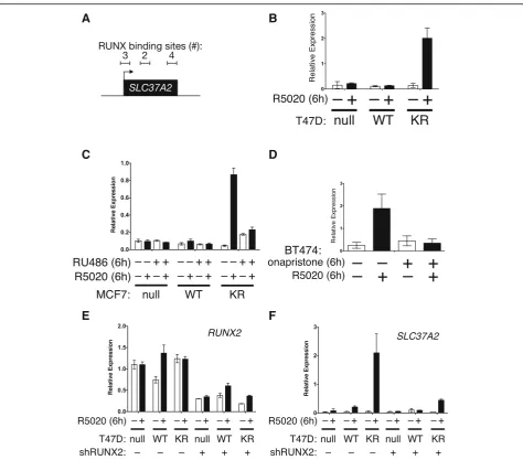
SLC37A2, a candidate PR target gene containing mul-tiple RUNX2 binding motifs immediately upstream and within the gene (Fig. 7a). SLC37A2 is a glucose-6-phosphate transporter expressed in monocytes as well as breast and cervical tissues. Although no studies have been conducted in cancer models,SLC37A2is associated with at least 17 other public datasets that define stem cell genes or proteins [68]. We thus measured PR/ progestin-dependent regulation of SLC37A2 mRNA expression in multiple cell line models. In T47D cell models, SLC37A2 expression was robustly stimulated by progestin in cells expressing SUMO-deficient PR, but not in cells expressing WT PR (Fig. 7b). Similarly, in MCF-7 cell line models overexpressing SUMO-deficient K388R PR, SLC37A2 expression was upregulated by progestin exposure but blocked by mifepristone (Fig. 7c). Interestingly however, as predicted from our gene array studies, mifepristone exhibited weak partial agonist activity in cells expressing KR PRs (compare RU486 treatments across cell lines). We previously showed that BT474 breast cancer cells (luminal B; ER +/PR+/ERBB2+) superinduce selected SUMO-sensitive (activated) PR target genes upon progestin treatment relative to other PR+ cell line models, presumably
A
B
because kinase pathways downstream of HER2 (i.e., MAPKs) input to persistent PR Ser294 phosphorylation [32]. In unmodified BT474 cells, progestin exposure resulted in highly phosphorylated PR that turned over rapidly, characteristic of PRs with heightened tran-scriptional activity [51]. In these cells, progestin treat-ment also stimulated robust SLC37A2 mRNA expression that was effectively blocked by treatment with onapristone (Fig. 7d). We then tested the re-quirement for RUNX2 expression in transcriptional responses to progestin by knocking down RUNX2 in T47D cells using shRNAs. Although T47D cells
remained relatively resistant to RUNX2 loss in our hands, RUNX2 expression was reproducibly reduced by approximately 50% upon expression of specific shRNAs relative to shRNA controls (Fig. 7e). Knock-down of RUNX2 greatly attenuated induction of
SLC37A2 expression in cells expressing KR-PR and treated with progestin relative to controls (Fig. 7d). These data demonstrate that PR cooperation with RUNX2 contributes to SLC37A2 expression as part of a unique phospho-Ser294 PR transcriptome in breast cancer cells and illustrate the impact of context-dependent cell signaling on PR actions.
A
B
C
D
E
F
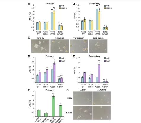
PR Ser294 phosphorylation is required for formation of secondary mammospheres
Interestingly, HER2, PAX2, AHR, AR, and RUNX factors have each been implicated in cancer stem cell biology ([27, 67, 69–71] and see “Discussion”). Further, these factors may cooperate; AR/RUNX2 complexes are im-portant drivers of prostate cancer stem cell expansion [72]. Mammosphere assays provide an assay of stem cell potential, wherein formation of secondary mammo-spheres (i.e., derived from dissociated and serially pas-saged primary mammospheres) is a definitive assay of the ability of minority breast cancer cell stem cells within a heterogeneous population to expand and rees-tablish as E-cadherin-positive spheres able to grow in suspension culture following long-term serial passage as non-adherent cells [73]. To demonstrate a role for phos-phorylated PRs in breast cancer stem cell biology, we performed mammosphere assays using our well-defined T47D cell model systems expressing either empty vector (EV PR-null), unmodified WT PR-B, point mutant KR PR-B (K388R), or point mutant S294A PR-B missing the consensus MAPK phosphorylation site Ser residue (Fig. 8). Equal numbers of T47D cells were inoculated into primary mammosphere assays (i.e., suspended cell culture) in defined media, and mammosphere numbers were scored after 2 weeks by manual counting using a uniformly scaled grid; primary mammospheres were gently dissociated and reseeded in order to form second-ary mammospheres (see “Methods”). Interestingly, cells expressing either EV or WT PR produced similar basal numbers of primary (~25) and secondary (~10) mam-mospheres, while cells expressing KR (phospho-mimic) PR consistently produced 55–70 primary mammo-spheres and 35–50 secondary mammospheres (Fig. 8a, b). Surprisingly, both primary and secondary mammosphere formation was greatly attenuated in cells expressing S294A PR relative to controls and cells expressing KR PR. Interestingly, PR-null (vector control) cells formed small
“flat”or“non-spheroid”clumps of loosely associated cells with raged or rough edges relative to cells expressing WT PR-B, which formed small round and smooth mammo-spheres (Fig. 8c). Notably, cells expressing KR PR formed larger mammospheres relative to cells expressing WT PR, in sharp contrast to cells expressing S294A PR, which formed few very small mammospheres (Fig. 8c). Addition of either estrogen (1 nM; not shown) or progesterone (10 nM; shown) to mammosphere culture media had no significant effect on total numbers in any condition. How-ever, removal (and add-back) of EGF to the mammo-sphere culture media demonstrated a clear requirement for growth factor signaling (Fig. 8d, e). These data suggest that formation of secondary mammospheres, a definitive assay of stem cell outgrowth, is largely dependent on the presence of signaling inputs (EGF) to phospho-Ser294 PRs
in T47D breast cancer cells but does not require exogen-ously added progesterone. Further, the finding that expres-sion of S294A PR attenuated mammosphere formation to levels below that of either PR-null or WT PR-containing cells in EGF-containing media suggests a dominant nega-tive effect of this mutant receptor, perhaps via interaction with other steroid receptors such as ER or AR (see
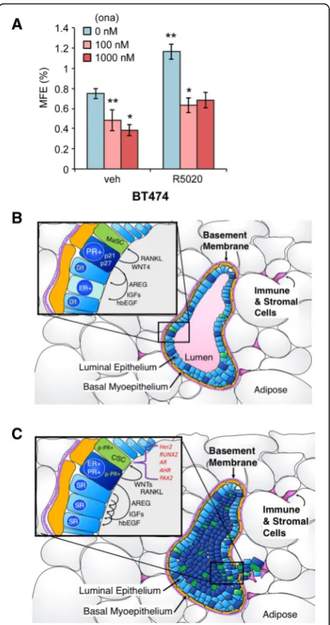
“Discussion”). We next tested the ability of PR-B+ T47D cells stably expressing either control shRNA (shGFP) or RUNX2 shRNA to form mammospheres (Fig. 8f, g). Again, cells expressing K388R PR-B formed larger and sig-nificantly greater numbers of primary mammospheres relative to cells expressing unmodified (WT) PR-B. Knockdown of RUNX2 greatly attenuated the formation of primary mammospheres in T47D cells expressing either WT PR-B or K388R PR-B, rendering the assay of second-ary mammospheres infeasible. Finally, in a similar set of experiments, we validated our results in unmodified ER +/PR+ BT474 cells. These cells express high levels of acti-vated HER2 and thus more closely resemble luminal B-type breast cancers, but express endogenous ER and both PR isoforms (PR-A and PR-B). In this “high kinase” context, PRs are readily phosphorylated on Ser294 (ref Knutson BCR 2012). Notably, BT474 cells exhibited a relatively high level of basal primary mammosphere formation that was further elevated in the presence of pro-gestin (Fig. 9a). Treatment with onapristone (an antipro-gestin that blocks PR Ser294 phosphorylation) effectively reduced both basal and progestin-stimulated primary mammosphere formation. As with RUNX2 knockdown studies, secondary mammospheres failed to form in the presence of onapristone (not shown).
Collectively, our data suggest that phospho-PRs are key gatekeepers that enable breast tumor progression via induction of multiple signaling pathways, including those required for outgrowth of breast cancer stem or progenitor cells (Fig. 9b, c). Identification of phosphory-lated receptors in human tumors and discovery of phospho-PR-regulated pathways (i.e., including HER2, RUNX2, AR, AHR, and PAX2) suggest novel ways to specifically target breast cancer stem cell (CSC) out-growth as part of durable breast cancer therapies. We conclude that antiprogestins that block PR Ser294 phos-phorylation should be included “up-front” as part of routine endocrine therapies for women undergoing long-term management of ER+ luminal breast cancer.
Discussion
transcriptional events), while stimulating proliferation in others (containing modest levels of phosphorylated and SUMO-deficient PRs that are active drivers of unique cancer transcriptomes). Additionally, our findings sup-port a growing body of evidence implicating PR as a master regulator of cell fate of both normal mammary epithelial and cancer stem/progenitor cell populations and reveal a key role for Ser294-phosphorylated PRs in this aspect of PR-driven cell biology. Ultimately, the transcriptional activity and biological actions of PRs are profoundly influenced by context. Herein, we identified
a subset of PR target genes that can be used as bio-markers reflective of activated PR expression (i.e., inde-pendently of clinically derived PR status as defined by IHC-based methods). Using breast cancer mRNA ex-pression data from the TCGA project, we determined that activated PR target genes were significantly upregu-lated in ILC as well as clinically determined PR-negative luminal patient samples (compared to gene sets specific-ally regulated by inactive or stabilized and abundant re-ceptors). These data suggest that a subset of breast cancer patients whose tumors are clinically classified as Fig. 8Mammosphere formation in T47D cells (empty vector, PR-B, PR-B K388R, and PR-B S294A).aPrimary andbsecondary mammospheres in T47D cells overexpressing empty vector, PR-B, PR-B K388R, or PR-B S294A and plotted as a percentage of mammosphere forming efficiency (MFE; see“Methods”). Cells were treated with vehicle (EtOH) or R5020 (10 nM).cImages of primary mammospheres (vehicle) froma.dPrimary and
esecondary mammospheres in T47D cells treated with vehicle (water) or EGF (20 ng/ml). Mammospheres were allowed to grow for 14 days prior to counting.fPrimary mammospheres in T47D cells (PR-B or K388R) expressing shGFP or shRUNX2.gImages of primary mammospheres from
PR-negative may have cancers driven in part by modest levels of highly transcriptionally active PRs that go un-detected by clinical standards. Alternatively, abundant phospho-PRs may reside in minority cancer cell popula-tions or “PR+ islands” within largely PR-null tumors (Figs. 1 and 2) capable of early dissemination [27]. Pa-tients harboring such tumors are strong candidates for antiprogestin therapy, including onapristone or similar agents that block PR Ser294 phosphorylation.
As an ER target gene product, PR is classically used as a biomarker of functional ER and thus indicative of a high likelihood of response to ER-targeted endocrine therapies [74, 75]. Tumors defined as ER+ PR+ HER2– are usually less aggressive and classified within the lu-minal A or B subtypes. Of these, ER+/PR-low or ER +/PR-null tumors (i.e., luminal B subtype) are more likely to become endocrine-resistant (Fig. 9c). The pres-ence of PR can profoundly modify ER behavior and cel-lular responses to estrogen, in part by direct ER/PR interactions [25, 76]. Modest levels of PR-B, but not pro-gesterone, were required for estrogen-induced changes in global gene expression associated with breast tumor progression to endocrine resistance and poor disease outcome [24]. In contrast, estrogenic responses were inhibited when ER+/PR+ breast cancer cells and breast tumor explants were exposed to both hormones; how-ever, relatively high hormone concentrations were used to demonstrate these effects [25, 26]. Like estrogen (alone), progesterone (alone) is a potent driver of breast cancer cell proliferation (Fig. 3a). PR+ but ER-null mam-mary gland progenitor cells exist, suggesting unique roles for PR that are independent of ER; PR+ bipotent progenitor cells are estrogen-insensitive, while estrogen regulates PR expression only in mature luminal cells [77]. Progesterone but not estrogen has emerged as a key mediator of both normal and neoplastic mammary gland stem cell expansion [78–81]. Our studies strongly implicate Ser294 phosphorylated PRs in this activity (Fig. 8). The finding that PR Ser294 phosphorylation is widely observed in breast tumors and is primarily found in premalignant regions suggests that this modification of PR is a relatively early event in tumor progression that is likely most relevant to luminal A to luminal B transition, wherein PR expression appears diminished as PR target gene expression peaks. Notably, expres-sion of the PR target gene, RANKL, also primarily oc-curs in early-stage premalignant epithelial layers (i.e., DCIS, and normal-like regions) [78, 82]. Recently, Hosseini et al. [27] showed that mRNA levels of pgr and progesterone-induced paracrine signals (PIPS;
rankl and wnt4) were upregulated in cells from early mammary lesions of BALB-NeuT mice. These PIPS were required for early lesion cell migration as well as sphere formation.
We found that commonly used PR ligands (agonists and antagonists alike) induce PR Ser294 phosphorylation and phospho-PR target gene expression (Figs. 4 and 5). Indeed, the partial agonist activity of antiprogestins ap-pears to map to SUMO-deficient/phosphorylated recep-tors. Only onapristone was effective in blocking Ser294 phosphorylation and gene expression in cells expressing either wild type (WT) PR or SUMO-deficient (KR) PR (Figs. 4 and 5). In breast cancer cells expressing KR PR, mifepristone and aglepristone stimulated considerable Ser294 phosphorylation and gene regulation, suggesting these antagonists may be less effective in cells that con-tain the highly transcriptionally active deSUMOylated PR. Antiprogestin therapies are being actively studied for breast (and other) cancers (reviewed in [19, 83]); there-fore, a more comprehensive understanding of the differ-ences in transcriptional regulation by these ligands (in relation to PR posttranslational modifications) will be critical. Our data (Fig. 5) demonstrate that different anti-progestins have unique gene regulatory action depending on the status of PR posttranslational modifications. Both mifepristone and progestin agonists (progesterone or R5020) upregulated similar genes in cells expressing SUMO-deficient (phospho-mimic) PRs, suggesting that mifepristone is a poor antiprogestin in that context. However, mifepristone treatment of cells containing WT PR (capable of SUMOylation) was far less likely to stimulate the expression of progestin-regulated PR target genes, making it a useful antagonist in that context. These results suggest that successful therapies for breast cancer patients using antiprogestins should consider the status of PR posttranslational events. Our data may ex-plain why mifepristone (RU486) has not been successful in clinical trials for breast cancer, considering that our TMA revealed that PRs in a majority of breast tumors are phosphorylated on Ser294, a posttranslational event predicted to confer partial agonist activity to ligands of this class. As such, alternative antiprogestin therapies (i.e., onapristone or SPRMs that block PR Ser294 phos-phorylation) may be more successful to silence the tran-scriptional action of activated phospho-PRs.
We previously defined phospho-Ser294 PRs as major transcriptional drivers of gene programs significantly as-sociated with HER2/ERBB2 signaling in breast cancers [32]. Related to our findings, PR and HER2 were shown to cooperate as required mediators of early breast cancer dissemination and metastasis [27]. Herein, our GSEA analyses confirmed that phospho-PRs significantly in-duce expression of HER2-associated gene sets and dem-onstrated that phospho-PR target genes also include key mediators of cancer stem cell biology, including PAX, AHR, AR, and RUNX family members (Additional file 3; Fig. 7). SUMO-deficient or phospho-Ser294 PR target genes may be co-regulated by one or more of these
transcription factor families. Notably, PAX2 is overex-pressed in >50% of breast cancers and was required for progesterone-stimulated lateral side-branching and lobu-lar development in a murine Pax2-knockout model [84]. Pax2 knockout in murine mammary glands phenotypic-ally resembled PR or Wnt4-knockout mice [21, 85]. These data indicate that PR-driven pathways important during mammary gland development may remain active during breast tumor progression. Aryl hydrocarbon re-ceptor (AHR) is a transcription factor member of the nuclear receptor (NR) superfamily expressed in female reproductive tissues that interacts with multiple environ-mental toxins as ligands, resulting in AHR translocation to the nucleus, where it dimerizes with AHR nuclear translocator (ARNT). This triggers upregulated expres-sion of cytochrome P450 enzymes that help metabolize a variety of compounds. Environmental toxins can modu-late reproductive functions and alter homeostasis of many endocrine functions in the reproductive tract largely because ligand-activated AHR can interfere with SR signaling. Thus, AHR is known to disrupt ER and AR target gene expression [86]. For example, ligand-bound AHR/ARNT binds specific motifs positioned near ER binding motifs in the promoters of multiple ER target genes, disrupting ER-mediated transcription [87, 88]. Similarly, our GSEA results demonstrate that activated PRs drive expression of important SR-regulated target genes that can also be disrupted by AHR/ARNT com-plexes (Additional file 3). Thus, phosphorylated and SUMO-deficient PR may interact preferentially with AHR/ARNT-repressed genes. Notably, like PR, which clearly contributes to maintenance of the adult mam-mary stem cell compartment ([80] and for review, see [89]), AHR signaling has recently been implicated in regulation of stem-like cell expansion in breast cancer models [90]. Similarly, AR is highly expressed (80%) across all breast cancer subtypes and also negatively and positively interacts with ER signaling [91–94].
DNA binding motifs in their promoters, suggesting that SUMO-deficient or phosphorylated PR and RUNX factors may cooperate in a way that WT PR (i.e., SUMOylated) does not. Both RUNX1 and RUNX3 have tumor suppressor roles in breast cancers, while RUNX2 is tumor-promoting (for review, see [95]). Namely, RUNX2 is an osteoblast differentiation transcription fac-tor expressed in developing breast epithelial cells that is enriched in the mammary stem cell population respon-sible for terminal end bud differentiation [96, 97]. As a proof of principle, we validated a requirement for RUNX2 in phospho-Ser294 PR target gene regulation (i.e., using the SLC37A2 gene identified in our arrays; Fig. 7) as well as in primary mammosphere formation (Fig. 8). Previous reports have shown that RUNX2 inter-acts with estrogen receptor (ER), androgen receptor (AR), and the glucocorticoid receptor (GR) to facilitate steroid hormone-mediated transcriptional activity. Curi-ously, we were unable to co-IP RUNX2 and PR from breast cancer cell whole cell lysates or detect them as co-associated factors at PRE sites by ChIP assays, sug-gesting that these factors function in the same pathway but may interact indirectly or may associate transiently or successively via binding to separate or distant sites in chromatin (a topic for further study). In mice, RUNX2 was required for proper alveolar differentiation [98], a property shared by PR-B [99]. Runx2 gene deletion re-sulted in disrupted alveolar progenitor cell populations, differentiated cell type histology, reduced levels of cell proliferation, reduced tumor burden, and longer overall survival in a mouse model of breast cancer [98]. In addition, deletion of RUNX2 in human breast cancer cells resulted in reduced tumorigenic properties, such as invasion and migration. Conversely, aberrant overex-pression of RUNX2 induced EMT-like changes in nor-mal mammary glands and also caused cells to remain in a less-differentiated state with elevated expression of cyclin D1, a well-known PR-B (and other SRs) target gene and transcriptional co-regulator of phospho-PR at cancer-relevant PR-B/cyclin D1 target genes [98, 100]. Our data show that RUNX2 is essential for mammo-sphere formation in PR-B+ cells (Fig. 8); we were unable to obtain sufficient cell numbers from primary mammo-spheres of RUNX2 knockdown models for assay of secondary mammospheres. Interestingly, while mammo-sphere formation was insensitive to added hormones, EGF was required for spheroid formation in breast cancer cells expressing K388R (phospho-mimic) PRs (Fig. 8d, e). These data suggest that cells growing in sus-pension no longer require exogenously added hormones but instead rely on growth factors to cue context-dependent (i.e., MAPK-context-dependent) phospho-PR actions, including gene expression of RUNX2. EGF-induced ster-oid hormone biosynthesis is a topic of further study as a
potential mechanism of SR action in breast cancer spheroids carried in media lacking exogenously added hormones (Fig. 8). A deeper understanding of RUNX2 as a mediator of PR actions in breast cancer also war-rants future study.
Conclusions
In sum, PR is emerging as a major mechanistic player that mediates early breast tumor progression in part via
“feeding” the stem cell compartment (i.e., via paracrine signals); our data support a requirement for phosphoryl-ation of PR Ser294 in this activity as an important gate-keeper of breast cancer cell fate and expanded tumor heterogeneity. Most notably, in addition to strongly ER +/PR+ lobular breast cancers, we observe expression of KR-specific target genes in human breast tumors clinic-ally determined to be PR-negative. We propose that this PR signature is an important biological“marker”of acti-vated phospho-PR species that undergo rapid protein loss due to turnover, an event that may precede loss of PR mRNA expression in more advanced and strongly HER2+ tumors [31–33]. Previous clinical trials using antiprogestins demonstrated poor response rates in PR+ tumors. These agents may have stimulated PR phosphor-ylation and unwanted target gene expression. Addition-ally, these early trials primarily targeted PR in strongly ER+/PR+ (luminal A) tumors. While this was a logical approach based on high expression of PR protein as a biomarker, our studies suggest a far more complex sce-nario in which luminal B (PR low) patients are the cor-rect cohort for antiprogestins. The recent finding that PR and HER2, a primary pathway induced by phospho-Ser294 PR [32], were requisite mediators of early breast cancer dissemination and metastasis [27] underscores the relevance of our work. Clearly, a paradigm shift to activated PR as measured by the presence of phospho-PR species or phospho-phospho-PR target gene sets (in addition to HER2) is urgently needed.
Additional files
Additional file 1:Gene sets derived from T47D cells expressing WT PR and KR PR. These gene sets were used in the analysis described in Fig. 5c, d. (A) 16 genes were discovered to be highly upregulated by progestin in cells expressing KR PR, compared to WT PR. These genes were also not regulated by other PR ligands. (B) 101 genes were discovered to be highly upregulated by progestin in cells expressing WT PR, compared to KR PR. These genes were also not regulated by other PR ligands. (XLSX 28 kb)
Additional file 2:PR Ser294 phosphorylation and total PR H-scores in only invasive lobular carcinoma (ILC) TMA tumor spots. H-scores for total PR expression and phospho-Ser294 PR were compared among individual tumors spots from our TMA study. A Pearson correlation was calculated (r= 0.041,R2= 0.0017). Tissue spots considered“positive”had
Additional file 3:Gene set enrichment analysis (GSEA) in T47D breast cancer cells comparing KR + progestin vs. WT + progestin treatment groups. GSEA identified significantly regulated gene sets in the KR + progestin samples when compared to the WT + progestin samples. Five gene sets from the c3 MSigDB collection and one from the c6 collection are shown: (A) genes containing PR DNA binding motifs, (B) genes upregulated after ERBB2 overexpression in MCF-7 cells, (C) genes containing androgen receptor DNA binding motifs, (D) genes containing PAX family DNA binding motifs, (E) genes containing AHR/ARNT DNA binding motifs, and (F) genes containing AML1/RUNX binding motifs. These upregulated gene sets contain (respective) DNA binding motifs (above) near their transcriptional start sites, suggesting that these factors are important co-transcriptional regulators with PR in T47D cells expressing Ser294/ SUMO-deficient PR (KR), compared to WT PR. (PDF 257 kb)
Abbreviations
ANOVA:Analysis of variance; AR: Androgen receptor; ER: Estrogen receptor; GR: Glucocorticoid receptor; GSEA: Gene set enrichment analysis; HER2: Human epidermal growth factor receptor 2; IDC: Invasive ductal carcinoma; IHC: Immunohistochemistry; ILC: Invasive lobular carcinoma; MAPK: Mitogen-activated protein kinase; NMF: Non-negative matrix factorization; PR: Progesterone receptor; SUMO: Small ubiquitin-like modifier; TCGA: The Cancer Genome Atlas
Acknowledgements
We would like to thank the University of Minnesota Genomics Center and the University of Minnesota Supercomputing Institute for assistance with our gene expression studies and software access. We also thank Arno Therapeutics for the reagents onapristone and aglepristone. We also thank Hsiangyu (David) Hu for technical assistance and Dr. Alpay Temiz for prereview of this manuscript with regard to the presentation of our bioinformatics results.
Funding
This work was supported by NIH/NCI R01 grants CA159712 (to C.A.L.) and CA132827 (to K.L.S.) and the Tickle Family Land Grant Endowed Chair in Breast Cancer Research (to C.A.L.)
Availability of data and materials
The datasets used and/or analyzed during the current study are available from the corresponding author on reasonable request. The gene expression datasets analyzed during the current study are available in the GEO repository (accession number: GSE94363).
Authors’contributions
TPK performed all experiments except where noted. THT performed all mammosphere experiments (Figs. 8 and 9), and SHM performed all breast tumor explant experiments (Fig. 3). MES scored the stained tissue microarray slides. NJB and KLS created and provided access to the breast cancer tissue microarray. GR supervised the tissue explant experiments, and CAL supervised the entire study. TPK and CAL conceived of the study and wrote the manuscript. All authors read and approved the final manuscript.
Competing interests
The authors declare that they have no competing interests.
Consent for publication
Not applicable.
Ethics approval and consent to participate
Fresh breast cancer tissue was acquired immediately after surgery for breast tumor explant culture. Samples were obtained after informed patient consent and in compliance with an institutional review board approved protocol at the hospitals of the University of Texas Southwestern Medical Center (Dallas, TX).
Publisher’s Note
Springer Nature remains neutral with regard to jurisdictional claims in published maps and institutional affiliations.
Author details
1Departments of Medicine (Division of Hematology, Oncology, and
Transplantation) and Pharmacology, Masonic Cancer Center, University of Minnesota, Delivery Code 2812, Cancer and Cardiovascular Research Building, 2231 6th St SE, Minneapolis, MN 55455, USA.2Department of Urology, UT Southwestern Medical Center at Dallas, 5323 Harry Hines Blvd, J8.130C, Dallas, TX 75390-9110, USA.3Microbiology, Immunology, and Cancer Biology Graduate Program, University of Minnesota, Minneapolis, MN 55455, USA. 4Department of Pathology, Evanston Hospital, University of Chicago,
NorthShore University HealthSystem, Evanston, IL 60201, USA.5Department of Laboratory Medicine and Pathology, Masonic Cancer Center, University of Minnesota, Minneapolis, MN 55455, USA.
Received: 13 February 2017 Accepted: 3 April 2017
References
1. Siegel RL, Miller KD, Jemal A. Cancer statistics, 2016. CA Cancer J Clin. 2016;66:7–30.
2. Banerji S, Cibulskis K, Rangel-Escareno C, Brown KK, Carter SL, Frederick AM, Lawrence MS, Sivachenko AY, Sougnez C, Zou L, Cortes ML, Fernandez-Lopez JC, Peng S, Ardlie KG, Auclair D, Bautista-Pina V, Duke F, Francis J, Jung J, Maffuz-Aziz A, Onofrio RC, Parkin M, Pho NH, Quintanar-Jurado V, Ramos AH, Rebollar-Vega R, Rodriguez-Cuevas S, Romero-Cordoba SL, Schumacher SE, Stransky N, et al. Sequence analysis of mutations and translocations across breast cancer subtypes. Nature. 2012;486:405–9. 3. Stephens PJ, Tarpey PS, Davies H, Van Loo P, Greenman C, Wedge DC,
Nik-Zainal S, Martin S, Varela I, Bignell GR, Yates LR, Papaemmanuil E, Beare D, Butler A, Cheverton A, Gamble J, Hinton J, Jia M, Jayakumar A, Jones D, Latimer C, Lau KW, McLaren S, McBride DJ, Menzies A, Mudie L, Raine K, Rad R, Chapman MS, Teague J, et al. The landscape of cancer genes and mutational processes in breast cancer. Nature. 2012;486:400–4.
4. Curtis C, Shah SP, Chin SF, Turashvili G, Rueda OM, Dunning MJ, Speed D, Lynch AG, Samarajiwa S, Yuan Y, Graf S, Ha G, Haffari G, Bashashati A, Russell R, McKinney S, Caldas C, Aparicio S, Curtis C, Shah SP, Caldas C, Aparicio S, Brenton JD, Ellis I, Huntsman D, Pinder S, Purushotham A, Murphy L, Caldas C, Aparicio S et al. The genomic and transcriptomic architecture of 2,000 breast tumours reveals novel subgroups. Nature. 2012;486(7403):346–52. 5. Network CGA. Comprehensive molecular portraits of human breast
tumours. Nature. 2012;490:61–70.
6. Shah SP, Roth A, Goya R, Oloumi A, Ha G, Zhao Y, Turashvili G, Ding J, Tse K, Haffari G, Bashashati A, Prentice LM, Khattra J, Burleigh A, Yap D, Bernard V, McPherson A, Shumansky K, Crisan A, Giuliany R, Heravi-Moussavi A, Rosner J, Lai D, Birol I, Varhol R, Tam A, Dhalla N, Zeng T, Ma K, Chan SK, et al. The clonal and mutational evolution spectrum of primary triple-negative breast cancers. Nature. 2012;486:395–9.
7. Ciriello G, Gatza ML, Beck AH, Wilkerson MD, Rhie SK, Pastore A, Zhang H, McLellan M, Yau C, Kandoth C, Bowlby R, Shen H, Hayat S, Fieldhouse R, Lester SC, Tse GM, Factor RE, Collins LC, Allison KH, Chen YY, Jensen K, Johnson NB, Oesterreich S, Mills GB, Cherniack AD, Robertson G, Benz C, Sander C, Laird PW, Hoadley KA, et al. Comprehensive molecular portraits of invasive lobular breast cancer. Cell. 2015;163:506–19.
8. Lippman ME, Allegra JC. Quantitative estrogen receptor analyses: the response to endocrine and cytotoxic chemotherapy in human breast cancer and the disease-free interval. Cancer. 1980;46:2829–34.
9. Paridaens R, Sylvester RJ, Ferrazzi E, Legros N, Leclercq G, Heuson JC. Clinical significance of the quantitative assessment of estrogen receptors in advanced breast cancer. Cancer. 1980;46:2889–95.
10. Campbell FC, Blamey RW, Elston CW, Morris AH, Nicholson RI, Griffiths K, Haybittle JL. Quantitative oestradiol receptor values in primary breast cancer and response of metastases to endocrine therapy. Lancet. 1981;2:1317–9. 11. Stewart J, King R, Hayward J, Rubens R. Estrogen and progesterone
receptors: correlation of response rates, site and timing of receptor analysis. Breast Cancer Res Treat. 1982;2:243–50.