R E S E A R C H
Open Access
An algorithm to predict phenotypic severity in
mucopolysaccharidosis type I in the first month
of life
Sandra DK Kingma
1,2†, Eveline J Langereis
1†, Clasine M de Klerk
1, Lida Zoetekouw
2, Tom Wagemans
1,2,
Lodewijk IJlst
2, Ronald JA Wanders
1,2, Frits A Wijburg
1*and Naomi van Vlies
1,2Abstract
Introduction:Mucopolysaccharidosis type I (MPS I) is a progressive multisystem lysosomal storage disease caused by deficiency of the enzymeα-L-iduronidase (IDUA). Patients present with a continuous spectrum of disease severity, and the most severely affected patients (Hurler phenotype; MPS I-H) develop progressive cognitive impairment. The treatment of choice for MPS I-H patients is haematopoietic stem cell transplantation, while patients with the more attenuated phenotypes benefit from enzyme replacement therapy.
The potential of newborn screening (NBS) for MPS I is currently studied in many countries. NBS for MPS I, however, necessitates early assessment of the phenotype, in order to decide on the appropriate treatment. In this study, we developed an algorithm to predict phenotypic severity in newborn MPS I patients.
Methods:Thirty patients were included in this study. Genotypes were collected from all patients and all patients were phenotypically categorized at an age of > 18 months based on the clinical course of the disease. In 18 patients, IDUA activity in fibroblast cultures was measured using an optimized IDUA assay. Clinical characteristics from the first month of life were collected from 23 patients.
Results:Homozygosity or compound heterozygosity for specific mutations which are associated with MPS I-H, discriminated a subset of patients with MPS I-H from patients with more attenuated phenotypes (specificity 100%, sensitivity 82%). Next, we found that enzymatic analysis of IDUA activity in fibroblasts allowed identification of patients affected by MPS I-H. Therefore, residual IDUA activity in fibroblasts was introduced as second step in the algorithm. Patients with an IDUA activity of < 0.32 nmol x mg-1× hr-1invariably were MPS I-H patients, while an IDUA activity of > 0.66 nmol × mg-1× hr-1was only observed in more attenuated patients. Patients with an intermediate IDUA activity could be further classified by the presence of differentiating clinical characteristics, resulting in a model with 100% sensitivity and specificity for this cohort of patients.
Conclusion:Using genetic, biochemical and clinical characteristics, all potentially available in the newborn period, an algorithm was developed to predict the MPS I phenotype, allowing timely initiation of the optimal treatment strategy after introduction of NBS.
Keywords:Mucopolysaccharidosis type I, Iduronidase, Classification, Phenotype, Haematopoietic stem cell transplantation, Residual enzyme activity, Newborn screening, Enzyme replacement therapy, Disease severity
* Correspondence:[email protected] †Equal contributors
1Department of Pediatrics and Amsterdam Lysosome Centre“Sphinx”,
Academic Medical Center, University Hospital of Amsterdam, Meibergdreef 9, 1105, AZ Amsterdam, The Netherlands
Full list of author information is available at the end of the article
Background
Mucopolysaccharidosis type I (MPS I, OMIM 252800) is a progressive multisystem lysosomal storage disease (LSD) caused by a deficiency of the lysosomal hydrolase
α-L-iduronidase (IDUA, [Genbank NG_008103]), re-sulting in the accumulation of the glycosaminoglycans heparan sulfate (HS) and dermatan sulfate (DS) in virtu-ally all body tissues [1]. MPS I encompasses a wide phenotypic spectrum, with at the severe end the Hurler phenotype (MPS I-H), which is the most prevalent phenotype, characterized by progressive central nervous system disease in addition to the most prominent som-atic manifestations: severe musculoskeletal, pulmonary and cardiac disease, inguinal and umbilical hernias and corneal clouding, all resulting in a significantly reduced life expectancy if left untreated [2-5]. Patients with the intermediate Hurler-Scheie phenotype (MPS I-H/S) are generally reported as having only mild or no cognitive impairment but relatively severe somatic symptoms that limit life expectancy to the 2ndor 3rd decade in the ab-sence of treatment, while the attenuated Scheie pheno-type (MPS I-S) is characterized by relatively milder somatic manifestations and a near normal life expect-ancy [5,6].
Two disease modifying treatment options are currently available in MPS I: hematopoietic stem cell transplant-ation (HSCT) and intravenous enzyme replacement therapy (ERT) [7]. HSCT can stabilize neurocognitive function, significantly ameliorate the course of several of the somatic symptoms and improve overall survival [8-11]. HSCT is the preferred treatment strategy for pa-tients with a presumed MPS I-H phenotype who are di-agnosed before the age of approximately 2.5 years of age [7]. HSCT may also be considered in patients with MPS I-H/S who display progressive neurocognitive involve-ment [7]. However, although outcomes have improved considerably [12], HSCT still carries a considerable risk for procedure-related morbidity and mortality. Weekly ERT with recombinant IDUA (Laronidase ®) is, therefore, the preferred treatment for patients with the more atten-uated phenotypes (MPS I-S and, in general, MPS I-H/S) and ERT was shown to improve respiratory and cardiac symptoms of MPS I and some of the skeletal and joint manifestations, reduce hepatosplenomegaly, and im-prove the overall quality of life [13-18].
As early initiation of the optimal treatment, either HSCT or ERT, is highly likely to improve clinical out-comes [8,9,11,19], early diagnosis is essential. However, the variable clinical expression and the nonspecific signs and symptoms, in combination with the rarity of the dis-order, often lead to a long diagnostic delay [3]. Popula-tion newborn screening (NBS), using dried blood spots for detection of MPS I [20-23], is probably the best ap-proach to identify patients at a very young age, thus
allowing timely initiation of treatment. The feasibility of inclusion of MPS I in NBS programs is currently studied in several countries [24-26]. Early diagnosis of MPS I through NBS, however, requires early prediction of the phenotype in each MPS I patient to guide decisions on the optimal treatment strategy. To date, assessment of the phenotype is generally based on signs and symptoms at clinical presentation and age of disease onset [15], as genotype is often not informative [27,28]. A recent study, initiated by our group, revealed a lack of consen-sus between experts on the assessment of phenotypic se-verity using a scale from 0 to 10, based solely on signs and symptoms at presentation [29]. This may be even more complicated within the scope of a NBS program, as patients may still lack many of the characteristic symptoms on which phenotyping is currently based. In addition to clinical and genetic characteristics, biochem-ical predictors have been sought to distinguish between phenotypes, but studies so far have been unsuccessful [30,31], except for one study by Fuller et al. [32]. The assay reported in this latter study, however, is rather complex and requires the availability of specific anti-bodies which renders it difficult to implement in other laboratories.
As NBS for MPS I may be implemented in several countries within the near future, there is an urgent need for a tool which allows reliable prediction of the pheno-type within the first months of life. Here we present an algorithm for early determination of phenotypic severity in patients with MPS I diagnosed through NBS, combin-ing mutational analysis, determination of residual en-zyme activity in cultured skin fibroblasts and clinical characteristics that are apparent within the first month of life. This algorithm may allow separation of those MPS I patients who will benefit from HSCT at an early age from those that will optimally benefit from an early start of ERT.
Methods
Outline of the prediction algorithm
Patients
Thirty patients with MPS I, who were diagnosed and treated in the Academic Medical Centre, Amsterdam, the Netherlands, were included in this study. All patients were classified into three categories (MPS I-H, MPS I-H/S and MPS I-S) by one of the authors (FAW), a clin-ician experienced in the diagnosis and treatment of lyso-somal storage disorders including MPS I. Classification was based on the clinical signs and symptoms at diag-nosis and the clinical course of the disease, and not on genotype, biochemical variables or clinical signs in early life.
Mutation analysis
Mutation analysis had been performed in all 30 patients by standard procedures within the scope of the normal diagnostic workup at our centre. Missense, nonsense, splice site mutations, insertions and deletions were iden-tified. Based on literature [10,27,33-41], potentially dis-criminating genotypes were identified.
Residual IDUA activity
Fibroblasts
Fibroblast cultures were available from 18 of the 30 pa-tients included in this study. Informed consent for the use of fibroblasts for these studies was obtained from all patients or parents.
Cell culture
To remove bovine IDUA activity from the Fetal Bovine Serum (FBS), FBS was inactivated by incubation at 56°C for 30 minutes before use. Patient and control fibroblasts were grown in Dulbecco’s Modified Eagle's Medium supplemented with 10% inactivated FBS and 100 U × ml-1 penicillin, 100μg × ml-1streptomycin and 250μg × ml-1 amphotericin in a humidified atmosphere containing 5% CO2 at 37°C. Fresh medium was added every 2 weeks. After culture for 1, 2, 4, 6, 8 or 10 weeks postconfluency, the medium was removed, cell layers were washed with phosphate buffered saline (PBS) and harvested. Cell pellets were washed once with PBS, twice with 0.9% NaCl and stored at−20°C until analysis.
IDUA activity analysis
The generally used method to measure residual IDUA ac-tivity, using 4-methylumbelliferyl-α-L-iduronide (Glycosynth Ltd., Warrington, Cheshire, England) as substrate [42,43], was optimized by varying the quantity of cell lysate, time of incubation and amount of substrate in order to accur-ately determine very low enzyme activities in MPS I pa-tient fibroblasts.
Cells were resuspended in PBS and disrupted by sonification using a Vibra Cell sonicator (Sonics & Materials Inc., Newtown, CT, USA). Protein concentration was
measured in whole cell lysates as described by Lowryet al. [44]. 20μl of cell lysate was added to
4-methylumbelliferyl-α-L-iduronide in 0.1 M sodium formiate buffer, with a final volume of 60μl, a final 4-methylumbelliferyl-α-L-iduronide concentration of 1mM and a final protein concentration of 0.5 mg × ml-1. After 2 hours of incubation at 37°C, the reac-tion was stopped by addireac-tion of 1440 μl 0.2 M sodium carbonate/glycine buffer, pH 10.5. Released 4-methylumbe-lliferone was measured fluorometrically with an excitation wavelength of 366 nm and an emission wavelength of 442 nm using a Perkin Elmer LS45 fluorescence spec-trometer (Perkin Elmer, Waltham, MA, USA). IDUA activity in each sample was calculated using a calibra-tion curve of 4-methylumbelliferone (Glycosynth Ltd., Warrington, Cheshire, England). All enzyme activity as-says were performed in duplicate and repeated at least once in independent cell cultures. Earlier experiments in our laboratory, using control fibroblasts, showed an intra-assay variation of 1.4% and an inter-intra-assay variation of 18.5%. To control for this relatively large inter-assay vari-ation, with each performed IDUA assay, IDUA activity was simultaneously determined in at least 4 other previ-ously analyzed cell lines, to make sure that the results from different experiments could be reliably compared.
Glycosaminoglycan (GAG) analysis in fibroblasts
Levels of HS and DS were determined as described pre-viously [45] with minor modifications. GAGs in 25μg of fibroblast lysate (prepared as described for the IDUA ac-tivity analysis) were enzymatically digested into disac-charides and as a final deproteination step samples were loaded on an Amicon Ultra 10 kD centrifugal filter (EMD Millipore, Billerica, MA, USA) (instead of a 30kD filter), and centrifuged at 14.000gfor 30 minutes on 25°C.
Clinical characteristics
Characteristics that showed a significant difference be-tween MPS I-H and MPS I-non Hurler patients were considered distinguishing and considered for the predic-tion algorithm.
Statistics
Statistical analysis was performed using the SPSS Statis-tics software version 19 (IBM Corp., Armonk, NY, USA). Nonparametric ranking statistics (Mann–Whitney-U tests) were used to analyze the relationship between the assigned MPS I phenotypes (MPS I-H versus non-MPSI-H) and residual IDUA activity and GAG levels in fibro-blasts. The most efficient cut-off values to discriminate between MPS I phenotypes based on IDUA activity were identified using receiver operating characteristic (ROC) curve analysis. True positive rates (sensitivity) were plotted against false positive rates (1-specificity) for all classifica-tion points, and p-values were calculated for the area under the curve.
Differences between the phenotypic groups (MPS I-H versus non-MPS I-H) in the frequency of specific muta-tions or clinical characteristics were assessed either by Fisher’s exact test (dichotomous variables) or Mann– Whitney-Utest (numeric variable).
A three-step algorithm was designed and discrimin-ation of the three phases in the flow chart was assessed separately by calculating sensitivity and specificity. This was also done for the algorithm as a whole, as a way to perform internal validation.
All p-values were based on two-sided testing and dif-ferences with p-values < 0.05 were considered statisti-cally significant.
Results
For all 30 patients with MPS I included in this study, in-formation on at least one potentially predictive criterion (genetic, biochemical or clinical) was available.
Mutation analysis
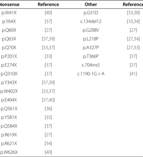
Based on the literature [10,27,33-41], a list of 25 muta-tions, which have been shown to reliably predict a Hurler phenotype when patients are homozygous or com-pound heterozygous for these mutations, was constructed (Table 3). In our cohort, the association of these mutations with MPS I-H could be confirmed for the mutations p. Q70X, p.W402X, p.L218P and c.134del12.
As mutations that have been associated with the more attenuated phenotypes [30,37,50] may be more suscep-tible to the effects of modifying polymorphisms in other genes [50], these mutations were not included in our al-gorithm. Using the list of predictive mutations in Table 3 in our group of 30 patients, a specificity of 100% for pre-diction of the MPS I-H phenotype and a sensitivity of 82% was calculated. The list of predictive mutations was integrated as first step in the prediction algorithm.
Biochemical analyses
Residual IDUA activity
Figure 1 shows that IDUA enzyme activity was linear up to at least 120 minutes of incubation time (Figure 1A) and 0.5 mg × ml-1 final protein concentration (Figure 1B). Based on these findings, we selected these conditions for subsequent studies. Substrate titrations were performed (Figure 1C) and although maximal enzyme activity was not reached, subsequent activity measurements were performed using a final substrate concentration of 1 mM. This concentration resulted in a 45% increase in activity, as compared to the commonly used substrate concentra-tion [42,43].
IDUA activity was determined in human skin fibro-blast cell lines from the 18 MPS I patients (Table 2 and Figure 2) of whom cell lines were available. Analyses were performed a week after the cells had reached confluency.
Residual IDUA activity in MPS I cell lines ranged from 0.23-2.43 nmol × mg-1 × hr-1 (Table 2), which is less than 2.5% of the activity found in control fibroblasts (control range: 101–270 nmol × mg-1× hr-1). MPS I-H
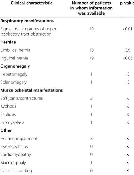
Table 1 Clinical characteristics scored in the patient cohort
Clinical characteristic Number of patients in whom information
was available
p-value
Respiratory manifestations
Signs and symptoms of upper respiratory tract obstruction
19 <0.01
Herniae
Umbilical hernia 18 0.6
Inguinal hernia 19 <0.05
Organomegaly
Hepatomegaly 1 X
Splenomegaly 1 X
Musculoskeletal manifestations
Stiff joints/contractures 2 X
Kyphosis 1 X
Scoliosis 1 X
Hip dysplasia 1 X
Other
Hearing impairment 3 X
Hydrocephalus 0 X
Cardiomyopathy 0 X
Macrocephaly 1 X
Corneal clouding 0 X
fibroblasts could be completely discriminated from the MPS I-S fibroblasts (p < 0.01) based on IDUA activity. Most MPS I-H/S fibroblasts had an intermediate IDUA activity (Figure 2). The diagnostic accuracy of the IDUA assay in differentiating MPS I-H from non-H MPS I pa-tients showed an area under the ROC curve of 0.951 (p < 0.001, Figure 3A), indicating a good discrimination. Two cut-off values were calculated, resulting in three cat-egories of enzyme activity: an IDUA activity of < 0.32 nmol × mg-1 × hr-1identified MPS I-H fibroblasts with a specificity of 100% (sensitivity 56%), as shown in Figure 3B.
This was regarded as the lower threshold, as only MPS I-H patients were found below this level of activity. Further-more, 100% sensitivity (specificity 89%) was reached at a cut-off value of 0.66 nmol x mg-1 × hr-1 enzymatic ac-tivity to discriminate MPS I-H fibroblasts from cell lines of non-H MPS I patients (Figure 3B). Subse-quently, this was set as the upper threshold; no MPS I-H fibroblasts had an enzyme activity higher than 0.66 nmol × mg-1 × hr-1.
The same sensitivity for discrimination of phenotypes was obtained when cells were cultured for 2, 4, 8 or
Table 2 Patient characteristics
General information Genetic characteristics Biochemical characteristics Clinical characteristics
Patient ID Phenotype Gestational age (weeks + days)
Mutation allele 1
Mutation allele 2
IDUA activity
(nmol x mg-1× hr-1) Upper respiratorytract involvement Inguinalhernia
1 H 37 + 0 p.W402X p.W402X 0.31 - +
2 H 38 + 1 p.W402X p.Q70X X - +
3 H 40 + 0 p.W402X p.Q70X X X X
4 H 39 + 6 p.W402X p.W402X 0.25 +
-5 H 37 + 5 p.W402X c.134del12 0.26 X
-6 H 33 + 6 p.Q70X p.L218P 0.47 X X
7 H 33 + 1 p.Q70X p.L218P 0.44 X X
8 H 38 + 0 p.Q70X p.L218P 0.58 + X
9 H 37 + 2 p.Q70X p.L218P X +
-10 H X p.W402X p.L218P X + X
11 H 33 + 0 p.L218P p.L218P X X X
12 H 40 + 0 p.L218P p.L218P X +
-13 H 38 + 3 p.W402X p.L218P 0.43 +
-14 H 41 + 1 p.A367E c.1650del117 X + +
15 H 38 + 1 c.494-1G > A c.494-1G > A 0.23 X +
16 H X p.H425fs p.H425fs 0.32 X X
17 H 40 + 0 p.W402X p.W402X X X X
18 H/S 40 + 0 p.W402X p.R505G 0.77 +
-191 H/S 38 + 0 p.W402X p.N348K 0.92 -
-201 H/S 37 + 1 p.W402X p.N348K 0.95 -
-21 H/S 41 + 2 p.L218P p.D315Y 0.35 -
-222 H/S 37 + 0 p.P533R p.P533R X -
-232 H/S 37 + 0 p.P533R p.P533R 2.43 -
-24 S 40 + 0 p.W402X p.R383H 1.05 -
-253 S 40 + 0 p.Q70X p.R383H 1.17 X X
263 S 40 + 0 p.Q70X p.R383H X X X
27 S X p.A327P p.R383H 1.70 X X
284 S 40 + 5 c.474-2A > G p.R383H X -
-294 S 40 + 5 c.474-2A > G p.R383H 1.61 -
-30 S 40 + 0 c.474-2A > G p.R383H X -
-H = -Hurler, -H/S = -Hurler/Scheie, S = Scheie.
+ = present, - = absent, X = data not available or excluded based on prematurity. 1,3,4
: siblings.2
10 weeks postconfluency. With increasing culture time, however, residual enzyme activity in all fibroblast cell lines decreased, as compared to cells cultured for 1 week postconfluency (results not shown).
Heparan sulfate and dermatan sulfate levels in fibroblasts No significant differences were seen between MPS I-H fibroblasts and non-MPS I-H cells in total HS and DS or in the levels of individual disaccharides (results not shown).
Clinical characteristics
Information on clinical signs and symptoms in the first month of life was available for 23 patients (Table 2). 3 patients, however, were excluded from the analysis
because they were born at a gestational age < 37 weeks. A significant difference between the incidence of signs and symptoms of upper respiratory tract obstruction (p = 0.005) and inguinal hernia (p = 0.033) was found be-tween MPS I-H patients and non-MPS I-H patients.
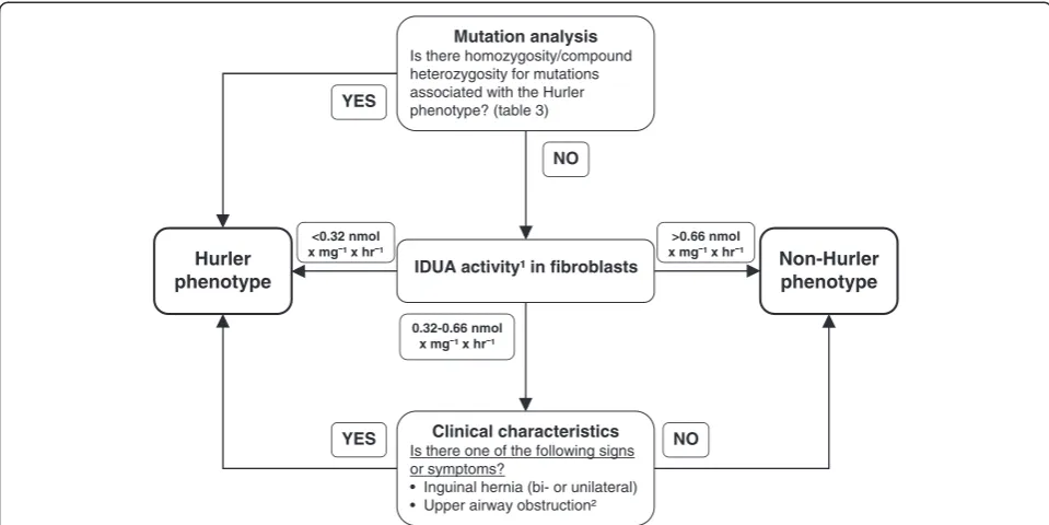
Prediction algorithm
Mutation analysis was integrated as the first step in the prediction algorithm (specificity 100%, sensitivity 82%) and IDUA activity was chosen as the second step. A cut off value of < 0.32 nmol × mg-1× hr-1was used to iden-tify MPS I-H patients. An IDUA activity of >0.66 nmol × mg-1 × hr-1 identified non-MPS I-H patients. Patients with IDUA activity between 0.32-0.66 nmol × mg-1× hr-1 were further classified by the presence of either one of the associated clinical characteristics (sensitivity 100%, specificity 100%).
This resulted in a sensitivity and specificity of 100% for the complete prediction algorithm. The flow chart for the prediction algorithm is shown in Figure 4.
Discussion
Here we present an algorithm, based on the combination of mutation analysis, residual IDUA activity and clinical signs and symptoms during the first month of life, which may allow early, sensitive and specific prediction of the phenotype in MPS I patients diagnosed through NBS. Such an algorithm can be essential as the decision to im-plement NBS for MPS I will depend, at least in a num-ber of countries, on the feasibility to decide on the optimal treatment strategy at an early age. NBS for MPS I is of high interest as early initiation of treatment, i.e. either HSCT for patients who will develop a MPS I-H phenotype and ERT for the non-MPS I-H patients, likely improves the disease outcome [8,9,12,19,51], and early diagnosis on clinical recognition can be very difficult.
To date, more than 200 different mutations in the IDUA gene have been reported [52], and this genetic heterogeneity partially explains the phenotypic variability
Table 3 Mutations described as MPS I-Hurler associated in literature
Nonsense Reference Other Reference
p.W41X [40] p.G51D [33,38]
p.Y64X [37] c.134del12 [10,34]
p.Q60X [27] p.G208V [27]
p.Q63X [37,39] p.L218P [27,34]
p.Q70X [33,37] p.A327P [27,33]
p.P201X [33] p.T366P [37]
p.E274X [37] c.704ins5 [37]
p.Q310X [37] c.1190-1G > A [41]
p.Y343X [37,39]
p.W402X [33,37]
p.E404X [37,40]
p.Q561X [36]
p.Y581X [35]
p.Q584X [37]
p.R619X [27]
p.R621X [34]
p.W626X [40]
Time (minutes)
0 20 40 60 80 100 120
0 200 400 600 800
Substrate concentration (mM)
0 0.2 0.4 0.6 0.8 1.0
0 50 100 150 200 250
0 0.1 0.2 0.3 0.4 0.5 0.6 0.7 0
2 4 6 8 10 12 14
IDU
A activity (nmol x mg )
-1
Protein (mg x ml )-1 -1
IDU
A activity (nmol x hr )
-1
IDU
A activity (nmol x mg x hr )
-1
-1
A
B
C
Figure 1IDUA activity analysis. (A)Time dependence, using 1 mM substrate and 0.17 mg × ml-1protein.(B)Protein dependence, using 1 mM
in MPS I. For most of the mutations no clear genotype-phenotype correlation is known. However, some muta-tions have been found to reliably predict a severe disease phenotype [10,27,33-41]. This was confirmed in our co-hort for the mutations p.Q70X, p.W402X, p.L218P and c.134del12. Therefore, mutation analysis was included as the first step in the algorithm to predict MPS I pheno-type. Several missense mutations, such as the p.R383H and p.R89Q mutations, are generally reported in associ-ation with more attenuated disease [30,37,50]. We did not include these latter mutations in our algorithm, however, because the effect of attenuated mutations might vary due to novel combinations of mutations, polymorphisms in other genes or environmental factors [50]. Other mutations present in our cohort were also not incorporated in the algorithm because of functional heterogeneity (e.g. the same mutations seem to have a different effect on phenotypic severity) in earlier
studies, such as the mutations p.P533R and c.474-2A > G [27,33,37]. Studies on genotype-phenotype correla-tions in large cohorts, focusing on allelic combinacorrela-tions of rarer mutations, could further improve the predictive power of this first step in our algorithm. Currently, rapid mutation analysis of the IDUA gene may not be available to all centers diagnosing MPS I. However, the fast technological advancements for gene sequencing will result in more universal access to mutation ana-lysis, allowing reliable results within 4 weeks after diag-nosis for most patients and applicability of the algorithm presented in this study.
It is highly likely that the introduction of NBS for MPS I will result in the identification of many novel mu-tations with unknown phenotypes. Therefore, a tool for prediction of phenotypic severity within the scope of NBS needs to include other variables. We found that the concentrations of HS and DS and of the individual disac-charides in cultured fibroblasts did not correlate with the phenotype. In contrast, Fuller et al. demonstrated that the levels of specific HS and DS derived trisaccha-rides in patient fibroblasts could discriminate between MPS I patients with and without neurological involve-ment [32]. In that study, only levels of short chain HS and DS oligosaccharides were measured, while the HS and DS derived disaccharides detected in our study ori-ginate predominantly from relatively larger HS and DS chains [45]. Possibly, fibroblasts from patients with neurological involvement store more short GAGs chains, as compared to patients without neurological symptoms, but similar levels of larger HS and DS oligosaccharides, which hinders discrimination between these phenotypes using our GAG analysis.
Analyses of IDUA activity in fibroblasts or leukocytes is generally used as the confirmatory step in MPS I diag-nosis. However, the most commonly used method, though sensitive for diagnostics [42,43], is not sensitive enough to reliably discriminate between the different
A
B
Figure 3ROC curve analysis. (A)ROC curve of IDUA activity for discrimination between MPS I-H and non MPS I-H.(B)Sensitivity and specificity for IDUA activity cut-off levels to discriminate between MPS I-H and non MPS I-H. Dashed lines represent chosen cut-off levels of 0.32 and 0.66 nmol × mg-1× hr-1IDUA activity.
MPS I-H MPS I-H/S MPS I-S 0
0.5 1.0 1.5 2.0 2.5
IDU
A activity (nmol x mg x hr )
-1
-1
MPS I phenotypes. A study in a cohort of 13 MPS I pa-tients [32], where the IDUA protein was first captured using antibodies followed by enzymatic studies, showed clear discrimination between patients with and without neurological involvement. This method, however, makes use of specific anti-IDUA antibodies which are not com-mercially available, making this assay difficult to imple-ment in other laboratories. In addition, specific putative mutations might result in a loss of epitopes, ob-structing capture of the protein and thus interfering with the analysis. For this reason, we optimized the 4-methylumbelliferyl-α-L-iduronide IDUA activity assay to provide a method that may be more generally applic-able. A higher concentration of substrate, independently reported by others to improve the accuracy of the IDUA assay in a recent study [53], combined with a longer incu-bation time and increased amount of protein, resulted in accurate measurement of very low enzyme activities, as seen in MPS I patients [43]. As these are minor changes to the commonly used IDUA activity analysis protocol, but very important to accurately determine very low IDUA activities, we feel that most laboratories will be able to im-plement this protocol after the necessary validation steps. Interestingly, a very narrow range of low IDUA activities is responsible for a broad range of clinical presentations in MPS I patients, as IDUA activity in all MPS I fibroblasts was less than 2.5% of the activity measured in healthy con-trol fibroblasts. Despite this small range of IDUA activities,
cut-off values could be calculated using ROC curve ana-lysis to differentiate between MPS I Hurler and non-Hurler fibroblasts.
Measurement of residual IDUA activity could not fully differentiate between phenotypes of patients with an ac-tivity in the range of 0.32-0.66 nmol × mg-1 × hr-1, as one MPS I-H/S cell line had an IDUA activity in this range. Although HSCT may be considered in some MPS I-H/S patients with neurocognitive involvement [7], this is not common practice. Therefore, the algorithm was improved by inclusion of potentially discriminating clin-ical characteristics early in life.
Of the 14 clinical characteristics studied, the presence of two were found to differ significantly between MPS I-H and non-MPS I-I-H MPS I patients: presence of in-guinal hernia and the presence of signs and symptoms of upper airway obstruction. Including clinical characteristics in the algorithm resulted in complete differentiation be-tween MPS I-H patients and patients with more attenu-ated phenotypes. Another clinical characteristic that may differentiate between MPS I-H patients and more attenu-ated patients is probably the severity of dysostosis multi-plex, a collection of radiographic abnormalities resulting from defective endochondral and membranous growth throughout the body seen in mucopolysaccharidoses. Es-pecially thoraco-lumbar kyphosis before the age of one month, might be a very sensitive and specific symptom for MPS I-H [54,55]. However, early kyphosis is often initially
IDUA activity¹ in fibroblasts
Hurler phenotype
Clinical characteristics
Is there one of the following signs or symptoms?
• Inguinal hernia (bi- or unilateral) • Upper airway obstruction²
NO YES
<0.32 nmol x mg ¹ x hr
0.32-0.66 nmol x mg ¹ x hr
>0.66 nmol x mg ¹ x hr
Non-Hurler phenotype
Mutation analysis
Is there homozygosity/compound heterozygosity for mutations associated with the Hurler phenotype? (table 3)
NO YES
not recognized by parents and caregivers and could there-fore not be included in this model, which is based on retrospective analysis of clinical data.
Our study has some limitations. Firstly, due to the ultra-orphan nature of the disease, the proposed algo-rithm is validated in only a relatively small number of patients (n = 30). Validation in other cohorts of patients needs to be performed to further determine its value. Secondly, our study includes a retrospective analysis of signs and symptoms during the first month of life. This may result in a recall bias, as both parents and investiga-tors knew the phenotype of the patients. To address this, only characteristics that could be clearly distinguished and are often well documented in the newborn period were used for this algorithm. Thirdly, the prevalence of mutations firmly associated with certain phenotypes dif-fers between regions around the world [37]. Therefore, positive and negative predicting values of the proposed algorithm may differ between countries and this needs to be further investigated. Also, as new mutations will be detected once NBS for MPS I has been introduced, a prediction algorithm including mutation analysis needs to be continuously adjusted and improved. Likewise, NBS will allow for further investigation on the predictive value of certain clinical signs such as early kyphosis, which could not be included in this study.
As a result of future studies, the algorithm might be adapted to also to differentiate between MPS I-H/S pa-tients with and without neurocognitive involvement. The improved outcome of HSCT, in combination with increasing knowledge on the risk for neurocognitive de-cline in a subset of MPS I-H/S patients, may result in a shift in treatment protocols, with HSCT as treatment of choice for this group of patients [7].
With the phenotypic prediction algorithm presented here, we hope to provide the basis for a tool to reliably predict phenotype in the majority of MPS I patients di-agnosed through NBS. Prospective studies could result in inclusion of additional predictive factors and improve-ment of the prediction algorithm.
Conclusion
Using genetic, biochemical and clinical characteristics, which can all be studied within the first month of life, an algorithm was developed for accurate prediction of the phenotype at an early age in MPS I patients. Such an algorithm allows timely initiation of the optimal treat-ment strategy, thus improving disease outcome. With the future launch of NBS programs for MPS I, patients will not have developed all characteristic signs and symptoms currently used for assessment of the pheno-type, making a prediction algorithm for early assessment of phenotypic severity indispensable.
Abbreviations
DS:Dermatan sulfate; ERT: Enzyme replacement therapy; FBS: Fetal bovine serum; GAG: Glycosaminoglycan; HS: Heparan sulfate; HSCT: Haematopoietic stem cell transplantation; IDUA:α-L-iduronidase; IRDS: Infant respiratory distress syndrome; LSD: Lysosomal storage disease; MPS I:
Mucopolysaccharidosis type I; MPS I-H: MPS I-Hurler; MPS I-H/S: MPS I-Hurler/ Scheie; MPS I-S: MPS I-Scheie; NaCl: Sodium chloride; NBS: Newborn screening; PBS: Phosphate buffered saline; ROC: Receiver operating characteristic.
Competing interests
The authors declare that they have no competing interests.
Authors’contributions
SDKK, EJL, NvV: Conception and design, data acquisition, analysis, and interpretation, manuscript draft and revision. CdK: data acquisition and analysis, manuscript draft. LZ: conception and design, manuscript revision. TW: data analysis, manuscript revision. LIJ, RJAW: conception and design, manuscript revision. FAW: Conception and design, manuscript draft, manuscript revision. All authors read and approved the final manuscript.
Acknowledgements
We thank W. Kulik and H. van Lenthe for helpful discussions and technical assistance.
Author details 1
Department of Pediatrics and Amsterdam Lysosome Centre“Sphinx”, Academic Medical Center, University Hospital of Amsterdam, Meibergdreef 9, 1105, AZ Amsterdam, The Netherlands.2Laboratory of Genetic Metabolic
Diseases, Department of Clinical Chemistry, Academic Medical Center, University Hospital of Amsterdam, Meibergdreef 9, 1105, AZ Amsterdam, The Netherlands.
Received: 3 May 2013 Accepted: 3 July 2013 Published: 9 July 2013
References
1. Wraith JE:The mucopolysaccharidoses: a clinical review and guide to management.Arch Dis Child1995,72:263–267.
2. Campos D, Monaga M:Mucopolysaccharidosis type I: current knowledge on its pathophysiological mechanisms.Metab Brain Dis2012,27:121–129. 3. D'Aco K, Underhill L, Rangachari L, Arn P, Cox GF, Giugliani R, Okuyama T,
Wijburg F, Kaplan P:Diagnosis and treatment trends in
mucopolysaccharidosis I: findings from the MPS I Registry.Eur J Pediatr
2011,171:911–919.
4. Hopkin RJ, Grabowski GA,et al:Lysosomal storage diseases. InHarrison's Principles of Internal Medicine.17th edition. Edited by Fauci A, Kasper D, Braunwald E. New York: McGraw Hill; 2005.
5. Roubicek M, Gehler J, Spranger J:The clinical spectrum of alpha-L-iduronidase deficiency.Am J Med Genet1985,20:471–481.
6. Vijay S, Wraith JE:Clinical presentation and follow-up of patients with the attenuated phenotype of mucopolysaccharidosis type I.Acta Paediatr
2005,94:872–877.
7. De Ru MH, Boelens JJ, Das AM, Jones SA, Van der Lee JH, Mahlaoui N, Mengel E, Offringa M, O'Meara A, Parini R, Rovelli A, Sykora KW, Vellodi A, Wynn RF, Wijburg FA:Enzyme replacement therapy and/or
hematopoietic stem cell transplantation at diagnosis in patients with mucopolysaccharidosis type I: results of a European consensus procedure.Orphanet J Rare Dis2012,6:1–9.
8. Peters C, Balthazor M, Shapiro EG, King RJ, Kollman C, Hegland JD, Henslee-Downey J, Trigg ME, Cowan MJ, Sanders J, Bunin N, Weinstein H, Lenarsky C, Falk P, Harris R, Bowen T, Williams TE, Grayson GH, Warkentin P, Sender L, Cool VA, Crittenden M, Packman S, Kaplan P, Lockman LA, Anderson J, Krivit W, Dusenbery K, Wagner J:Outcome of unrelated donor bone marrow transplantation in 40 children with Hurler syndrome.Blood1996,
87:4894–4902.
transplantation in fifty-four children. The Storage Disease Collaborative Study Group.Blood1998,91:2601–2608.
10. Souillet G, Guffon N, Maire I, Pujol M, Taylor P, Sevin F, Bleyzac N, Mulier C, Durin A, Kebaili K, Galambrun C, Bertrand Y, Froissart R, Dorche C, Gebuhrer L, Garin C, Berard J, Guibaud P:Outcome of 27 patients with Hurler's syndrome transplanted from either related or unrelated haematopoietic stem cell sources.Bone Marrow Transplant2003,31:1105–1117. 11. Vellodi A, Young EP, Cooper A, Wraith JE, Winchester B, Meaney C,
Ramaswami U, Will A:Bone marrow transplantation for
mucopolysaccharidosis type I: experience of two British centres.Arch Dis Child1997,76:92–99.
12. Boelens JJ, Aldenhoven M, Purtill D, Ruggeri A, Defor T, Wynn R, Wraith E, Cavazzana-Calvo M, Rovelli A, Fischer A, Tolar J, Prasad VK, Escolar M, Gluckman E, O'Meara A, Orchard PJ, Veys P, Eapen M, Kurtzberg J, Rocha V:
Outcomes of transplantation using various hematopoietic cell sources in children with Hurler's syndrome after myeloablative conditioning.Blood
2013,121:3981–3987.
13. Clarke LA, Wraith JE, Beck M, Kolodny EH, Pastores GM, Muenzer J, Rapoport DM, Berger KI, Sidman M, Kakkis ED, Cox GF:Long-term efficacy and safety of laronidase in the treatment of mucopolysaccharidosis I.Pediatrics
2009,123:229–240.
14. Kakkis ED, Muenzer J, Tiller GE, Waber L, Belmont J, Passage M, Izykowski B, Phillips J, Doroshow R, Walot I, Hoft R, Neufeld EF:
Enzyme-replacement therapy in mucopolysaccharidosis I.N Engl J Med2001,344:182–188.
15. Muenzer J, Wraith JE, Clarke LA:Mucopolysaccharidosis I: Management and Treatment Guidelines.Pediatrics2009,123:19–29.
16. Sifuentes M, Doroshow R, Hoft R, Mason G, Walot I, Diament M, Okazaki S, Huff K, Cox GF, Swiedler SJ, Kakkis ED:A follow-up study of MPS I patients treated with laronidase enzyme replacement therapy for 6 years.Mol Genet Metab2007,90:171–180.
17. Wraith JE, Clarke LA, Beck M, Kolodny EH, Pastores GM, Muenzer J, Rapoport DM, Berger KI, Swiedler SJ, Kakkis ED, Braakman T, Chadbourne E, Walton-Bowen K, Cox GF:Enzyme replacement therapy for
mucopolysaccharidosis I: a randomized, double-blinded, placebo-controlled, multinational study of recombinant human alpha-L-iduronidase (laronidase).J Pediatr2004,144:581–588.
18. Wraith JE, Beck M, Lane R, van der Van der Ploeg A, Shapiro E, Xue Y, Kakkis ED, Guffon N:Enzyme replacement therapy in patients who have mucopolysaccharidosis I and are younger than 5 years: results of a multinational study of recombinant human alpha-L-iduronidase (laronidase).Pediatrics2007,120:e37–e46.
19. Gabrielli O, Clarke LA, Bruni S, Coppa GV:Enzyme-Replacement Therapy in a 5-Month-Old Boy With Attenuated Presymptomatic MPS I: 5-Year Follow-up.Pediatrics2010,125:183–187.
20. Blanchard S, Sadilek M, Scott CR, Turecek F, Gelb MH:Tandem mass spectrometry for the direct assay of lysosomal enzymes in dried blood spots: application to screening newborns for mucopolysaccharidosis I.
Clin Chem2008,54:2067–2070.
21. De Ruijter J, De Ru MH, Wagemans T, Ijlst L, Lund AM, Orchard PJ, Schaefer PB, Wijburg FA, Van Vlies N:Heparan sulfate and dermatan sulfate derived disaccharides are sensitive markers for newborn screening for mucopolysaccharidoses types I, II and III.Mol Genet Metab2012,
107:705–710.
22. Chamoles NA, Blanco M, Gaggioli D:Diagnosis of á-L-iduronidase deficiency in dried blood spots on filter paper: the possibility of newborn diagnosis.Clin Chem2001,47:780–781.
23. Li Y, Scott CR, Chamoles NA, Ghavami A, Pinto BM, Turecek F, Gelb MH:
Direct multiplex assay of lysosomal enzymes in dried blood spots for newborn screening.Clin Chem2004,50:1785–1796.
24. Burton B, Charrow J, Angle B, Widera S, Waggoner D:A pilot newborn screening program for lysosomal storage disorders (LSD) in Illinois.
Mol Genet Metab2012,105:S23–S24.
25. De Ru MH, Bouwman MG, Wijburg FA, Van Zwieten MCB:Experiences of parents and patients with the timing of Mucopolysaccharidosis type I (MPS I) diagnoses and its relevance to the ethical debate on newborn screening.Mol Genet Metab2012,107:501–507.
26. Scott CR, Elliott S, Buroker N, Thomas LI, Keutzer J, Glass M, Gelb MH, Turecek F:Identification of Infants at Risk for Developing Fabry, Pompe, or Mucopolysaccharidosis-I from Newborn Blood Spots by Tandem Mass Spectrometry.J Pediatr2013. doi:10.1016/j.jpeds.2013.01.031.
27. Beesley CE, Meaney CA, Greenland G, Adams V, Vellodi A, Young EP, Winchester BG:Mutational analysis of 85 mucopolysaccharidosis type I families: frequency of known mutations, identification of 17 novel mutations and in vitro expression of missense mutations.Hum Genet
2001,109:503–511.
28. Li P, Wood T, Thompson JN:Diversity of mutations and distribution of single nucleotide polymorphic alleles in the human alpha-L-iduronidase (IDUA) gene.Genet Med2002,4:420–426.
29. De Ru MH, Teunissen QGA, Van der Lee JH, Beck M, Bodamer OA, Clarke LA, Hollak CE, Lin SP, Munoz Rojas MV, Pastores GM, Raiman JA, Scarpa M, Treacy EP, Tylki-Szymanska A, Wraith JE, Zeman J, Wijburg FA:Capturing phenotypic heterogeneity in MPS I: results of an international consensus procedure.Orphanet J Rare Dis2012,7:1–9.
30. Bunge S, Clemets PR, Byers S, Kleijer WJ, Brooks DA, Hopwood JJ:
Genotype-phenotype correlations in mucopolysaccharidosis type I using enzyme kinetics, immunoquantification and in vitro turnover studies.
Biochim Biophys Acta1998,1407:249–256.
31. Yogalingam G, Guo XH, Muller VJ, Brooks DA, Clements PR, Kakkis ED, Hopwood JJ:Identification and molecular characterization of alpha-L-iduronidase mutations present in mucopolysaccharidosis type I patients undergoing enzyme replacement therapy.Hum Mutat2004,24:199–207. 32. Fuller M, Brooks DA, Evangelista M, Hein LK, Hopwood JJ, Meikle PJ:
Prediction of neuropathology in mucopolysaccharidosis I patients.
Mol Genet Metab2005,84:18–24.
33. Bertola F, Filocamo M, Casati G, Mort M, Rosano C, Tylki-Szymanska A, Tuysuz B, Gabrielli O, Grossi S, Scarpa M, Parenti G, Antuzzi D, Dalmau J, Rocco MD, Vici CD, Okur I, Rosell J, Rovelli A, Furlan F, Rigoldi M, Biondi A, Cooper DN, Parini R:IDUA mutational profiling of a cohort of 102 European patients with Mucopolysaccharidosis type I: identification and characterization of 35 novel á-L-iduronidase (IDUA) alleles.Hum Mutat
2011,32:e2189–e2210.
34. Bunge S, Kleijer WJ, Steglich C, Beck M, Zuther C, Morris CP, Schwinger E, Hopwood JJ, Scott HS, Gal A:Mucopolysaccharidosis type I: identification of 8 novel mutations and determination of the frequency of the two common alpha-L-iduronidase mutations (W402X and Q70X) among European patients.Hum Mol Genet1994,3:861–866.
35. Chkioua L, Khedhiri S, Turkia HB, Tcheng R, Froissart R, Chahed H, Ferchichi S, Ben Dridi MF, Vianey-Saban C, Laradi S, Miled A:Mucopolysaccharidosis type I: molecular characteristics of two novel alpha-L-iduronidase mutations in Tunisian patients.Diagn Pathol2011,6:47.
36. Matte U, Yogalingam G, Brooks D, Leistner S, Schwartz I, Lima L, Norato DY, Brum JM, Beesley C, Winchester B, Giugliani R, Hopwood JJ:Identification and characterization of 13 new mutations in mucopolysaccharidosis type I patients.Mol Genet Metab2003,78:37–43.
37. Terlato NJ, Cox GF:Can mucopolysaccharidosis type I disease severity be predicted based on a patient's genotype?Genet Med2003,5:286–294. 38. Venturi N, Rovelli A, Parini R, Menni F, Brambillasca F, Bertagnolio F, Uziel G,
Gatti R, Filocamo M, Donati MA, Biondi A, Goldwurm S:Molecular analysis of 30 mucopolysaccharidosis type I patients: evaluation of the mutational spectrum in Italian population and identification of 13 novel mutations.Hum Mutat2002,20:231.
39. Voskoboeva EY, Krasnopolskaya XD, Mirenburg TV, Weber B, Hopwood JJ:
Molecular genetics of mucopolysaccharidosis type I: mutation analysis among the patients of the former Soviet Union.Mol Genet Metab1998,
65:174–180.
40. Wang X, Zhang W, Shi H, Qiu Z, Meng Y, Yao F, Wei M:
Mucopolysaccharidosis I mutations in Chinese patients: identification of 27 novel mutations and 6 cases involving prenatal diagnosis.Clin Genet
2012,81:443–452.
41. Sun L, Li C, Song X, Zheng N, Zhang H, Dong G:Three novel alpha-L-iduronidase mutations in 10 unrelated Chinese mucopolysaccharidosis type I families.Genet Mol Biol2011,34:195–200.
42. Hopwood JJ, Muller V, Smithson A, Baggett N:A fluorometric assay using 4-methylumbelliferyl L-iduronide for the estimation of alpha-L-iduronidase activity and the detection of Hurler and Scheie syndromes.Clin Chim Acta1979,92:257–265.
43. Stirling JL, Robinson D, Fensom AH, Benson PF, Baker JE:Fluorimetric assay for prenatal detection of Hurler and Scheie homozygotes or
heterozygotes.Lancet1978,1:147.
45. De Ru MH, Van der Tol L, Van VN, Bigger BW, Hollak CE, Ijlst L, Kulik W, Van LH, Saif MA, Wagemans T, Van der Wal WM, Wanders RJ, Wijburg FA:
Plasma and urinary levels of dermatan sulfate and heparan sulfate derived disaccharides after long-term enzyme replacement therapy (ERT) in MPS I: correlation with the timing of ERT and with total urinary excretion of glycosaminoglycans.J Inherit Metab Dis2013,36:247–255. 46. Cleary MA, Wraith JE:The presenting features of mucopolysaccharidosis
type IH (Hurler syndrome).Acta Paediatr1995,84:337–339. 47. Colville GA, Bax MA:Early presentation in the mucopolysaccharide
disorders.Child Care Health Dev1996,22:31–36.
48. Donaldson MD, Pennock CA, Berry PJ, Duncan AW, Cawdery JE, Leonard JV:
Hurler syndrome with cardiomyopathy in infancy.J Pediatr1989,
114:430–432.
49. Watts RW, Spellacy E, Adams JH:Neuropathological and clinical correlations in Hurler disease.J Inherit Metab Dis1986,9:261–272. 50. Scott HS, Litjens T, Nelson PV, Thompson PR, Brooks DA, Hopwood JJ,
Morris CP:Identification of mutations in the alpha-L-iduronidase gene (IDUA) that cause Hurler and Scheie syndromes.Am J Hum Genet1993,
53:973–986.
51. Hobbs JR, Hugh-Jones K, Barrett AJ, Byrom N, Cahmber D, Henry K, James DC, Lucas CF, Rogers TR, Benson PF, Tansley LR, Patrick AD, Mossman J, Young EP:Reversal of clinical features of Hurler's disease and biochemical improvement after treatment by bone-marrow transplantation.Lancet1981,2:709–712.
52. HGMD®.http://www.hgmd.cf.ac.uk/.
53. Herzog T, Ou L, Whitley C:Increased substrate concentration boosts enzyme activity levels of fluorometric a-L-iduronidase enzyme activity assay.Mol Genet Metab2013,108:S48.
54. Aldenhoven M, Sakkers RJ, Boelens J, De Koning TJ, Wulffraat NM:
Musculoskeletal manifestations of lysosomal storage disorders.
Ann Rheum Dis2009,68:1659–1665.
55. White KK:Orthopaedic aspects of mucopolysaccharidoses.Rheumatology (Oxford)2011,50(Suppl 5):v26–v33.
doi:10.1186/1750-1172-8-99
Cite this article as:Kingmaet al.:An algorithm to predict phenotypic severity in mucopolysaccharidosis type I in the first month of life.
Orphanet Journal of Rare Diseases20138:99.
Submit your next manuscript to BioMed Central and take full advantage of:
• Convenient online submission
• Thorough peer review
• No space constraints or color figure charges
• Immediate publication on acceptance
• Inclusion in PubMed, CAS, Scopus and Google Scholar
• Research which is freely available for redistribution