R E S E A R C H
Open Access
Comparative evaluation of Icon® resin
infiltration and Clinpro
™
XT varnish on
colour and fluorescence changes of white
spot lesions: a randomized controlled trial
Annapurna Kannan
*and Sridevi Padmanabhan
Abstract
Background:The aim of this trial was to comparatively evaluate Icon® resin infiltration and Clinpro™XT varnish in restoring aesthetics of white spot lesions (WSLs) present post-orthodontic treatment.
Materials and methods:Two hundred forty WSLs were detected in 193 teeth of 12 patients. The participants were
analysed—before intervention (T0), immediately after intervention (T1), 3 months later (T2) and 6 months later (T3), with a 1:1 allocation ratio for the application of Icon® resin infiltration and Clinpro™XT varnish. Using a computer-generated allocation sequence, block randomization was done. A spectrophotometer was used to assess the colour of WSLs and the adjacent enamel, while a DIAGNOdent® was used to assess the fluorescence loss.
Results:Immediately after the intervention, Icon® resin infiltration showed statistically significant better
improvement than Clinpro™XT varnish in restoring the colour (p= 0.000); however, at 3 (p= 0.001) and 6 months (p= 0.000), this was reversed. Except at 3 months, the fluorescence loss sequentially reduced more for Icon® resin infiltration (4.48 ± 1.42 at T0to 1.48 ± 0.81 at T3) and was not statistically significant.
Conclusions:Clinpro™XT varnish showed significantly better improvement than Icon® resin infiltration in restoring the colour and lightness of the WSLs at 3 and 6 months. The fluorescence loss significantly recovered with both intervention methods between immediate application and at 6 months. However, Clinpro™XT varnish-treated WSLs showed a statistically significant difference compared to the adjacent sound enamel at 6 months.
Keywords:White spot lesions, Icon® resin infiltration, Clinpro™XT varnish
Background
Aesthetics is one of the cornerstone objectives of orthodon-tic treatment. One of the deleterious effects of orthodonorthodon-tic treatment, primarily caused due to poor oral hygiene main-tenance, is the occurrence of white spot lesions (WSLs) [1].
The management of WSLs can be divided into preven-tion (prior to the formapreven-tion of WSLs), intervenpreven-tion (dur-ing the course of orthodontic treatment) and treatment (after the completion of orthodontic treatment). In the prevention and intervention categories, fluorides in the form of varnish, toothpaste, mouthwash, sealant and
miswaks [2]; CPP-ACP, NovaMin®; laser therapy; silver
nanoparticles; and ozone have been used extensively [3–
7]. However, the clinicians often encounter WSLs post-debonding. The effects of the prevention methods are inadequate due to their effectiveness being restricted to only remineralizing the superficial surface of the lesion and not the body of the lesion [8].
The treatment of the WSLs depends on its severity. The mild forms of WSLs can be left to natural remineralization
which occurs over a period of 1 year [1, 9] and through
other means such as CPP-ACP [3, 10] and fluoride
var-nishes [11]. The moderate and severe forms can be treated using bleaching, microabrasion, resin infiltration and res-torations [12,13].
© The Author(s). 2019, corrected publication 2019.Open AccessThis article is distributed under the terms of the Creative Commons Attribution 4.0 International License (http://creativecommons.org/licenses/by/4.0/), which permits unrestricted use, distribution, and reproduction in any medium, provided you give appropriate credit to the original author(s) and the source, provide a link to the Creative Commons license, and indicate if changes were made.
* Correspondence:[email protected]
The recent emphasis on the mini aesthetics of tooth such as its colour, texture and enamel translucency has created more concern among patients and orthodontists. This augurs an immediate solution.
With Icon® resin infiltration (DMG America,
Engle-wood, NJ, USA), its acid etchant—15% hydrochloric
acid—removes the surface layer of the decalcified area
due to its penetration depth of 58 ± 37μm. [14,15]. This
opens up access to the body of the lesion which allows the resin to occlude the pores. The body of the lesion is rendered watertight by means of the resin which has a refractive index (RI Icon® = 1.44) close to that of healthy enamel (RI = 1.63) [14] and also helps in stopping the diffusion of acids by creating a barrier within the lesion and not on the surface [16,17].
Icon® resin infiltration has been proved to provide im-mediate restoration of aesthetics of mild WSLs present post-orthodontic treatment to match that of the adjacent
sound enamel [18,19]. It has been shown to remain
dur-able for 6 months [18] with no significant changes at 12 and 24 months [20]. With moderate lesions, a sequential improvement was seen to occur over a period of time [19]. The fluorescence loss of the WSLs significantly re-covered immediately with resin infiltration and remained unchanged at the end of 6 weeks in artificially created WSLs [21]. These factors establish Icon® resin infiltration as a gold standard intervention method in aesthetic res-toration of white spot lesions.
Resin-modified glass ionomer cement contains
fluoroaluminosilicate glass. The fluoride reacts at the surface to provide an immediate release, while the bulk of the glass matrix offers a reservoir of fluoride
for sustained release [22, 23] which is said to
pro-vide remineralization.
Clinpro™ XT varnish (3M ESPE, Pymble, New South
Wales, Australia), an RMGIC product, has been widely used previously for treating dentinal
hypersen-sitivity [24, 25]. In orthodontics, it is shown to be
ef-fective in preventing WSLs from occurring during
orthodontic treatment [23, 26, 27] and treating
artifi-cially induced demineralized areas [28]. However, its efficacy in treating WSLs present post-orthodontic treatment has not yet been tested.
Specific objective
With both Icon® resin infiltration and Clinpro™XT
var-nish, being minimally invasive and site-specific in office single sitting procedures containing composite resin, the aim of this randomized clinical trial was to
compara-tively evaluate the efficacy of Clinpro™ XT varnish
against the ‘gold standard’ Icon® resin infiltration in re-storing the aesthetics of the enamel affected by white spot lesions post-orthodontic treatment.
Materials and methods
The study was conducted at the Department of Ortho-dontics, Sri Ramachandra Institute of Higher Education and Research after the approval from the University’s In-stitutional Ethics Committee [number: CSP/16/SEP/51/ 280 dated October 4, 2016].
Trial design
This was a parallel-group, randomized, active-controlled trial with a 1:1 allocation ratio for the application of Icon® resin infiltration and Clinpro™XT varnish.
Participants, eligibility criteria and settings
Fifty patients who had fixed orthodontic appliances re-moved during April 2017 were screened for WSLs using Gorelick’s scale [29], and only mild and moderate lesions were considered. This was confirmed using a DIAGNO-dent® (KAVO Dental Corporation, Lake, Zurich, IL,
USA). Only WSLs having fluorescence loss (Q) scores of
2–9 were considered.
Inclusion criteria
Patients within 14–30 years of age
Symmetrical number of permanent teeth in each arch (mesial to second molars)
Exclusion criteria
Active carious lesions
Facial surface restorations
Deciduous teeth
Fluorosis
Intrinsic and extrinsic strains
Enrollment
A consent form was obtained from those participants willing to be part of the trial.
Sample calculation
Calculations [colour change (ΔE) threshold value = 3.0,
d= 0.5,α error = 0.05 and power of study = 85%] were
based on the study by Knosel et al. [18] (219 WSLs; 111 control, 108 treatment) to detect a clinically relevant dif-ference between the two trial arms. This indicated that 115 non-cavitated, unrestored WSLs were required in each arm.
Randomization
Using a computer-generated programme (www.randomi zation.com), block randomization allocation sequence [AABB; ABBA; BBAA; BAAB (A-Icon® resin infiltration,
B-Clinpro™ XT varnish)] was generated to equally
A total of 240 non-cavitated, unrestored WSLs after multibracket treatment were detected in 193 teeth of 12 patients [7 females (18 years ± 2 months), 5 males (20 years ± 6 months)] and formed a part of the study.
Allocation concealment and blinding
The participants were blinded until the allocation of the intervention method. Further blinding was not possible as the intervention methods had different techniques of material application.
Intervention
All the procedures were performed by the same clin-ician. On completion of orthodontic treatment, residual composite cleanup and polishing were done.
The VITA Easyshade® advance spectrophotometer (VITA Zahnfabrik, Bad Sackingen, Germany) was used to objectively assess the colour of WSLs and the adja-cent sound enamel, using the Munsell system: CIE colour parameters (ΔE):L,aandb.
ΔE¼ ΔL2þΔa2þΔb2ð1=2Þ
L* refers to the lightness coordinate, and its value ranges from 0 for perfect black to 100 for perfect white.a* andb* are the chromaticity coordinates in the red-green axis and yellow-blue axis, respectively [30,31].
Similarly, the laser fluorescence method, DIAGNOdent®,
was used to assess the fluorescence loss (Q) of WSLs and
the adjacent sound enamel present around the lesions. The DIAGNOdent® illuminates the teeth with blue laser light. The tooth dentin contains atoms called fluor-ophores which fluoresce green when illuminated with blue laser light. When a WSL is present, it appears as a black area surrounded by green reflected colour and this is seen as fluorescence loss [32].
Intervention methods
Icon® resin infiltration
First, Icon® Etch, DMG, was applied over the WSLs for 2 min. This was followed by water rinsing and air blow-ing. Then, Icon® Dry, DMG, was applied for 30 s and air blown. Using the provided sponge applicator, Icon® Infil-trant, DMG, was rubbed on and left for 3 min. Subse-quently, it was light-cured for 60 s.
CLINPRO™XT varnish
Thirty-seven percent orthophosphoric acid gel was ap-plied for 15 s. On a paper pad, the paste and liquid
com-ponents were mixed for 10–15 s using an agate spatula
and applied over the WSLs. Subsequently, it was light-cured for 20 s.
Both the treatment methods were assessed before intervention (baseline) [T0], immediately after
interven-tion [T1], 3 months later [T2] and 6 months later [T3].
Photographs were taken during each time period, and
the size and location of the WSLs were mapped at T0
for the exactness of the readings at each time interval.
Outcomes
The primary outcome measure was the difference in colour and fluorescence values between the treatment groups over a period of 6 months, and the secondary outcome measure was the difference within the treat-ment groups.
Statistical analysis
The data was analysed with IBM.SPSS statistics software,
version 23.0. Kolmogorov-Smirnov’s test was used to
check the normality of the data. To find the significant dif-ference within the groups, at different time intervals, a one-way repeated measures ANOVA was used followed by post hoc Tukey test. Between the groups, independent Student’sttest was used for the intergroup comparison.
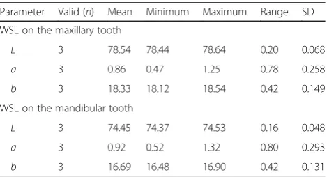
Error of method (Table1)
The L*a*b* values of one white spot lesion each in the upper and lower arch of one patient were recorded in vivo three times on three consecutive days. The ranges
of variance due to measurement error for Lvalue were
0.20 units for the WSL on the maxillary tooth and 0.16 units for WSL on the mandibular tooth. This indicates precision in measurements recorded with spectropho-tometer when compared to previous results [33]. All measurements were carried out under normalized condi-tions to make certain their accuracy and reproducibility.
Results
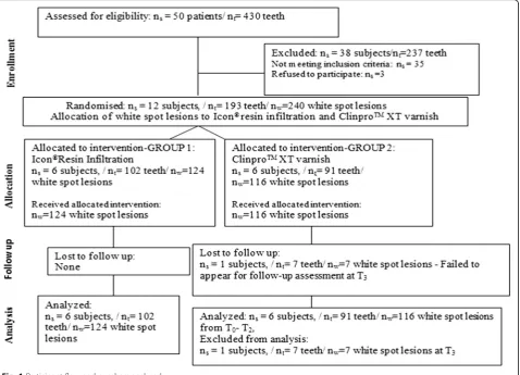
Baseline data and recruitment (Fig.1)
One hundred twenty-four white spot lesions present in 102 teeth of 6 patients received the allocated intervention method of Icon® resin infiltration, while 116 white spot lesions present in 91 teeth of 6
Table 1Error assessment results
Parameter Valid (n) Mean Minimum Maximum Range SD
WSL on the maxillary tooth
L 3 78.54 78.44 78.64 0.20 0.068
a 3 0.86 0.47 1.25 0.78 0.258
b 3 18.33 18.12 18.54 0.42 0.149
WSL on the mandibular tooth
L 3 74.45 74.37 74.53 0.16 0.048
a 3 0.92 0.52 1.32 0.80 0.293
b 3 16.69 16.48 16.90 0.42 0.131
patients received the allocated intervention method
of Clinpro™ XT varnish. At T3, 1 patient under the
Clinpro™ XT varnish intervention method was lost
to follow-up.
Outcomes and estimation
Analysis of lightness (L) values
Intergroup comparison (Table2).
Clinpro™ XT varnish showed more increase in the
L values compared with Icon® resin infiltration, which was statistically significant at T2 and T3.
Intragroup comparison (Table2)
In Icon® resin infiltration and Clinpro™XT varnish, a statistically significant increase in theLvalue was seen from T0to T1.
In both groups, a statistically significant overall improvement was seen in theLvalue from T0to T3.
Fig. 1Participant flow and numbers analysed
Table 2Intergroup and intragroup comparison of lightness values at various time intervals
Lvalues of Icon® resin infiltration
l values of Clinpro™ XT varnish
Intergroup comparison
Intragroup comparison
Icon® resin infiltration Clinpro™XT varnish
T0 73.60 ± 7.71 74.62 ± 7.33 p= 0.295 T0vs T1,p= 0.000** T0vs T1,p= 0.000**
T1 78.37 ± 5.94 78.93 ± 5.40 p= 0.446 T1vs T2,p= 0.211 T1vs T2,p= 0.003**
T2 80.08 ± 6.46 81.68 ± 5.55 p= 0.041* T2vs T3,p= 0.168 T2vs T3,p= 0.069
T3 81.88 ± 6.27 83.69 ± 5.29 p= 0.017* T0vs T3,p= 0.000** T0vs T3,p= 0.000**
Adjacent sound enamel 81.67 ± 4.69 82.73 ± 4.66 p= 0.117 T3vs adjacent sound enamel,p= 0.999 T3vs adjacent sound enamel,p= 0.761
Statistical Tests: intergroup comparison—independent Student’sttest; intragroup comparison—one-way repeated measures ANOVA Llightness,T0before intervention,T1immediate after intervention,T23 months,T36 months
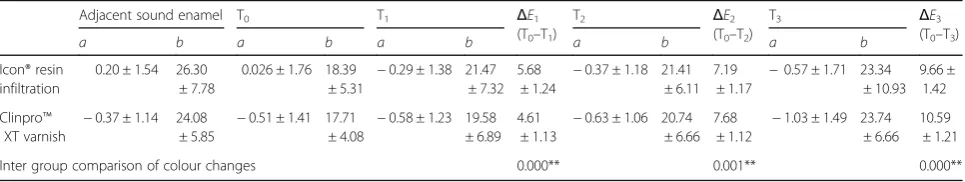
Analysis of colour changes (ΔE) (Table3)
Icon® resin infiltration showed a statistically significant colour change value from T0to T1(ΔE1)
than Clinpro™XT varnish.
Clinpro™XT varnish showed a statistically significant colour change value from T0to T2(ΔE2) and from T0
to T3(ΔE3) than Icon® resin infiltration.
Analysis of fluorescence loss (Q)
Intergroup comparison (Table4).
At all other time points except T2,Qvalue
reduction was seen more with Icon® resin infiltration when compared to Clinpro™XT varnish. However, on comparison, it was not significant.
Intragroup comparison (Table4).
In both groups, a statistically significant fluorescence loss amelioration was seen from T0to T1to T2to T3.
At T3, in both groups, the amelioration of the
fluorescence loss brought the values close to that of the adjacent sound enamel, which was not
statistically significant for Icon® resin infiltration but statistically significant for Clinpro™XT varnish.
Discussion
A parallel-group trial as opposed to a split study design was carried out to avoid any treatment bias to mitigate
any carry-across effect of fluorides present in Clinpro™
XT varnish. [34–36]
The difference in the refractive index of the enamel and lesions’crystals contributes to the whitish nature of the
WSLs [37]. WSLs have lower L* values due to the large
portions of transmitted light being absorbed and scattered
within the micropores of the body of the lesion [38–40].
Hence, the lightness value is of vital importance.
As expected, following the application of Icon® resin infil-tration, a significant increase was seen immediately in the lightness of the WSLs, suggestive of an immediate increase in enamel reflectivity (T1). The subsequent increase from
T1to T3was not statistically significant, showing the
dur-able nature of the material (Tdur-able2).
Surprisingly with Clinpro™XT varnish, a significant in-crease was seen immediately in the lightness of the WSLs with its application. It can be hypothesized that the usage of 37% orthophosphoric acid would have pen-etrated and removed the surface layer of the WSL, open-ing up access to the body of the lesion. The PC would have further increased with the usage of low viscous resin HEMA. The presence of Bis-GMA could have en-hanced the resin reactivity and reflectivity, aiding in the immediate improvement in the optical properties of the
WSLs. At T2, a significant increase was further seen in
the lightness and this could be attributed to the release of fluoride from the fluoroaluminosilicate glass particles.
The small amount of increase seen from T2 to T3 was
probably due to the sustained release of fluoride and cal-cium glycerophosphate from the reservoir of the glass
matrix. Further, the overall change seen with Clinpro™
XT was significantly greater than Icon® resin infiltration (Table2).
The resultantΔEvalues calculated at all time intervals
for both groups were more than 3.7 ΔE units, which is
the critical value for clinical detection [41] and were sta-tistically significant. However, the comparison between the groups showed that Icon® resin infiltration demon-strated a significant immediate improvement, whereas the long-term improvement was more significant with
Clinpro™XT varnish (Table3).
Icon® resin infiltration showed more revival of the
lost fluorescence at T1 than Clinpro™ XT, probably
due to the creation of an immediate diffusion barrier
within the body of the lesion. At T2, a comparable
improvement was seen with both groups. At T3,
Icon® resin infiltration showed further amelioration
of the fluorescence loss than Clinpro™ XT, though
not statistically significant. Both groups at T3had aQ
value lesser than that of the incipient demineralization value range of 2–9 [42]. Yet, the results have to be cau-tiously interpreted, as Icon® resin infiltration only occludes
the acid pathways [15,43] and does not remineralize the
WSLs per se (Table4).
Table 3aandbvalues and intergroup comparison of colour changes at various time intervals
Adjacent sound enamel T0 T1 ΔE1
(T0–T1)
T2 ΔE2
(T0–T2)
T3 ΔE3
(T0–T3)
a b a b a b a b a b
Icon® resin infiltration
0.20 ± 1.54 26.30 ± 7.78
0.026 ± 1.76 18.39 ± 5.31
−0.29 ± 1.38 21.47 ± 7.32
5.68 ± 1.24
−0.37 ± 1.18 21.41 ± 6.11
7.19 ± 1.17
−0.57 ± 1.71 23.34 ± 10.93
9.66 ± 1.42
Clinpro™ XT varnish
−0.37 ± 1.14 24.08 ± 5.85
−0.51 ± 1.41 17.71 ± 4.08
−0.58 ± 1.23 19.58 ± 6.89
4.61 ± 1.13
−0.63 ± 1.06 20.74 ± 6.66
7.68 ± 1.12
−1.03 ± 1.49 23.74 ± 6.66
10.59 ± 1.21
Inter group comparison of colour changes 0.000** 0.001** 0.000**
Statistical tests: intergroup comparison—independent Student’sttest
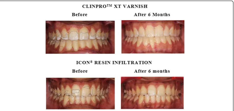
Clinically, repeated etching was required with Icon® resin infiltration for moderate lesions to provide a pre-view of the aesthetic result to be expected after
infiltra-tion. Both Icon® resin infiltration and Clinpro™ XT
varnish provided immediate aesthetic results with mild lesions. However, with moderate lesions, though pro-gressive improvements were seen in both groups,
clinic-ally, visible improvements were seen more with Clinpro™
XT at T3than with Icon® resin infiltration (Fig.2).
While both methods showed a significant increase in improving the aesthetics of the WSLs, the enamel
in response to Clinpro™ XT was more comparable to
that of the adjacent sound enamel. Icon® resin infiltra-tion has already been proven in restoring the
aesthet-ics of the enamel, and this study offers Clinpro™ XT
varnish as an alternative with gratifying short- as well as long-term results.
Further, long-term follow-up is required to see the
ef-fects of Clinpro™ XT varnish on moderate WSLs over a
year’s period to see if the colour and fluorescence are
completely restored.
Conclusion
Immediately after the intervention, Icon® resin infiltration demonstrated a significantly better improvement than Clinpro™XT varnish in restoring the colour; however, at 3 and 6 months, this was reversed.
At the end of the study period, the fluorescence loss significantly ameliorated in response to both Icon® resin infiltration between immediate intervention and at 6 months.
However, at 6 months, the fluorescence of the white spot lesions infiltrated with Icon® resin was
comparable to that of the adjacent sound enamel while those treated with Clinpro™XT varnish showed a statistically significant difference.
Table 4Intergroup and intragroup comparison of fluorescence loss (Q) values at various time intervals
Qvalues of Icon® resin infiltration
Qvalues of Clinpro™ XT varnish
Intergroup comparison
Intragroup comparison
Icon® resin infiltration Clinpro™XT varnish
T0 4.48 ± 1.42 4.60 ± 1.29 p= 0.498 T0vs T1,p= 0.000** T0vs T1,p= 0.000**
T1 2.84 ± 1.53 3.14 ± 1.20 p= 0.094 T1vs T2,p= 0.000** T1vs T2,p= 0.000**
T2 2.06 ± 1.05 2.05 ± 0.83 p= 0.935 T2vs T3,p= 0.000** T2vs T3,p= 0.000**
T3 1.48 ± 0.81 1.51 ± 0.72 p= 0.767 T0vs T3,p= 0.000** T0vs T3,p= 0.000**
Adjacent Sound Enamel 1.06 ± 0.58 1.08 ± 0.51 p= 0.80 T3adjacent sound enamel,p= 0.050 T3adjacent sound enamel,p= 0.016*
Statistical tests: intergroup comparison—independent Student’sttest; intragroup comparison—one-way repeated measures ANOVA Qfluorescence loss,T0before intervention,T1immediate after intervention,T23 months,T36 months
*p< 0.05, **p< 0.01
Abbreviations
ΔE:Colour changes;L: Lightness;Q: Fluorescence loss; WSLs: White spot lesions
Acknowledgements
The authors would like to acknowledge the university for providing the opportunity to perform the research.
Funding Not applicable
Availability of data and materials
The datasets used and/or analysed during the current study are available from the corresponding author on reasonable request.
Authors’contributions
AK performed the intervention methods on the patients and was a major contributor in writing the manuscript. SP analysed and interpreted the patient data and contributed to the writing of the manuscript. Both authors read and approved the final manuscript.
Ethics approval and consent to participate
University’s Institutional Ethics Committee [number: CSP/16/SEP/51/280 dated October 4, 2016]. Institution’s consent form was obtained from those participants willing to be part of the trial.
Consent for publication
Permission has been obtained from the participant (or legal parent or guardian for children) to report individual patient data.
Competing interests
The authors declare that they have no competing interests.
Publisher’s Note
Springer Nature remains neutral with regard to jurisdictional claims in published maps and institutional affiliations.
Received: 16 March 2019 Accepted: 9 May 2019
References
1. Øgaard B, Rølla G, Arends J. Orthodontic appliances and enamel demineralization: Part 1. Lesion development. Am J Orthod Dentofac Orthop. 1988;94(1):68–73.
2. Nascimento PL, Fernandes MT, Figueiredo FE, Faria-e-Silva AL. Fluoride-releasing materials to prevent white spot lesions around orthodontic brackets: a systematic review. Braz Dent J. 2016;27(1):101–7.
3. Lopatiene K, Borisovaite M, Lapenaite E. Prevention and treatment of white spot lesions during and after treatment with fixed orthodontic appliances: a systematic literature review. J Oral Maxillofac Res. 2016;7(2):e1.
4. Hoffman DA, Clark AE, Rody WJ, McGorray SP, Wheeler TT. A prospective randomized clinical trial into the capacity of a toothpaste containing NovaMin to prevent white spot lesions and gingivitis during orthodontic treatment. Prog Orthod. 2015;16(1):25.
5. Hsu CY, Jordan TH, Dederich DN, Wefel JS. Effects of low-energy CO2 laser irradiation and the organic matrix on inhibition of enamel demineralization. J Dent Res. 2000;79(9):1725–30.
6. Kronenberg O, Lussi A, Ruf S. Preventive effect of ozone on the development of white spot lesions during multibracket appliance therapy. Angle Orthod. 2009;79(1):64–9.
7. Borzabadi-Farahani A, Borzabadi E, Lynch E. Nanoparticles in orthodontics, a review of antimicrobial and anti-caries applications. Acta Odontol Scand. 2014;72(6):413–7.
8. Yamazaki H, Litman A, Margolis HC. Effect of fluoride on artificial caries lesion progression and repair in human enamel: regulation of mineral deposition and dissolution under in vivo-like conditions. Arch Oral Biol. 2007;52(2):110–20.
9. Bishara SE, Ostby AW. White spot lesions: formation, prevention, and treatment. Semin Orthod. 2008;30(3):174–82.
10. Bröchner A, Christensen C, Kristensen B, Tranæus S, Karlsson L, Sonnesen L, Twetman S. Treatment of post-orthodontic white spot lesions with casein
phosphopeptide-stabilised amorphous calcium phosphate. Clin Oral Investig. 2011;15(3):369–73.
11. Featherstone JD. Prevention and reversal of dental caries: role of low level fluoride. Community Dent Oral Epidemiol. 1999;27(1):31–40.
12. Sonesson M, Bergstrand F, Gizani S, Twetman S. Management of post-orthodontic white spot lesions: an updated systematic review. Eur J Orthod. 2016;39(2):116–21.
13. Yetkiner E, Wegehaupt F, Wiegand A, Attin R, Attin T. Colour improvement and stability of white spot lesions following infiltration, micro-abrasion, or fluoride treatments in vitro. Eur J Orthod. 2014;36(5):595–602.
14. Paris S, Meyer-Lueckel H, Kielbassa AM. Resin infiltration of natural caries lesions. J Dent Res. 2007;86(7):662–6.
15. Meyer-Lueckel H, Paris S, Kielbassa AM. Surface layer erosion of natural caries lesions with phosphoric and hydrochloric acid gels in preparation for resin infiltration. Caries Res. 2007;41(3):223–30.
16. Paris S, Hopfenmuller W, Meyer-Lueckel H. Resin infiltration of caries lesions: an efficacy randomized trial. J Dent Res. 2010;89(8):823–6.
17. Doméjean S, Ducamp R, Léger S, Holmgren C. Resin infiltration of non-cavitated caries lesions: a systematic review. Med Princ Pract. 2015;24(3):216–21.
18. Knösel M, Eckstein A, Helms HJ. Durability of esthetic improvement following Icon resin infiltration of multibracket-induced white spot lesions compared with no therapy over 6 months: a single-center, split-mouth, randomized clinical trial. Am J Orthod Dentofac Orthop. 2013;144(1):86–96. 19. Kim S, Kim EY, Jeong TS, Kim JW. The evaluation of resin infiltration
for masking labial enamel white spot lesions. Int J Paediatr Dent. 2011;21(4):241–8.
20. Knösel M, Eckstein A, Helms HJ. Long-term follow-up of camouflage effects following resin infiltration of post orthodontic white-spot lesions in vivo. Angle Orthod. 2019;89(1):33–9.
21. Yuan H, Li J, Chen L, Cheng L, Cannon RD, Mei L. Esthetic comparison of white-spot lesion treatment modalities using spectrometry and fluorescence. Angle Orthod. 2013;84(2):343–9.
22. Justus R. Prevention of white spot lesions. In: iatrogenic effects of orthodontic treatment: decision- making in prevention, diagnosis and treatment. 1st ed. Heidelberg: Springer; 2015. p. 1–35.
23. Kumar Jena A, Pal Singh S, Kumar Utreja A. Efficacy of resin-modified glass ionomer cement varnish in the prevention of white spot lesions during comprehensive orthodontic treatment: a split-mouth study. J Orthod. 2015;42(3):200–7.
24. Sohn S, Yi K, Son HH, Chang J. Caries-preventive activity of fluoride-containing resin-based desensitizers. Oper Dent. 2012;37(3):306–15. 25. Bhandary S, Hegde MN. A clinical comparison of in-office management of
dentin hypersensitivity in a short term treatment period. Int J Biomed Adv Res. 2012;3:169–74.
26. Shailesh S, Nayak US, Joseph S, Saidath K. To evaluate the clinical efficiency of resin modified glass ionomer varnish in preventing enamel
demineralisation during orthodontie treatment. J Pierre Fauchard Acad (India Section). 2012;26(3):95–107.
27. Mehta A, Paramshivam G, Chugh VK, Singh S, Halkai S, Kumar S. Effect of light-curable fluoride varnish on enamel demineralization adjacent to orthodontic brackets: an in-vivo study. Am J Orthod Dentofac Orthop. 2015;148(5):814–20.
28. Zhou SL, Zhou J, Watanabe S, Watanabe K, Wen LY, Xuan K. In vitro study of the effects of fluoride-releasing dental materials on remineralization in an enamel erosion model. J Dent. 2012;40(3):255–63.
29. Gorelick L, Geiger AM, Gwinnett AJ. Incidence of white spot formation after bonding and banding. Am J Orthod. 1982;81(2):93–8.
30. McLaren K. XIII—The development of the CIE 1976 (L* a* b*) uniform colour space and colour-difference formula. J Soc Dye Colour. 1976;92:338–41. 31. Knösel M, Reus M, Rosenberger A, Ziebolz D. A novel method for testing
the veridicality of dental colour assessments. Eur J Orthod. 2011;34(1):19–24. 32. Pianotti RS, Ambrozaitis JD, Mc Namara TF. Cariostatic activity of
calcium glycerophosphate in hamsters: topical vs dietary administration. J Dent Res. 1976;55(6):1092–6.
33. Sluzker A, Knosel M, Athanasiou AE. Sensitivity of digital dental photo CIE L*a*b* analysis compared to spectrophotometer clinical assessments over 6 months. Am J Dent. 2011;24:300–4.
35. Lesaffre E, Philstrom B, Needleman I, Worthington H. The design and analysis of split-mouth studies: what statisticians and clinicians should know. Stat Med. 2009;28:3470–82.
36. Hujoel PP. Design and analysis issues in split mouth clinical trials. Community Dent Oral Epidemiol. 1998;26:85–6.
37. Benson P. Evaluation of white spot lesions on teeth with orthodontic brackets. Semin Orthod. 2008;14(3):200–8.
38. Fondriest J. Shade matching in restorative dentistry: the science and strategies. Int J Periodontics Restorative Dent. 2003;23:467–79.
39. Darling CL, Huynh GD, Fried D. Light scattering properties of natural and artificially demineralized dental enamel at 1310nm. J Biomed Optics. 2006;11(3):1–11. 40. Ko CC, Tantbirojn D, Want T, Douglas WH. Optical scattering power for
characterization of mineral loss. J Dent Res. 2000;79:1584–9. 41. Johnston WM, Kao EC. Assessment of appearance match by visual
observation and colorimetry. J Dent Res. 1989;68(5):819–22.
42. Matheus P, Demito CF, Scheibel PC, Bowman SJ, Ramos AL. Correlation between the microscopic evaluation and reading by laser fluorescence of enamel decalcification in bovine teeth: In vitro study. Odonto. 2010;18:31–9. 43. Meyer-Lueckel H, Paris S. Progression of artificial enamel caries lesions after