1482
Phytocompounds Investigation, Isolation Of
Flavan-3-Ol From The Stem Of Manilkara
Hexandra (Roxb.) Dubard And Its Potential In
Anti-Oxidant
S.Irudaya Monisha, J.Rosaline Vimala
Abstract: The plant poly phenols are present adequately which has high impact on human drugs. Flavan- 3- ol consist in the stem of Manilkara hexandra and it has rich medicinal value like anti-oxidant. The physical and chemical properties were investigated from the stem of Manilkara hexandra. Proximate, histochemical, fluorescence, mineral analysis were examined by using standard procedures. The flavan-3-ol was isolated using chromatographics method such as coloumn chromatography and thin layer chromatography. Then it was characterized by UV, FT-IR, LC-Ms, NMR (1H & 13 C) and it is further extended to in-vitro anti-oxidant (DPPH method) potential. The flavanoid was identified by preliminary investigation after then flavan-3-ol was isolated and it gives good response for anti-oxidant activity. The isolated catechin has a unique biological behaviours, and act as a metal chelating agent. So it can be used for further research from this plant.
Index Terms: Anti-Oxidant, Fluorescence , Histochemical, Isolation, Manilkara Hexandra , Mineral Analysis ,Proximate.
1 INTRODUCTION
MEDICINAL plants are used in all the cultures as a basic source for medicines.[1], [2], [3]. Plant posses’ secondary metabolities which contain pharmacological and chemical properties it is also encouraging tool for human health [4], [5]. Rapid development of new biological and chemical science has brought forth a new variance in the infinite capable of the plant products from nature [6]. Flavonoids are widely present in plants and edible of plant source which is present as phenolic compounds [7], [8], [9], [10]. Phytoconstituents are belongs to the flavonoids, terpenoids and alkaloids acts as a drug for many diseases [4]. Espeacially, few compounds has effective to prevent and inhibit different type of cancer [5] and it also depends on the state of 15 carbon skeleton [8]. Plant antioxidant contains polyphenols and it posses pharmacological behaviour due to action of anti-oxidant [11].
Since, the poly phenol present in plant has a various potential for anti-oxidant such as catechins,morin, gallic acid derivatives, tannins, quercetin, rutin, carnosol, eugenol and thymol. The above plant derived anti-oxidant has enormous applications in preservation of food, dietary supplementation, and used in free radical mechanism [12]. The countries in south asia used to ancient herbal systems such as unani and ayurveda[13]. In this system, plant compounds have rich in anti-oxidant property. In this report, we have included 49 Bangladeshi herbal plants 36 families and claimed to have strong anti-oxidative
properties,such as sapotaceae [14]. The genus Manilkara includes 135 plants that are distributed worldwide. Manilkara hexandra (Roxb.) and Manilkara zapota(L) are native to south Asia*15+. Plant’s minerals have nutritional importance [16] and it thus finds its applications in curing diseases related to liver, kidney, hepatitis and cancer diseases [17]. The aim of the present research is thus to carry out histochemical, fluorescence, proximate and mineral studies to understand the nutritional value of the plant material(Manilkara hexandra stem) , finally isolate , characterize the active principle by using modern techniques (FT-IR, UV, LC-MS, Proton and Carbon NMR) and evaluate the anti-oxidant efficasy of the compound by in-vitro method.
2 EXPERIMENTAL SECTIONS:
Manilkara hexandra (Sapotaceae) stems were collected on January 2016 at Jayangkondam, Ariyalur district and it was authenticated by Rapinet Herbarium of St. Joseph’s College (Autonomous), Trichy. The Voucher No. of this plant is 00I1.
2.1 Preparation of the Stem Extract:
The collected stems were washed, dried and grounded by using pestle mortar. Stem (1kg) was percolated with 10L EtOAc at room temperature in 48 hours and it was filtered through whatman filter paper no.1. This extract (750ml) was collected and stored in a refrigerator.
2.2 Physical Characterizations:
The qualitative, quantitative and the yield reported for the stem of Manilkara hexandra.[18] led to the determination of physiochemical parameters such as total ash, moisture content, acid insoluble ash, water soluble ash, sulfated ash, dry matter content and pH by adopting WHO guidelines and Indian pharmacopeia. Further analysis was done in
S.Irudaya Monisha is currently pursuing P.hD (Research Scholar)in Department of Chemistry, Holy Cross College(Autonomous),India. Mobile: +918190918082 E-mail: [email protected]
J.Rosaline Vimala is currently work as a Assistant Professor, Department of Chemistry, Holy Cross College (Autonomous),India.
1483 IJSTR©2019
this part of the plant such as histochemical, fluorescence and mineral analysis [19].
2.3 Isolation of flavan-3-ol from this plant:
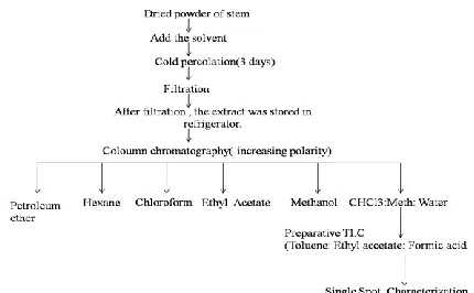
Fig.1. The method of isolation for flavan-3-ol is clearly shown in the flowchart.
The stationary phase of silica gel (60-120 mesh) was used in a coloumn apparatus (15x 4 cm). Low polar to high polar solvents (Mobile phase) were used to separate the following fractions such as petroleum ether, hexane, chloroform, ethyl acetate, methanol and water. Finally, the CHCl3: Methanol: Water (7:1:3) ratio was added in the coloumn tube. A clear green coloured band separated is spotted in precoated TLC in Toluene: Ethyl acetate: Formic acid (4:5:1) for single compound. Flowchart displayed in Figure 1.
2.4 Confirmation test for flavan-3-ol:
2.4.1 Match stick test:
The match stick dipped in the sample and also in concentrated HCl and dried. It is then shown in the flame to identify the presence of phloroglucinal (Flavan-3-ol modification) when red or pink flame colouration is noticed.
2.4.2 Vanillin in Ethanol (Contains H2SO4):
The production of pink or red colour on the addition of vanillin to the isolated compound indicates the presence of phloroglucinal.
3 Characterization Section:
3.1 UV-Visible Spectral analysis:
The compound was identified by Lambda 35 UV-Visible double beam scanning spectrophotometer. The normal wavelength range starts from 200-400 nm and visible range from 400-800 nm. This technique has highly sensitive for the detection of organic compound.
3.2 Fourier Transform –Infra Red analysis:
The isolated flavanol compound from ethyl acetate extract of Manilkara hexandra were characterized by using IR
spectrophotometer. The solid material (1mg) was moulded with (10 to 100 mg) KBr to provide a thin small disc with hydrous conditions. The spectral interval range taken is between 4000 and 400 cm-1. It took three minutes for recording process.
3.3 Liquid Chromatography – Mass Spectroscophy analysis: ( LC/ESI/MS/MS)
500 μL of the sample was analyzed by LC/ESI/MS/MS using UHPLC (Ultra high performance liquid chromatography) and reverse phase column C18, (120 Å, size 150 x 2.1 mm Acclaim 120) ( 3000 series, Dionex) in liquid chromatography joined with mass spectrometer (micrOTOF-Q II, Bruker- Germany). The mobile systems a and b (acidified to 0.1% of HCOOH) were used which is represented by water (MiliQ) and methanol respectively. The gradient elution is as follows: 75%A and 25%B (0-8.5 min); 0%A and 100%B (8.6-18.9 min); 75%A and 25%B (19 - 24 min). ; Sample injection volume - 10 μL . ESIMSMS peaks were generated by using certain parameters such as 250 nm(wavelength by UV detector), flow rate was 0.2 mL/min mode of ionization was negative, nebulizer pressure was 24.7 psi, dry gas (N2) 6.0 L/min, 4500 V of capillary voltage, heater range at 280oC. The Msn mode range was between 50- 1500 m/z and it was analyzed for all peaks. The Ms/Ms fragmentation was done by using collision 350 RF Vpp and energy was at 10.0 eV.
3.4 Nuclear Magnetic Resonance (NMR):
The structure of isolated compound was analyzed by nuclear magnetic resonance (BRUKER 300 MHz for 1H and 75 MHz for 13C ). AVANCE II type (Ultra Shield Magnet) was used. The scanning range of proton is 1-10 ppm and carbon is 20-200 ppm.
4 Application:
4.1 In-vitro anti-oxidant DPPH assay for isolated compound:
0.5 ml of isolated compound was mixed with 2ml of DPPH (2,2-Diphenyl-1-picrylhydrazyl) in methanol (25 μg/ ml) at different concentrations such as 0.1,0.2, 0.3, and 0.4 ml. This mixture was shaked and kept at room temperature for 30 minutes. After some time, the absorbance was noted at particular wavelength (517 nm) by using UV spectrophotometer. L-ascarbic acid was used as a standard for radical scavenging activity and it was calculated by the following equation.
Radical Scavenging Activity (%) =
Ac = Control; As = Sample
4.1.1 Statistical status:
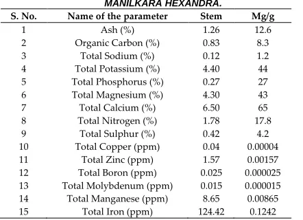
1484 TABLE 2 MINERAL COMPOUNDS PRESENT IN THE STEM OF
MANILKARA HEXANDRA.
S. No. Name of the parameter Stem Mg/g
1 Ash (%) 1.26 12.6
2 Organic Carbon (%) 0.83 8.3
3 Total Sodium (%) 0.12 1.2
4 Total Potassium (%) 4.40 44
5 Total Phosphorus (%) 0.27 27
6 Total Magnesium (%) 4.30 43
7 Total Calcium (%) 6.50 65
8 Total Nitrogen (%) 1.78 17.8
9 Total Sulphur (%) 0.42 4.2
10 Total Copper (ppm) 0.04 0.00004
11 Total Zinc (ppm) 1.57 0.00157
12 Total Boron (ppm) 0.025 0.000025
13 Total Molybdenum (ppm) 0.015 0.000015
14 Total Manganese (ppm) 8.65 0.00865
15 Total Iron (ppm) 124.42 0.1242
TABLE 3 HISTOCHEMICAL REPORT FOR STEM OF MANILKARA HEXANDRA
Test Reagents Observation Compounds Stem
Phloroglucinol + concentrate
HCl
Red/Pink Lignin
++
Dilute Ammonia +
H2SO4
Yellow Flavonoids ++
Mayers
Reagent Reddish Brown Alkaloids ---
Liberman (5 drops of
acetic anhydride + 5 drops of H2SO4
Violet to Blue
(or) Green Steroids +
Toludine blue Blue green/Red Polyphenol ++
TABLE 4 FLUORESCENCE REPORT FOR STEM OF MANILKARA HEXANDRA
S.No Chemicals Visible Light Short UV
(254nm)
Long UV (365 nm) 1 Plant powder (PP) Whitish Brown Light Green Black
2 Plant powder with
water Dark Green Dark Green
Brown
3 PP with Hexane Whitish Yellow Light Green Black
4 PP with
Chloroform Light Yellow Light Green
Brown
5 PP with Methanol Green Black Black
6 PP with Acetone Yellow Black Black
7
PP with 1N Sodium hydroxide
in water
Reddish Brown Greenish
Brown Black
8 PP with 1N
Hydrochloric acid Greenish Yellow Dark Green Brown
9 PP with
1NSulphuric acid Light Green Dark Green
Brown
10 PP with1N Nitric
acid Yellowish Brown Dark Black Dark Brown sample was drawn by linear regression method in
Ms-Windows based on Graph Pad Instat (Version 3) software.
5 Discuss about the results:
5.1 Proximate analysis:
Table 1 shows the physical parameters such as total ash, moisture content, acid insoluble ash, water soluble ash, sulphated ash, dry matter and pH. The total ash value shows the amount of inorganic matter present, the amount of soil in the compound is estimated from the acid insoluble ash value, pharmaceutical impurities(natural origin) mixed with inorganic matters were analyzed by sulphated ash, water content in extraction is found through the water soluble ash, and stability and susceptibility of microbial contamination were determined by moisture content which means the high moisture value posses more effective in water soluble enzymes and coenzymes metabolic functions[20]. These reports were provided without crossing the limits of pharmacopeia [21].
5.2 Mineral analysis:
The ingredients of herbal drugs depends upon the mineral composition[22]. This report is shown in table. Ca,K,Mg, and P were highly present in the stem of this plant. They play important role in our body system. Calcium is used for development of teeth, nerves and bones. In citric acid cycle, it involves a cofactor of three dehydrogenase enzymes such as pyruvate, isocitrate and α-ketoglutarate[23]. Magnesium element has vital role in dietary disorder. Ca and Mg are common deficient mineral in most developing and developed countries [24].
Potassium is the significant element to regulate water and fluid balance in the body. It develops the muscle paralysis due to low amount of potassium. Phosphorous is also major element for the formation of DNA and RNA in respiration process. The rickets appeared from the insufficient growth of bones and teeth due to deficiency of P [25]. N, S and Na were present in small amount in this plant. Fe, Mn, Zn, Cu, B and Mo were present in trace amount as illustrated in Table 2.
5.3 Histochemical and Fluorescence analysis:
The Histochemical study (Figure 2) shows characteristic colours for the phytoconstituents viz., Lignin (Pink), Flavanoids(Yellow), Steroids(Green) and Polyphenols (Blue). The essential observation was obtained in Table 3. Fluoresence light is the significant tool for the standardization of the herbal drug. The plant powder contains some chemical compounds mixed with chemical reagents to reach fluorescence [26]. Since, these microscopic reports reveals the presence of flavanoids in stem is the best proof of this isolation process carried. They are shown in Figure 2 and Table 4.
TABLE 1 PROXIMATE ANALYSIS FOR THE STEM OF MANILKARA HEXANDRA.
S. No. Proximate Tests Stem
1 Total ash 4.91±0.04
2 Moisture content 8.74±0.07
3 Acid soluble ash 7.46±0.08
4 Acid insoluble ash 0.15±0.03
5 Water soluble ash 10.52±0.07
6 Water insoluble ash 1.14±0.38
7 Sulphated ash 5.04±0.07
8 Dry Matter 93.81±0.11
1485 IJSTR©2019
TABLE 5 THIN LAYER CHROMATOGRAPHY SOLVENT SYSTEM FOR ISOLATED CATECHIN
Solvent System Ratio Retention Factor (Rf)
Compound
1. Toluene: Ethyl
acetate: Formic acid [27].
4:5:1 0.26
Isolated flavan -3 – ol
2. Chloroform: Ethyl acetate: Formic acid [28]
5:4:1 0.96
3. Hexane:Ethyl acetate:Methanol[29]
5:4:1 0.24
4. Chloroform :
Methanol [30]
3:1 0.54
Fig 2. The histochemical image proof for fllavanoids.
5.4 Chromatogaphic analysis:
Silica gel column chromatography is used for the purification of bioactive compound of flavan-3-ol.The isolated compound appears greenish yellow by passing through developing liquids viz., as shown in Figure 3a and 3b. Chloroform: Methanol: Water (7:1:3) fraction and other fractions as shown in Figure 4. In TLC, the Rf value of the pure compound is noted and it projects characteristic colour before and after vanillin sulphuric acid is sprayed as shown in table and fig. Different solvent systems were used and the retention factor matches with the literature data for isolated compound flavan-3-ol. Hence, this report confirmes the flavan-3-ol . The solvent systems were mentioned in Table 5.
Fig 3. a) Isolated fraction in coloumn b) Collection of fractions (petroleumether, hexane, chloroform, ethyl acetate, methanol and
water)
Fig 4. Thin layer Chromatography for isolated catechin
5.4.1 Confirmation test for flavan-3-ol: 1. Match stick test:
The match stick gives red colour in the flame which shows the presence of phloroglucinal. 2. Vanillin in Ethanol (Contains H2SO4):
Vanillin produced pink colour which indicates the presence of phloroglucinal as mentioned in Figure 5.
Fig 5. Qualitative confirmation test for catechin (flavan-3-ol)
6 Spectral Analysis:
6.1 UV-Visible Spectroscopy:
The UV-Visible spectrum of flavan-3-ol exhibits (Figure 6) two maximum adsorption at 283 nm and 374nm. Literature studies shows that peaks appearing at the range of 200-600 nm indicates the presence of phenolic compounds [31]. These adsorption peaks were allotted for A ring (benzene system) and B ring (cinnomyl system) for flavonoid in UV-Visible region at 240- 285 nm and 300 to 400 nm respecteively [32].
Fig 6. UV-Visible spectra for presence of flavanoid ring.
6.2 FT-IR Spectroscopy:
1486 adsorption band3325 cm-1 which indicates the presence of
aliphatic and aromatic C-H phenolic and alcoholic O-H stretching [28]. A peak around the range of 1619-1492 cm-1 appears might be due to presence of aromatic C=C which is the general frequency range noticed for flavan-3-ol [33].
Fig 7. FT-IR spectra obtained a flavanoid frequency
6.3 LC/ESI/MS/MS):
Samples were ionized using negative mode to the formation of m/z ratio and it was dissociated. This was classified into two fragment ions (i) primary and (ii) secondary ions. Primary ions of this compound (flavan-3-ol) [M-H]- is 289.1574 m/z. Secondary ions were obtained in the range of 124.7 and 159.2, which correlates with the literature data [34] of 123.1001-125.1001 and 159.1001-165.1001 respectively. These values indicate the loss of pholoroglucinol (heterocyclic ring fission cleavage) and the formation of benzofuran. This range is also composed of A-ring and an O (Oxygen) which explains that the sugar was not attached to C5 and C7 position. 159.1001-165.1001 resulted from RDA (Retro-Diels Alder) cleavage of 289.1574 and it was composed B-ring(C2,C3 and C4). The another fragment of m/z 205 resembles with LC/MS/MS report for flavan-3-ol, which tells the loss flavonoid ring –A [35]. From this successful evidence declares that the compound is 2,3-trans-3,4-trans flavan-3-ol (catechin) [33]at RT=15.7.It was shown in and also included total ion chromatogram Figure 8a and b.
Fig 8. a) Fragmentation of flavanoid ring proved by LC/MS/MS analysis. b) Total ion chromatogram image for flavan-3-ol.
6.4 NMR:
The structure of compound was illustrated from the proton and carbon nuclear magnetic resonance datas as shown in Figure 9a and b. 1H NMR reveals that the –CH2 and aromatic group regions from 2.50 – 2.90 ppm and 5.0 – 6.20 ppm for this compound [36]. This stereocompound has a trans orientation between H2 and H3 at δ 4.59 and 4.04 *37+. Three aromatic 1H signals appear in ring-B such as 1d at δ 6.82 for H-6′, 1d at δ 6.73 for H-5′, and 1d at δ 6.7 for H-2′. For C-ring obtained 1d at δ4.57 for H-2 and multiplet at δ3.99 for H-3 [38]. Then 13C NMR shows the pholoroglucinol ring – A signal provide a similar peak of catechol ring – B, but pyran ring – C slightly varied from ring A and B (C2 - δ82.56, C3 – δ68.33) *39+. If the chemical shift occurred at more than 80 ppm means it indicates that C2-is in trans orientation [40]. The important view for this compound is C2 and C3 position. The chemical shift at δ66.75 correlates with the literature data for this compound at δ 62.07.
Fig 9. a) Proton NMR shows isolated catechin from the stem of Manilkara hexandra
Fig 9. b) Carbon NMR interprets the proof for isolated compound.
7 Anti-Oxidant:
1487 IJSTR©2019
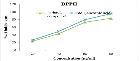
aromatic ring attached with the benzopyran, which denotes the presence of anti-oxidant and anti-inflammatory behaviour [44]. This trans isolated compound has more scavenging property than cis compound [45]. This activity depends upon the presence of OH groups in B-ring and also the addition of metal chelation potential. The properties (interacting molecules & chemical structure) and quantity of anti-oxidant are analyzed by this activity [46]. This trans molecule has benzene A & B ring and linked to heterocyclic pyran C-ring at middle [47]. Catechin contains a phenyl and hydroxy group linked through the position 2 and 3 [48]. Finally, this activity increased by the attachment of catechol present in B-ring [49]. This DPPH assay is very useful to determine the polyphenols in plant tissues [50], [51], [52] and transfer of H atom for protection of antioxidant [53].
Fig 10. In-vitro anti-oxidant DPPH assay for the isolated compound from the stem of Manilkara hexandra
8 Conclusion:
The stem of Manilkara hexandra consist of poly phenols which are primarly examined by basic evaluations such as screening, histochemical, fluorescence, minerals, physiochemical studies and further flavan-3-ol is being isolated by chromatographic analysis. It is then characterized by UV, FTIR, NMR and LCMS analysis. The isolated compound belongs to flavanol and it enhances to precede metal chelation process. The flavan-3-ol has pharmacological applications such as antioxidant property, anti-inflammatory, etc. due to this reason it can be implied for weight loss to improve human health. So, the plant twigs, stems can be extracted and consumed as like that of tea extract.
REFERENCES
[1]. L. Hoareau and E.J. silva, ‚Medicinal plants: A re-emerging health aid,‛ Electronic J Biotechnol, vol.2, no.2, pp.56-70, 1999. [2]. M.G. Cragg and D.J. Newman ‚Natural product drug discovery
in the next millennium,‛ Pharm Biol, vol.39, no.1, pp.8-17, 2001. [3]. D.S. Fabricant and N.R.Fransworth (2001): ‚The value of plants
used in traditional medicine for drug discovery,‛ Environ
Health Perspec, Suppl 1: 69-75, March. 2011.
https://doi.org/10.1289/ehp.01109s169
[4]. I. Raskin, D.M. Ribnicky,S. Komarnytsky,N. Ilic, A. Poulev,N. Borinker, D.A. Moreno,C. Ripoll, N. Yakoby, ‚Plants and human health in the twenty-first century,‛ Trends Biotechnol, vol.20, no.12, pp.522-531, Dec. 2002.
[5]. L. Reddy,B. Odhav, K.D. Bhoola, ‚Natural products for cancer prevention: a global perspective,‛ Pharmacol Ther, vol.99,no.1, pp.1-13.Jul.2003.
[6]. N.R. Krishnaswamy, ‚Chemistry of Natural Products: A Unified Approach,‛ University Press (India) Ltd., Orient Longman Limited, Hyderabad, 1999.
[7]. B. Argaez, B.A. Flowers, A. Gimenez-Turba, G. Ruiz, P.G. Waterman, L.M. Pena-Rodriguez, ‚Cytotoxic and antiprotozoal activity of flavonoids from Lonchocarpus spp,‛ Phytomedicine, vol.14, no.7-8, pp.530-533. August. 2007.
[8]. J.B. Harborne, Phytochemical Methods: A guide to Modern Techniques of Plant Analysis. Chapman and Hall, London, pp. 1-74, 1973.
[9]. K.R. Markham, Techniques of Flavonoid Identification. Academic Press, London, pp. 1-60, 1982.
[10]. A.M. Nuutila, K.M. Kammiovirta, K.M. Oksman-Caldentey, ‛Comparison of methods for the hydrolysis of flavonoids and phenolic acids from onion and spinach for HPLC analysis,‛ Food Chemistry, Vol.76, no.4, pp.519-525. April. 2002. doi: 10.1016/S0308-8146(01)00305-3
[11]. B.N. Ames, L.S. Gold, and W.C. Willett, ‚The causes and prevention of cancer,‛ Proc Natl Acad Sci U S A., Vol. 92, no.12, pp.5258-5265.Jun.1995. doi: 10.1073/pnas.92.12.5258
[12]. B. Halliwell, R. Aeschbach, J. Loliger, O.I. Aruoma, ‚The characterization of antioxidants,‛ Food Chem Toxic, Vol.33, no.7, pp.601-617.Jul. 1995.
[13]. P. Scartezzini , E. Speroni, ‚Review on some plants of Indian traditional medicine with antioxidant activity,‛ J Ethnopharmacol, vol.71, no. 1-2, pp. 23-43. Jul. 2000.
[14]. R. Govindarajan, M. Vijayakumar, P. Pushpangadan,
‚Antioxidant approach to disease management and the role of ‘Rasayana’ herbs of Ayurveda,‛ J Ethnopharmacol, vol. 99, no.2, pp.165-178. Jun. 2005.
[15]. Anjali, Vandana Garg, Anju Dhiman, Rohit Dutt and Sweety Ranga, ‚The genus Manilkara: An update,‛ The Pharma Innovation Journal, vol.7, no.1, pp. 316-318. 2018.
[16]. P.J. White and R. M. Broadley, ‚Biofortification of crops with seven mineral elements often lacking in human diets - iron, zinc, copper,calcium, magnesium, selenium and iodine,‛ New Phytologist, vol.182, no.1, pp.49-84. 2009. doi: 10.1111/j.1469-8137.2008.02738.x
[17]. M.A.M. Sajib, M.M. Hoque, S. Yeasmin, and M.H.A. Khatun, ‚Minerals and heavy metals concentration in selected tropical fruits of Bangladesh,‛ International Food Research Journal, vol. 21, no.5, pp.1731-1736.2014.
[18]. S. Irudaya Monisha, G Dayana Jeya Leela, A Anitha Immaculate and J Rosaline Vimala, ‚Comparative studies on yield and the phytochemical appraisal (Quality and Quantity) of Manilkara hexandra (Roxb) Dubarb using leaf, stem, and bark,‛ Journal of Pharmacognosy and Phytochemistry, vol.6, no.4, pp. 2052-2058. 2017.
[19]. S. Irudaya Monisha and J. Rosaline Vimala, ‚Extraction, Identification and Pharmacological Evaluation of Phyto-Active Compound in Manilkara Hexandra (Roxb.) Dubard Stem Bark,‛ Biosciences Biotechnology Research Asia, vol.15, no.3, pp. 687-698. September.2018.
[20]. David Febrilliant Susanto, Hakun Wirawasista Aparamarta, Arief Widjaja, Setiyo Gunawan, ‚Identification of phytochemical compounds in Calophyllum inophyllum leaves,‛ Asian Pacific Journal of Tropical Biomedicine, vol.7, no.9, pp.773-781. September 2017.
1488
[22]. R. Venkataraman, and S. Gopalakrishnan, ‚Trace elemental profile in medicinal plants traditionally used for Jaundice,‛ J Current Sci, vol.2, no.2. pp. 305-308. 2002.
[23]. B. Claude, and S. Paule, S, The Manual of Natural Living (1st ed.). Biddles Ltd., Guildford, Surrey, pp.98-101, 1979.
[24]. T.D. Thacher, P.R. Fischer,M.A. Strand, J.M. Pettifor, ‚Nutritional rickets around the world: causes and future directions,‛ Ann Trop Paediatr, vol.26, no.1, pp.1-16. March. 2006. doi: 10.1179/146532806X90556.
[25]. W.K. Michael, Medical Biochemistry. 3rd ed., Queen Mary Publishers London, pp. 13-17, 2007.
[26]. D. Janchen, H.J. Issaq. ‚Modern thin layer chromatography: Advances and perspectives,‛ J Liquid Chromatogr, vol.11, no.9-10, pp.1941-1965, 1988.
[27]. Muhammad Yanis Musdja, Mustikaning Ayu Hapsari, and Andria Agusta,‛Comparison of Activity and Inhibitory Mechanism between (+)-Catechin and Water Extract of Gambier (Uncaria Gambir Roxb.) Against Some Bacteria,‛ Scientific Journal of PPI-UKM, vol.4, no.2, pp.55-60. 2017. doi: 10.27512/sjppi-ukm/se/a29012018
[28]. M.A. Hye, M.A. Taher, M.Y. Ali, M.U. Ali, S. Zaman, ‚Isolation of (+)-catechin from Acacia catechu (Cutch tree) by a convenient method,‛ J Sci Res, vol.1, no.2, pp. 300-305. 2009. doi: 10.3329/jsr.v1i2.1635
[29]. Dartini, Hazli Nurdin, Afrizal, M. Taufik Ekaprasada and Dwimaryam Suciati, ‚Isolation and characterization Flavonoids from The Bark of Toona Sureni (Blume) Merr,‛ J. Chem. Pharm. Res, vol.8, no.6, pp. 156-159. 2016.
[30]. R. V. Malbaša, E. S. Lončar and L. A. Kolarov, ‚Tlc Analysis Of Some Phenolic Compounds In Kombucha Beverage,‛ APTEFF, vol. 35,pp. 1-280. 2004.
[31]. M.N. Matos, ‚Chemical and Sensory Characterization of Fortified Muscatel Wines,‛ M.Sc.Thesis, Faculty of Pharmacy, University of Lisbon, 2001.
[32]. Shahabuddin Memon, Abdul Hafeez Laghari, Aftab A. Kandhro, Fakhar N. Memon and Aisha Nelofar, ‚Purification of flavonoid metal complexes from Alhagi camelorum with calix*4+arene based impregnated resin,‛ Anal. Methods, vol.6, pp.6332-6336. 2014.doi: 10.1039/c4ay00167b.
[33]. Sushma Kainsa1 , Randhir Singh,‛Flavan-3-ol Isomers Isolated from Euphorbia Thymifolia Linn,‛ Pharmacogn. Commn, vol.6, no.1,pp.28-33. March. 2016.
[34]. Seyit Yuzuak , James Ballington and De-Yu Xie,
‚HPLC-qTOF-MS/MS-Based Profiling of Flavan-3-ols and Dimeric
Proanthocyanidins in Berries of Two Muscadine Grape Hybrids FLH 13-11 and FLH 17-66,‛ Metabolites, vol.8,no.57, pp.1-24. 2018. doi:10.3390/metabo8040057.
[35]. M.N. Bravo , S. Silva , A.V. Coelho , L. Vilas Boas , M.R. Bronze, ‚Analysis of phenolic compounds in Muscatel wines produced in Portugal,‛ Analytica Chimica Acta, vol.563, pp. 84-92. 2006. [36]. Givaldo Souza da Silvaa, Lorena Mara Alexandre e Silvab,
Elenilson de Godoy Alves Filhob, Kirley Marques Canutob, Edy Sousa deBritob and Raildo Mota de Jesus, ‛1H quantitative nuclear magnetic resonance and principal component analysis as tool for discrimination of guarana seeds from different geographic regions of Brazil,‛ Proceedings of the XIII International Conference on the Applications of Magnetic Resonance in Food Science ,2016. doi: 10.1255/mrfs.5.
[37]. L.Y. Foo, and J. J. Karchesy,‛Procyanidin dimers and trimers from douglas fir inner bark,‛ Phytochemistry, vol. 28, pp. 1743-1747. 1989. doi:10.1016/ 0031-9422(89)80303-6.
[38]. Devan Elumalai, Maduraiveeran Hemavathi, Periaswamy
Hemalatha, Chandrasekar Vijayalakshmi Deepaa, Patheri Kunyil Kaleena, ‚Larvicidal activity of catechin isolated from
Leucas aspera against Aedes aegypti, Anopheles stephensi, and Culex quinquefasciatus (Diptera: Culicidae) ,‛ Parasitol Res vol.115, no.3, pp. 1203-1212. March. 2016. doi :10.1007/s00436-015-4856-6.
[39]. Yinrong Lu, L. Yeap Foo, ‚The polyphenol constituents of grape pomace,‛ Food Chemistry vol.65, no.1, pp.1-8.April. 1999. doi: 10.1016/S0308-8146(98)00245-3.
[40]. Xiaolan Jiang, Yajun Liu, Yahui Wu, Huarong Tan, Fei Meng, Yun sheng Wang, Mingzhuo Li, Lei Zhao, Li Liu, Yumei Qian, Liping Gao and Tao Xia, ‚Analysis of accumulation patterns and preliminary study on the condensation mechanism of proanthocyanidins in the tea plant *Camellia sinensis+,‛ Scientific Reports, vol.5, no.8742, pp.1-15, March.2015. doi: 10.1038/srep08742
[41]. D. Krishnaiah, R. Sarbatly, R. Nithyanandam, ‛A review of the antioxidant potential of medicinal plant species,‛ Food and Bioproducts Processing,vol.89, pp. 217-233. 2011. doi: 10.1016/j.fbp.2010.04.008.
[42]. A. Faridi, E. Seid, M. Jafari, ‚Biopolymer nano-particles and natural nano-carriers for nano-encapsulation of phenolic compounds,‛ Colloids Surf B Biointerfaces, vol.146, pp. 532-543. October. 2016. doi: 10.1016/j.colsurfb.2016.06.053
[43]. N. Jahan, M.S. Parvin, N. Das, M.S. Islam, M.E. Islam,‛Studies on the antioxidant activity of ethanol extract and its fractions from Pterygota alata leaves,‛ Journal of Acute Medicine,vol.4, pp.103-108. 2014. doi: 10.1016/j.jacme.2014.05.001
[44]. J. Kanwar, M. Taskeen, L. Mohammad, C. Huo,T. Chan,Q. Dou,‛Recent Advances on Tea Polyphenols,‛ Front. Biosci. (Elite Ed.) vol. 4, pp. 111-131. 2012.
[45]. Q. Guo, B. Zhao,S. Shen, J. Hou, J. Hu,W. Xin, ‛ESR study on the structure-antioxidant activity relationship of tea catechins and their epimers,‛ Biochim. Biophys. Acta vol.1427, no.1, pp.13-23. March. 1999. doi: 10.1016/s0304-4165(98)00168-8
[46]. O. Blokhina, E. Virolainen, K.V. Fagerstedt, ‚Antioxidant, oxidative damage and oxygen deprivation stress: A review,‛
Ann. Bot.vol.91, pp.179-194. January 2003. doi:
10.1093/aob/mcf118
[47]. L.R. Juneja, M.P. Kapoor, T. Okubo, T. Rao, ‚Green Tea Polyphenols: Nutraceuticals of Modern Life,‛ CRC Press: Boca Raton, FL, USA, pp. 19-23. 2013.
[48]. D. Chen, S.B Wan, H. Yang, J. Yuan, T.H. Chan, Q.P. Dou, ‚EGCG, green tea polyphenols and their synthetic analogs and prodrugs for human cancer prevention and treatment,‛ Adv. Clin. Chem, vol.53. pp.155-177. 2011.
[49]. E. Tripoli,M. La Guardia, S. Giammanco,D. di Majo, M. Giammanco, ‚Citrus flavonoids: Molecular structure, biological activity and nutritional properties: A review,‛ Food Chem, vol.104, pp. 466-479. 2007. doi: 10.1016/j.foodchem.2006.11.054 [50]. Y.C. Chen, Y. Sugiyama, N. Abe, R. Kuruto-Nima, R. Nozawa,
A. Hirota, ‚DPPH radical scavenging compounds from Dou-Chi, a soybean fermented food,‛ Biosci. Biotechnol. Biochem, vol.69, pp.999-1006. 2005.
[51]. F. Shahidi, P.K. Janitha, P.D. Wanasundara, ‛Phenolic antioxidants,‛ Crit Rev Food Sci Nutr, vol.32.pp.67-103. 1992. doi: 10.1080/10408399209527581.
[52]. P. Kefalas, S. Kllithraka, L. Parejo,D.P. Makris, ‚A Comparative Study on the in Vitro Antiradical Activity and Hydroxyl Free Radical Scavenging Activity in Aged Red Wines,‛ Food Sci. Technol. Int, vol.9. pp.383-387. December. 2003. doi: 10.1177/1082013203040080