Volume: 4 Issue: 3 Dec- 2019 48
Survey on Cancer Detection using Machine
Learning
Vrushali U. Uttarwar, Aditi A. Kalia, Mr. R. K. Ghotekar Department of computer Engineering
D. Y. Patil College Of Engineering, Akurdi, Pune, India
ABSTRACT
In counties such as India, where the population is on the rise and living conditions are improving, the focus was on the medical field. In the AI era, Machine Learning is becoming prominent in a variety of areas as it provides a precise solution to a wide range of medical problems. Carcinoma can be a disease that poses a health challenge for women across the globe. The worldwide scenario shows that carcinoma ranks second when it comes to causing death among women when it involves cancer. However, if it is always diagnosed early, carcinoma can be a curable disease. Many fatalities of women between 40 and 55 years of age are attributed to carcinoma. According to the WHO study, everywhere on the world, about 1.2 million people are affected by carcinoma per year. This article seeks to address women's cancer and a variety of other machine learning methods suggested by researchers to identify the disorder.
Keywords: machine learning, melanoma, cancer.
I.
INTRODUCTION
Melanoma is the worst form of skin cancer and is responsible for approximately 75% of carcinoma-related deaths [1]. Accurate early-stage detection of melanoma may improve patients ' survival rates significantly. The manual diagnosis of melanoma, however, creates an immense demand for well-trained experts and suffers from differences between findings. An automatic efficient system for detecting melanoma should be built to improve pathologists ' accuracy and performance. The technique of dermoscopy has been developed to improve melanoma diagnosis. Dermoscopy may
be a noninvasive skin imaging process that enhances the visual effect of a skin lesion by removing surface reflection to gain a magnified and luminous image of a skin region for greater clarity of points [2]. However, it remains a difficult task to recognize automatically the melanoma in dermoscopic images because of its many challenges. First, it is hard to classify correct lesion areas because of the poor contrast between skin lesions and normal skin layer. Second, a high level of visual resemblance may exist in the lesions of Melanoma and non-Melanoma, contributing to differentiating between the lesions of Non-Melanoma melanoma. Shift between individuals in the presentation of melanoma, e.g. colour and shape, natural hair or veins, and others; thirdly, the consistency in skin conditions. Segmentation of skin lesions is the key to many approaches to classification. There is often a recent review of automatic algorithms for segmentation of skin lesions. The accuracy of the following lesion classification may benefit from accurate segmentation.
Stages of carcinoma
Volume: 4 Issue: 3 Dec- 2019 49 epidermis into the next layer of the skin (dermis)
but are tiny and have no effect, such as ulceration that places them at a high risk of spreading to or beyond tightly connected lymph hubs. Stage II tumors are, albeit small, larger (1 mm thick or greater) and may, in turn, possess different properties, such as ulceration, which place them in
high risk of spreading into or beyond closely related lymph hubs. These are known as melanomas in transformation or "high chance." Further melanomas (steps III and IV) have metastasized into various parts of the body. There are additionally subdivisions inside stages.
II.
LITREATURE SURVEY
Table: Survey TablePaper Name Author Technique
ABCD rule based detection
Hardian et.al
ABCD rule of dermoscopy for extracting the skin lesion. Feature extraction is performed on the pre- processed image wherever the four options asymmetry (A), Border (B), Colour(C), and D (Diameter)
square measure extracted in the following method.
Backpropagation Neural
Networks based
identification
Pratik et al. The projected model makes use of Back propagation neural networks. However, the projected methodology as certain drawbacks. The major drawbacks of this methodology square measure slow convergence
rates and trapping in native minima.
Convolution Neural
Network based
identification
Mobeen et al
The advantage of this projected system is that CNN does not need any extra classifier like SVM, KNN since three fully-connected layers were used for training the classification model. This sort of classification brings its own unique advantages, like it is feasible to use back-propagation algorithm that adjusts the parameters of neurons altogether layers to get higher
Classification model.
Detection using
Delaunay Triangulation
A. Pennisi The proposed approach is very correct once handling benign lesions, whereas the detection accuracy con- siderably decreases once malignant melanoma pictures are divided. Additionally, this rule is very sensitive with respect to images containing irregular borders, multiple reminder pigmentation, and ranging structure and so, it presents a lesion space that’s
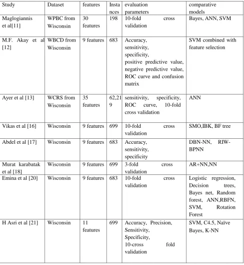
Volume: 4 Issue: 3 Dec- 2019 49 Table 2: Survey on different Models for melanoma detection
Study Dataset features Insta
nces
evaluation parameters
comparative models Maglogiannis
et al[11]
WPBC from Wisconsin
30 features
198 10-fold cross
validation
Bayes, ANN, SVM
M.F. Akay et al [12]
WBCD from Wisconsin
9 features 683 Accuracy, sensitivity, specificity,
positive predictive value, negative predictive value, ROC curve and confusion matrix
SVM combined with feature selection
Ayer et al [13] WCRS from Wisconsin
35 features
62,21 9
sensitivity, specificity, ROC curve, 10-fold cross validation
ANN
Vikas et al [16] Wisconsin 9 features 699 10-fold cross validation
SMO,IBK, BF tree
Abdel et al [17] Wisconsin 9 features 683 Accuracy, sensitivity, specificity
DBN-NN,
RIW-BPNN
Murat karabatak et al [18]
Wisconsin 9 features 699 3-fold cross validation
AR+NN,NN
Emina et al [20] Wisconsin 9 features 683 10-fold cross validation
Logistic regression, Decision trees, Bayes net, Random forest, ANN,RBFN,
SVM, Rotation
Forest H Asri et al [21] Wisconsin 11
features
699 Accuracy, Precision, Sensitivity,
Specificity,
10-cross fold
validation
SVM, C4.5, Naïve Bayes, K-NN
Artificial Neural Network for Skin Cancer Detection [2]: Sarika Choudhari and Seema Biday presented a carcinoma detection method focused on a neural network algorithm (NN) in 2014. Selection of dermoscopic images, filtering of hair and noise reduction images, segmentation of images using Maximum Entropy Threshold,
Volume: 4 Issue: 3 Dec- 2019 49 analyzed by pre-processing. Dull Razor and
Median Filter are usually taken from Dermoscopy images to remove hair, air bubbles, etc. Using the highest entropy procedure, photographs are segmented during preprocessing. Total Entropy Thresholding is used to check for the Interest Area. Using function extraction methods, the specific characteristics of the segmented images are removed. This system has a precision of 86.66%. Through changing ANN's image processing methods and training algorithms, the precision of this method is increased and the photographs are therefore marked as cancerous or non-cancerous, and they are also ready to find the type of cancer and cancer level.
Detection and Analysis of carcinoma in Skin Lesions by using Segmentation[1] In 2015, Amruta M. Gajbar et al. demonstrated a totally unique measurement to determine the non-appearance or proximity of streaks in skin injuries by dissecting the presence of distinct streak lines and performing a three-collection sequence for streaks, Absent, Normal, and Unpredictable, during an extremely pigmented skin injury. In fact, the spatial case of identified lines is analyzed to find alternatives to their implementation to consider the elementary example. The method uses a graphical delineation to show the legal streak geometric illustration and the structure's conveyance and distance along these axes. Mistreatment of these organized alternatives of the extensive streaks opposite the coloring and design options of the whole skin sores, consistency is achieved for characterizing dermatoscopy pictures in streaks Missing General or Unpredictable
Paper [3] recommends that you use a deep learning algorithm for pixel-specific labeling based on an automated process of lesion detection. Tested on public data and used ISIC database for Network Learning and PH2 database to prove that the process is not compatible with the Daten Collection. The analysis was carried out in two network architectures. The experimental results show that the method presented is very specific and segmented, even in the presence of bubbles of hair and oil. The additional contribution of this paper is the data annotation, which produces many
test images and the implementation of a semi-automatic GUI.
Article [4] suggests melanoma treatment and early detection of skin cancer. A method of research is introduced for lesion diagnosis and melanoma prevention. A real-time system has been developed to prevent people from skin burns and warn users. Using a long period of time and when the rays are damaging, a warning reached the user. The alarm has been put into an equation.
In this review, elements of a compact, non-invasive skin lesion detection device within real time are suggested to help prevent and diagnose melanoma. The first item is a realtime alarm which helps users prevent sunlight-caused skin burn, thus introducing a new equation which calculates the time for skin to burn. The second part consists of a robotic image test, including image acquisition, hair recognition and prohibition, injury division, extraction of highlight, and characterisation. In a request for PDA, the structure was made. The results of the test show that the framework proposed is effective, with high grouping accuracy [4].
Paper [5] attempts to recognize and differentiate skin sores from pictures taken by cameras as being kind-hearted or dangerous. The images are grouped into parts, highlights are differentiated by the ABCD rule and a neural network prepares for high-precision injuries A standard accuracy of 76.9 per cent on a dataset of 463 images, separated into six sections, was achieved by the neural Network.
Through designing the device neural on a much wider and diverse data set with strong intraclass shifts, the overall accuracy performance and execution of the frame can be increased. This would reduce the error and have a major influence on the accuracy rate. The amount of highlights removed from pictures is increased by an option as opposed to expansion of the dataset [5].
Volume: 4 Issue: 3 Dec- 2019 50 processing is used to clear the clamor. Images are
the threshold portion. There are certain different components in the field of skin development. Such products use the highlight extraction method separately. For the highlight extraction method, multi-level 2-D wavelet disintegration is used. The data hubs of the neural system are provided these highlights. For characteristics which classify the picture to destructive or uncarcinogenic, the neural system back and outdone the essential neural system are used.
Smartphones take on real e-wellbeing work to ensure m-wellbeing plays a critical role in the human services industry. Image management systems are key to distinguishing irregularities in the human body in the medical services industry. Skin development (Melanoma) is a major disease, but can be recovered very well in the early study. Reports indicate that over millions of people have died due to the Skin tumour itself.
This paper[7] examined how the disease of the skin can be distinguished by dissecting the disorder, asymmetry, boundary and variation of colour, diameter and extension (ABCDE) using a cell phone application in the initial periods. Dissected with distinctive techniques of processing images, such as changes in the gray scale, segmentation, form and histogram analysis.
Distinctive advanced images break down in this paper [ 8] in view of uncontrolled methods of division. On these portioned videos, highlight extraction techniques are related. Despite this, a broad exchange was analyzed based on the results received. Automatic skin lesion treatment is based on the dermatic spectrum of the so-called ABCD statute. ABCD describes the so-called lesion diameter and asymmetry, boundary shape, color variance and derma scope composition, which determines the foundation of a dermatologist's diagnosis
Detection of Melanoma carcinoma Using camera Images [9]: In 2015, with standard and anomalous sections, V. Jeya Ramya et al. suggested an automated system for skin malignancy identification. The preprocessing of
the picture was done by the Wiener channel to begin with. A successful execution of the division is accomplished by aggressive segmentation of the contour. The modules used as a framework cluster are extracted using GLCM. A 90 % affectability, 95% accuracy and 85 % specificity are observed in an order approach with two classifications (threatening and favorable sores). Often the surface parameters are included in the list of capabilities to support the overall implementation of the system, the lesion boundary and external texture descriptors are not yet included in the list of capabilities, and could provide a truthful starting stage to validate the discriminative details within the list of capabilities.
III.
CONCLUSION
Volume: 4 Issue: 3 Dec- 2019 51
REFERENCES
[1] Gajbar, Amruta & Deshpande, Anuradha. (2015). Detection and Analysis of Skin Cancer in Skin Lesions. IJARCCE. 285-291. 10.17148/IJARCCE.2015.4263.
[2] Sarika Choudhari, Seema Biday , " Artificial Neural Network for SkinCancer Detection" , International Journal of Emerging Trends & Technology in Computer Science (IJETTCS) , Volume 3, Issue 5, September - October 2014 , pp. 147-153 , ISSN 2278-6856.
[3] Deep Convolution Pixel-wise Labeling for skin lesion Image Segmentation ,Ali Youssef ;Domenico D. Bloisi ;Mario Muscio ; Andrea Pennisi ; Daniele Nardi ; Antonio Facchiano IEEE International Symposium on Medical Measurements and Applications (MeMeA) Year: 2018
[4] Early Detection of Melanoma Skin Cancer Using Classifiers, VS. Sabeera, P. Vamsi Krishna PG Scholar, Dept. of ECE, R. K. College of Engineering, Vijayawada, Andhra Pradesh Assistant Professor, Dept. of ECE, R.K. College of Engineering, Vijayawada, Andhra Pradesh, Year-2016
[5] Skin Cancer Detection and classification Pratik Dubal, Sankirtan Bhatt, Chaitanya Joglekar, Dr. Sonali Patil, Department of Information Technology K. J. Somaiya College of Engineering Vidyavihar, Year-2017
[6] Skin Cancer Detection using Artificial Neural Network, Ekta Singhal M.Tech II Year, Dept of Computer Science Engineering, MUST – FET, Lakshmangarh, India Shamik Tiwari, Assistant Professor, Dept of Computer Science Engineering, MUST – FET, Lakshmangarh, India, Year-2015
[7] Detection of Skin Cancer Using Image Processing Techniques, Chandrahasa M, Varun Vadigeri and Dixit Salecha, Computer Science and Engineering, The National Institute of Engineering, Year-2016
[8] Image Processing for Skin Cancer Features Extraction Md.Amran Hossen Bhuiyan, Ibrahim Azad, Md.Kamal Uddin, Department of Computer Science & Telecommunication Engineering, Noakhali Science & Technology University, Bangladesh, International Journal of Scientific & Engineering Research Volume 4, Issue 2, February-2013
[9]V. Jeya Ramya, J. Navarajan, R. Prathipa and L. Ashok Kumar, DETECTION OF MELANOMA SKIN
CANCER USING DIGITAL CAMERA IMAGES, ARPN Journal of Engineering and Applied Sciences, VOL. 10, NO. 7, APRIL 2015
[11]. Maglogiannis, I., Zafiropoulos, E., &
Anagnostopoulos, I. (2009). An intelligent system for automated breast cancer diagnosis and prognosis using SVM based classifiers. Applied intelligence, 30(1), 24-36.
[12.] Akay, M. F. (2009). Support vector machines combined with feature selection for breast cancer diagnosis. Expert systems with applications, 36(2), 3240-3247.
[13]. Ayer, T., Alagoz, O., Chhatwal, J., Shavlik, J. W., Kahn Jr, C. E., & Burnside, E. S. (2010). Breast cancer risk estimation with artificial neural networks revisited: discrimination and calibration. Cancer, 116(14), 3310-3321.
[14]. Kim, W., Kim, K. S., Lee, J. E., Noh, D. Y., Kim, S. W., Jung, Y. S., ... & Park, R. W. (2012). Development of novel breast cancer recurrence prediction model using support vector machine. Journal of breast cancer, 15(2), 230-238.
[15]. Pritom, A. I., Munshi, M. A. R., Sabab, S. A., & Shihab, S. (2016, December). Predicting breast cancer recurrence using effective classification and feature
selection technique. In 2016 19th International
Conference on Computer and Information Technology (ICCIT) (pp. 310-314). IEEE.
[16.] Chaurasia, V., & Pal, S. (2017). A novel approach for breast cancer detection using data mining techniques.
[17]. Abdel-Zaher, A. M., & Eldeib, A. M. (2016). Breast cancerclassification using deep belief networks. Expert Systems with Applications, 46, 139-144.
[18]. Karabatak, M., & Ince, M. C. (2009). An expert system for detection of breast cancer based on association rules and neural network. Expert systems with Applications, 36(2), 3465-3469.
[19]. Ahmad, L. G., Eshlaghy, A. T., Poorebrahimi, A., Ebrahimi, M., & Razavi, A. R. (2013). Using three machine learning techniques for predicting breast cancer recurrence. J Health Med Inform, 4(124), 3.
Volume: 4 Issue: 3 Dec- 2019 52
Forest. Neural Computing and Applications, 28(4), 753-763.