ISSN (e): 2250-3021, ISSN (p): 2278-8719
Vol. 07, Issue 09 (September. 2017), ||V1|| PP 57-65
Improve Image Segmentation Techniques of FCM, HMF,
FCM-HMRF and ARK-FCM
*
Yogesh Kumar Gupta, Nirupama Tiwari
Dept. of (CSE/IT)) S.R.C.E.M College Banmore,Gwalior (India) PROF. Dept. of . (CSE/IT) S.R.C.E.M College Banmore, Gwalior (India)Corresponding Author: Yogesh Kumar Gupta
Abstract
: Image segmentation is one of the essential tasks in the field of pc vision. This paper proposes another image segmentation strategy in view of Fuzzy C Means (FCM) and MRF. FCM has the ability to represent ambiguous information in a more robust way. HMRF and HMF have the ability to find optimal parameters in search spaces. These characteristics of FCM and ALO have been utilized in this paper for improving image Segmentation. The proposed hybrid FCM-HMRF based However the problem of image segmentation speed is still an important problem in image processing. Therefore, how to improve the segmentation speed of different algorithms is an indispensable topic. In this paper apply Adaptively Regularized Kernel-Based Fuzzy C-Means Clustering strategy for enhances image resultKeywords
: IS; Fuzzy C Means; MRF; HMRF-FCM; ARK-FCM.--- --- Date of Submission: 24-08-2017 Date of acceptance: 08-09-2017
--- ---
I.
INTRODUCTION
DIP assumes a basic part in numerous applications to retrieve required realities from the given picture in a way that it has not have an effect on the opposite capabilities of the image. Images are the vital medium of conveying data and via knowledge image the conveying data can be used for many responsibilities. A digital photograph (DI) consists with the aid of finite range of things or pixels and the acquisition of pix is called as imaging. DIP is a multidisciplinary operation and it has considered one of sort styles of framework which incorporates photograph representation, segmentation, pressure and change.
IS is a classical and basic issue in many bundles which incorporates therapeutic IP, bio measurements, thing checking and popularity, video and lPc Vision applications. IS has been an critical and tough trouble inside the discipline of IP and it performs a vital position for maximum picture evaluation responsibilities which encompass object popularity, object-based image compression and content material based totally indexing.
It has been various one of a kind interpretation for distinct styles of application toward content material evaluation and picture know-how. Many IS methods have been developed, but there's still no excellent in level of performance measure by means of purpose of image segments consequences relies upon on type of pictures.[1].
In this paper, we aggregate up customary fuzzy c-means algorithm and MRF mode, going for image segmentation at that point we propose an enhanced strategy to illuminate the vulnerability problem in gray scale images [2]. In order to achieve the purpose of anti-noise, the MRF spatial constraint field is introduced into FCM algorithm. The image segmentation algorithm based on MRF which can effectively resist noise uses the related information of domain space as priori information, and applies Gibbs field. first combined MRF with FCM, and overcame their respective shortcomings.
Nevertheless, these proposed algorithms obtain unsatisfactory segmentation results in the case of low noise-signal ratio of an image.
IMAGE SEGMENTATION
IS is characterized as the technique in which a image is divided into numerous added substances, with the end goal that a image is portrayed into something. that is easy to particular and smooth to have a take a look at. It is vital for meaningful analysis and interpretation of medical pix.
There are a few calculations and strategies that have been built up for segmenting image. Modern medical imaging modalities like MRI and CT examines create enormous images which can't be considered physically. This develops the requirement for more effective and robust image determination approaches, tailored to the problems met in medical images [3].
II.
USING
TECHNIQUES
A. Fuzzy C Means MethodBy and huge there are types of clustering viz., difficult clustering and tender clustering. In hard clustering the image pixels are separated into various clusters where every pixel is placed in exactly one cluster. On the other hand, in fuzzy clustering the image pixels are fitted into many number of clusters based on the individual pixel’s membership value. The membership value shows the strength of connection between the individual pixel with a particular cluster [4].
Fuzzy C Means (FCM) is an eminent and partitioning based method which follows the unsupervised learning mechanism. The FCM method processes the original image pixels and partitions them into one or more distinctive clusters based on the membership function which utilizes the cluster centroid (distance) calculation function and FCM objective function.
The predominant footstep of FCM is an iterative technique which revises the club feature values and centroid positions and their values. In FCM, the high membership value specifies that the partitioned pixels are nearer to the centroid and the low membership value represents the far-flung pixels from the centroid position.
Markov Random Fields (MRF)
segmentation based on MRF is called as Model based segmentation An built in region smoothness constraint is used in MRF which is used for color segmentation. Some portion of the color pixel tuples are assumed as impartial - random arbitrary factors for besides preparing. With part location MRF is blended for distinguishing the edges effectively. Markov Random Field (MRF) has limitation of spatial locale smoothness and some of the shading segments there are connections [5].
HMRF-FCM
To acquire an estimate of the HMRF version parameters, given a model fitting dataset, the usage of the HMRF-FCM algorithm, we ought to iteratively limit the fuzzy objective feature Qλ (Ψ), given through (22),
over R, θ, and β, in a coordinate descent fashion. Let V(k ) stand for the estimate of the amount V got on the kth
cycle of the calculation. Let us consider the (k + 1)th iteration of the algorithm; as a result, we assume that the present day value of the model parameters set estimate Ψˆ is equal to the cost obtained from the kth iteration of the algorithm Ψ(k) , i.e., Ψˆ = Ψ(k) .[6]
ARK-FCM
An adaptively regularized kernel-based totally fuzzy - means clustering system is proposed for segmentation of brain magnetic resonance images. The structure might be as three calculations for the area normal grayscale being changed through the grayscale of the normal get out, median filter out, and devised weighted images, separately. [7].
Initialize threshold = 0.001, =2, loop counter =0,V, and (0). Calculate the adaptive regularization parameter
Calculate for ARKFCM1 and ARKFCM2 or for ARKFCM . Calculate cluster centers V( ) using ( )
Calculate the membership function ( +1) .
If max ‖( +1) − ( )‖ 100 then stop; otherwise, update = +1and go to step (4). 1. The main advantages are:
a. Adaptive to local context,
B. Enhanced robustness to hold image details.
C. Independent of clustering parameters, and with decreased computational costs.
III.
LITERATURE
SURVAY
Dingsheng Hu, et.al. [9] This represents one of the most advanced PolSAR unsupervised statistical segmentation set of rules and uses the doubly flexible, two parameter, -distribution model for the PolSAR data. However complexity of the danger density function ends in excessive time intake. These papers look into the important aspect structured variable inside the distribution version and discover a new parameter location in which the PDFs are easy. Then a one-dimensional appearance-up desk is prepared on this place with nodes wide variety determined via corresponding Fourier spectrum and is followed to keep avoid re- evaluating the numerical basic in PDF to compute style posteriori probabilities for each sample. The proposed technique is joined in the segmentation calculation. Prototype check has been done to approve the productivity of the proposed technique.
Marek Wdowiak, et.al. [10] This paper gives change of traditional watershed calculation for cell segmentation in microscopic images of desmoglein-three recolored example. Exhibited approach joins color deconvolution for ihc marker detachment and GVF for watershed segmentation. Conventional watershed is extraordinarily noise sensitive, which often takes place in microscopy images. Suggested solution drastically reduces over segmentation hassle (80- 90% cells segmented efficiently) and permits similarly image analysis.
Maithili Lawankar, et.Al. [11] In this paper, Watershed Transform segmentation Algorithm is used because it produces absolute partition of images in separate area despite the fact that contrast is poor. In this way this technique could be accomplished 92.1% accuracy.
Samah Bouzidi, et.al. [12] In this paper, we build up another semi- automated segmentation technique to wipe out the turbulent blood float sign inside the left ventricle (LV) in cardiovascular magnetic resonance (MR) images with parallel imaging. The segmentation is achieved the use of a deformable version driven by a brand new outside power based on estimated probability density function (pdf) of the MR sign inside the LV. The utilization of noise distribution through the insights permits us both to pull the form towards the myocardium edges and to guarantee the smoothness of the curve.Since measurements for each cut are gotten with the GRAPPA parallel imaging strategy, the spatial segmentation is trailed by utilizing a worldly spread to make strides. the convergence in phrases of satisfactory and rapidity. Experiments display that the proposed model gives higher consequences than those received from the usual Active Contour, which must facilitate the use of the method for clinical functions.
Renjun Shuai, et.Al. [13] MRI segmentation by using K means clustering is finished in this unique paper. Schemes of MRI vicinity supported segmentation that can significantly differentiate between normal and abnormal tissue. MRI would not want contact to radiation. Magnetic Resonance Imaging can be an intense approach to help for assessment of illness, or to pursue disease development. At the procedure finishing the tumor is taken out from the MRI picture and its specific region and the shape also decided. The step of the tumor is displayed depending upon quantity of region measured from the cluster.
IV.
PROPOSE
WORK
Proposed Methodology
According to using adaptive weight which can adaptively change the weight value in different conditions. The experiment results show that our method adequately considers about the characteristics of poor anti-noise performance caused by spatial information, and our approach has better performance than different techniques within the aspect of edge records processing. However, the trouble of image segmentation velocity continues to be an important problem in image processing. Therefore, the way to enhance the segmentation speed of various algorithms is an essential topic. In this paper look at adaptively Regularized Kernel-Based Fuzzy C-Means clustering strategy for enhances image result.
PROPOSED ALGORITHM
Input: SAR Images of variable length. Output: Segmented zone.
1) Start.
2) Taken a SAR images. 3) Consider a 3X3 window.
4) Calculate the Mean, Variance and Entropy of That SAR Images
5) Store the color function because the coloration depth of 3 number one colour Red, Green and Blue with recognize to Threshold fee.
6) Segmentation is received the use of K-means clustering Algorithm and GSA set of rules. k-means is one of the handiest unsupervised learning algorithms that solve the well regarded clustering problem. The system follows a easy and easy way to classify a given data set via a certain range of clusters (expect k clusters) constant apriori. The essential idea is to define k centers, one for every cluster. GSA optimization set of rules based on the law of gravity.
Fig.1. Flow chart of Propose Work
V.
RESULT
ANALYSIS
This approach in view of considering a 3X3 window and figures the men, variance and Entropy of that SAR Images at that point store the shade highlight in light of the fact that the color intensity with appreciate to color model Red, Green and Blue. Next, Segmentation is received using K-means clustering Algorithm and the use of GSA it become a fast segmentation manner.
Presently SAR images are utilized to test proposed calculation. In the first place mean, change, entropy ascertained from possibility for unique image. Images had been tried for analyzing window size of 3x3 and 5x5 to find entropy. At long last K-implies clustering Algorithm wound up noticeably utilized on entropy image for segmentation. In GSA, each mass (agent) has 4 specs: part, inertial mass, dynamic gravitational mass, and passive gravitational mass.
Fig. 2. Image data set
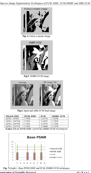
Fig. 4. Fuzzy-c-means image
Fig.5. HMRF-FCM image
Fig.6. Input and ARK-FCM final image
IMAGE SIZE PSNR HMF FCM HMRF- FCM LENA 64*64 5.7994 11.6774 5.8196 LENA 128*128 5.7823 11.8353 5.7839 LENA 256*256 5.7441 11.9576 5.7396 LENA 512*512 5.7455 11.9880 5.7570
TABLE 1BASE PSNRHMF AND FCM,HMRF-FCM TECHNIQUES
IMAGE SIZE SNR HMF
SNR FCM
SNR HMRF- FCM
LENA 64*64 233 118 232
LENA 128*128 234 117 236
LENA 256*256 236 116 237
LENA 512*512 250 116 248
TABLE 2BASE SNRHMF AND FCM,HMRF-FCM TECHNIQUES
Fig.8. Graph2. Base PSNR HMF and FCM, HMRF-FCM techniques
IMAGE SIZE SPEED HMF
SPEED FCM
SPEED HMRF- FCM
LENA 64*64 0.4133 0.2932 0.3464 LENA 128*128 1.1063 0.9923 1.1813 LENA 256*256 1.1102 3.5519 4.3481 LENA 512*512 4.5745 14.3034 17.8063
TABLE 3BASE SPEED OF HMF AND FCM,HMRF-FCM TECHNIQUES
Fig.9. Graph 3.Base SPEED HMF and FCM, HMRF-FCM techniques
Image size PSNR HMF PROPOSED
PSNR FCM PROPOSED
PSNR HMRF –FCM PROPOSED
PSNR ARK- FCM PROPOSED
Lena 64*64 5.8115 11.6774 5.8196 14.2827
Lena 128*128 5.7841 11.8353 5.7823 14.2811
Lena 256*256 5.7438 11.9576 5.7396 14.2804
Lena 512*512 5.7455 11.9880 5.7570 14.2801
TABLE 4PROPOSE PSNRHMF AND FCM,HMRF-FCMAND ARK-FCM TECHNIQUES 0 50 100 150 200 250 300
1 2 3 4 5 6 7
Base SNR
IMAGE SIZE SNR HMF SNR FCM SNR HMRF- FCM
0 2 4 6 8 10 12 14 16 18 20
1 2 3 4 5 6 7 8
Base SPEED
IMAGE SIZE
SPEED HMF
SPEED FCM
Fig.10. Graph.4. Propose PSNR HMF and FCM, HMRF-FCM and ARK-FCM techniques
IMAGE SIZE SNR HMF PROP OSED SNR FCM PROP OSED SNR HMRF-FCM PROPOSED SNR ARK-FCM PROPOSED
Lena 64*64 233 118 232 231
Lena 128*128 234 117 234 233
Lena 256*256 236 116 237 237
Lena 512*512 250 116 248 247
TABLE 5PROPOSE SNRHMF AND FCM,HMRF-FCM AND ARK-FCM TECHNIQUES
Fig.11. Graph.5. Propose SNR HMF and FCM, HMRF-FCM and ARK-FCM techniques
Image size SPEED HMF PROPOSED SPEED FCM PROPOSED SPEED HMRF-FCM PROPOSED SPEED ARK-FCM PROPOSED
Lena 64*64 1.7937 0.2147 0.3188 0.7778
Lena 128*128 0.6451 0.9279 1.5152 1.6193
Lena 256*256 1.1394 3.6599 4.4717 8.7742
Lena 512*512 4.2260 14.7652 18.1561 38.8345
TABLE 6PROPOSE SPEED OF HMF AND FCM,HMRF-FCM AND ARK-FCM TECHNIQUES 0 2 4 6 8 10 12 14 16
Lena 64*64 Lena 128*128 Lena 256*256 Lena 512*512
Propose
PSNR
PSNR HMF PROPOSED
PSNR FCM PROPOSED
PSNR HMRF –FCM PROPOSED
PSNR ARK- FCM PROPOSED 0 50 100 150 200 250 300
PROPOSED PROPOSED PROPOSED PROPOSED SNR HMF SNR FCM SNR
Fig.12. Graph 6.Propose PSNR HMF and FCM, HMRF-FCM and ARK-FCM techniques
VI.
CONCLUSION
During the process of image segmentation, we will meet several problems, and we always make mistakes because of uneven illumination, influence of image noise, indistinct parts in an image, shadow, et al. Aiming at these several problems, after we analyzing the traditional FCM and MRF method, we propose a novel algorithm based on improved FCM and MRF mode. This method primarily based on considering a 3X3 window and calculates the men, variance and Entropy of that SAR Images then keep the shade function because the colour intensity with respect to color model Red, Green and Blue. Next, Segmentation is obtained using K-means clustering Algorithm and using GSA it become a fast segmentation process. Therefore, how to improve the segmentation speed of different algorithms is an indispensable topic. In this paper observe adaptively Regularized Kernel-Based Fuzzy C-Means Clustering technique for improves image result However, the hassle of image segmentation velocity is still an important problem in image processing. Therefore, how to improve the segmentation speed of different algorithms is an indispensable topic. In our future work, we will focus on the IS velocity.
REFERENCES
[1]. P. Sivakumar1 , Dr. S.Meenakshi, “A REVIEW ON IMAGE SEGMENTATION TECHNIQUES”. International Journal of Advanced Research in Computer Engineering & Technology (IJARCET) Volume 5 Issue 3, March 2016 ISSN: 2278 – 1323
[2]. Hossam M. Moftah, Ahmad TaherAzar, EimanTamah Al-Shammari , Neveen I. Ghali , Aboul Ella Hassanien and Mahmoud Shoman,” Adaptive k-means clustering algorithm for MR breast image segmentation”, Neural Comput&Applic (2014) 24:1917–1928
[3]. Shashi Bala and Ajaya Kumar, “A brief review of image segmentation techniques”. 2016 IJARECE [4]. P. Parvathi, R.Rajeswari, “A Hybrid FCM-ALO based Technique for Image Segmentation”.
978-1-5090-3770-4/16/$31.00©2016 IEEE
[5]. Shashi Bala and Ajaya Kumar, “A brief review of image segmentation techniques”. International Journal of Advanced Research in Electronics and Communication Engineering (IJARECE) Volume 5, Issue 5, May 2016 ISSN: 2278 – 909X
[6]. Sotirios P. Chatzis, and Theodora A. Varvarigou,, “A Fuzzy Clustering Approach Toward Hidden Markov Random Field Models for Enhanced Spatially Constrained Image Segmentation” 1063-6706/$25.00 © 2008 IEEE
[7]. A. SRUTHI1 , M. RAVI KISHORE, “AR Kernel-Based Fuzzy C Method for Brain Tumour Segmentation of MR Images A. SRUTHI1 , M. RAVI KISHORE”. ISSN 2319-8885 Vol.05,Issue.19 July-2016, Pages:3812-38162016 IJSETR.
[8]. Ming Yan, Zilu Wang “A Novel Natural Image Segmentation Algorithm based on Markov Random Field and Improved Fuzzy C-Means Clustering Method”.IEEE
[9]. Dingsheng Hu, Anthony P. Doulgeris, Xiaolan Qiu, Bin Lei, A FAST AUTOMATIC U-DISTRIBUTION SEGMENTATION ALGORITHM FOR POLSAR”. 978-1-5090-3332-4/16/$31.00 ©2016 IEEE
[10]. Marek Wdowiak, Janina Slodkowska, Tomasz Markiewicz, “Cell segmentation in Desmoglein-3 Stained Specimen Microscopic Images using GVF and watershed algorithm”. 978-1-5090-2800-9/16/$31.00 c 2016 IEEE
0 5 10 15 20 25 30 35 40 45
PROPOSED PROPOSED PROPOSED PROPOSED
SPEED HMF
SPEED FCM
SPEED HMRF-FCM
SPEED ARK-FCM
Propose SPEED
Lena 64*64
Lena 128*128
Lena 256*256
[11]. Maithili Lawankar, ShraddhaSangewar and SomuluGugulothu, “Segmentation of Liver using Marker Watershed Transform Algorithm for CT Scan Images”. 978-1-5090-0396-9/16/$31.00 ©2016 IEEE [12]. Samah Bouzidi, Aurélie Emilien, Bruno Quesson, Chokri Ben Amar, Jenny Benois-Pineau and Pascal
Desbarats, “Segmentation of left ventricle on MRI sequences for blood flow cancelation in Thermotherapy”. 978-1-4799-8637-8/15/$31.00 ©2015 IEEE
[13]. Renjun Shuai, Yang Shen, Jing Pan, “An Algorithm for Medical Imagining Compression That Is Oriented to ROI-Characteristics Protection”. 2015