Case Report
Collapsing Focal Segmental Glomerulosclerosis in
a Patient with Acute Malaria
Najamus Sehar, Emad Gobran, and Suzanne Elsayegh
Staten Island University Hospital, 475 Seaview Avenue, Staten Island, NY 10305, USA
Correspondence should be addressed to Najamus Sehar; sehar.najamus@gmail.com
Received 11 May 2015; Revised 18 June 2015; Accepted 21 June 2015
Academic Editor: Masahiro Kohzuki
Copyright © 2015 Najamus Sehar et al. This is an open access article distributed under the Creative Commons Attribution License, which permits unrestricted use, distribution, and reproduction in any medium, provided the original work is properly cited.
Introduction. Collapsing focal segmental glomerulosclerosis (FSGS) is most commonly seen in association with HIV infection.
Rare data is available about the association between collapsing FSGS and malaria.Case Description. A 72-year-old African male
patient presented to the hospital for generalized body aches, fatigue, fever, and night sweats for three days. He had history of recent travel to Ghana. Patient looked in acute distress and was shivering. Laboratory tests showed elevated serum creatinine (Cr) of 2.09 mg/dL (baseline was 1.5 mg/dL in 2012). Hospital course was significant for rapid elevation of Cr to 9.5 mg/dL and proteinuria of
7.9 grams. Autoimmune studies resulted negative. Blood smear resulted positive forPlasmodium falciparumand patient was treated
with Artemether/Lumefantrine. Patient’s fever and pain improved, but kidney function continued to deteriorate and he became oliguric. On day seven, he was started on Hemodialysis. Tests for different causes of glomerular pathology were also negative. He
underwent left kidney biopsy which resulted in findings consistent with severe collapsing glomerulopathy.Discussion. This case
illustrates a biopsy proven collapsing FSGS likely secondary to malarial infection requiring renal replacement therapy. Literature review revealed only few case reports that suggested the possible association of malaria with secondary form of FSGS.
1. Introduction
Collapsing glomerulopathy is a morphologic variant of FSGS characterized by segmental and global collapse of the glomerular capillaries, marked hypertrophy and hyper-plasia of podocytes, and severe tubulointerstitial disease [1]. It is most commonly seen in association with HIV, but other numerous etiologies have been reported as well. These include infections like tuberculosis, cytomegalovirus, hepatitis B, and hepatitis C. Other causes include drugs such as bisphosphonates, steroids, opioids, and interferons; autoimmune diseases like SLE, adult Still’s disease, and mixed connective tissue disease; and hematologic abnormalities including multiple myeloma, hemophagocytic syndrome, and acute monoblastic leukemia [1–5]. There have been very few cases of malaria associated collapsing glomerulopathy. Most of such cases are reported in Africa and Southeast Asia where malaria is more prevalent or amongst the immigrants from these areas. Malaria caused byPlasmodium malariae
andPlasmodium falciparumis more commonly involved in acute renal failure as compared to other strains. Very few
studies have been published, showing association ofP. falci-parumwith collapsing FSGS.
2. Case Description
A 72-year-old man was admitted to hospital for right upper quadrant abdominal pain, generalized body aches, night sweats, chills, subjective fever at night, and weakness for three days.
One month before presenting, patient travelled to his native African country Ghana for a two-week trip. Ten days after returning back to the United States, he developed the symptoms. Initially, he took over the counter common cold medication without any relief. He denied sick contacts, vomiting diarrhea, shortness of breath, chest pain, or any other symptoms.
Patient was also taking mefloquine for malaria prophy-laxis during his visit.
Patient’s past medical history included hypertension treated for five years, high cholesterol, gout, and chronic kidney disease secondary to hypertension. The patient had
2 Case Reports in Medicine
no surgeries in past and had no known allergies. He denied smoking, alcohol intake, or drug abuse. Patient’s home medi-cations included allopurinol, losartan, amlodipine, labetalol, furosemide, and simvastatin.
On physical examination, temperature was 39.3 degrees of Celsius, blood pressure was 184/86 mm of Hg, respiratory rate was 18 breaths per minute, and heart rate was 76 beats per minute. Patient looked in acute distress and was shivering. He was awake, alert, and oriented to time, place, and person. Neck was supple. Lungs were clear and heart sounds regular on auscultation. No murmur, rubs, or gallop was appreciated. Abdomen was soft and nontender and bowel sounds were audible in all four quadrants. No leg edema was noted.
Initial blood tests in the emergency room showed a normal complete blood picture including white count with differential, platelet count and a hemoglobin of 12.3 grams/dL. On metabolic profile, patient’s creatinine was found to be 2.09 mg/dL that was higher than baseline of 1.5 mg/dL recorded six months earlier. Urea nitrogen was 30 mg/dL with glomerular filtration rate of 38. Liver function tests were normal except for albumin of 2.6 g/dL and total bilirubin of 1.6 mg/dL. Creatinine kinase was 514 IU/L. Amylase was 54 U/L and lipase 35 U/L.
Initially, patient was admitted to medical floor and started on broad spectrum empiric antibiotics. Blood culture, urine cultures, and nasal swab for influenza were sent. Because of the acute kidney injury, furosemide and losartan were stopped and intravenous fluids were given. A urinary catheter was placed and urinalysis along with urine electrolytes was ordered.
On the second day of hospitalization, patient continued to have abdominal pain and fever. An abdominal computerized tomography scan without contrast showed findings concern-ing acute pancreatitis or acalculous cholecystitis.
On the third day, his serum creatinine rapidly increased to 4.17 mg/dL with GFR of 17. Urine analysis showed dark colored, cloudy, and concentrated urine with small amount of bilirubin, large blood, large protein, no white blood cells, and red blood cells of 3–5/hpf. No casts were seen on urine sediment. Urine electrolytes showed a fractional excretion of sodium of 1.2% and urine creatinine of 157 mg/dL. A urine protein to creatinine ratio of 7929.41 was calculated. Patient’s urinalysis one year ago showed trace protein and total protein of 34 mg/dL and creatinine of 144 mg/dL.
An abdominal ultrasound was done that was suggestive of acalculous cholecystitis. There was newly increased renal echogenicity but no hydronephrosis.
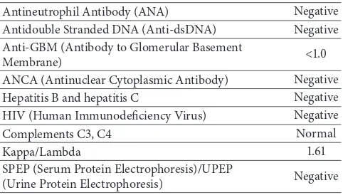
From days three to five, patient’s urine output declined and serum creatinine continued to rise from 4.75 mg/dL up to 8.74 mg/dL and patient was started on Hemodialysis. Laboratory tests for different glomerulopathy causes resulted negative (Table 1).
In view of recent travel, a blood smear for malaria was done that showed crescentic shaped gametocyte which is usually the shape ofPlasmodium falciparumand parasitemia of 1.64% (Figure 1).
Patient was then started on Coartem (Artemether: 20 mg and Lumefantrine: 120 mg) for malaria. This is considered as the treatment of acute malarial infections due toPlasmodium
Table 1: Glomerulopathy laboratory tests.
Antineutrophil Antibody (ANA) Negative
Antidouble Stranded DNA (Anti-dsDNA) Negative
Anti-GBM (Antibody to Glomerular Basement
Membrane) <1.0
ANCA (Antinuclear Cytoplasmic Antibody) Negative
Hepatitis B and hepatitis C Negative
HIV (Human Immunodeficiency Virus) Negative
Complements C3, C4 Normal
Kappa/Lambda 1.61
SPEP (Serum Protein Electrophoresis)/UPEP
(Urine Protein Electrophoresis) Negative
Figure 1: Blood peripheral smear showing crescentic shaped game-tocytes.
falciparumin geographical regions where chloroquine resis-tance has been reported.
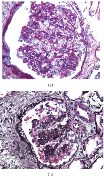
Finally, the kidney biopsy was done (Figure 2) that was consistent with severe collapsing glomerulopathy. It showed collapse of the glomerular tufts and prominent hyperplasia of overlying epithelial cells, acute injury of the tubules, interstitial fibrosis and arteriosclerosis, complete foot process effacement, and tubular degenerative changes.
Patient’s symptoms improved after starting the antimalar-ials and he was discharged after few days, but his kidney function never returned to his baseline and he continues to use renal replacement therapy.
3. Discussion
We presented an interesting case of acute renal failure secondary to falciparum malaria showing histopathology of collapsing FSGS. We briefly discuss the pathogenesis, treat-ment, and prognosis of this case in light of the current literature review.
(a)
(b)
Figure 2: Light microscopy of the kidney biopsy. Glomeruli dis-played lesions of collapsing glomerulopathy which ranged from segmental to global are shown by periodic acid-Schiff (a) and methenamine silver staining (b).
in terms of age, sex, serum creatinine, proteinuria, and glomerular findings [8].
So far, no defined treatment exists for malaria associated FSGS. According to a study by Albaqumi et al. performed in 2006, FSGS did not respond to corticosteroid treatment in 72 % of the cases [1]. Glucocorticoid and immunosuppressive agent therapies are recommended for patients based on anecdotal evidence only, and the only proven intervention for suppressing the progression of disease process is the treat-ment of the underlying cause. Malaria-induced kidney injury is usually managed by antimalarials, supportive therapy, and Hemodialysis (HD), but as said before no specific treatment exists for FSGS caused by malarial infection [9].
The prognosis of FSGS not associated with HIV infection was reported to be comparatively poor especially in African patients and a progression to end-stage renal disease (ESRD) is seen despite treatment [10,11].
Ehrich and Eke performed a meta-analysis using African data and literature on Afro-Americans about the malaria infected children undergoing renal failure in Africa. They concluded that FSGS and minimal change disease were found to be a more frequent and unusual cause of nephrotic syndrome in West Africans as compared to the rest of African regions and Europe, where membranoproliferative glomeru-lonephritis (MPGN) and quartan malarial nephropathy are
the usual culprits [12]. This is supported by similar studies amongst African children in Ghana [13, 14]. Our patient was also born in Ghana and hence the finding of collapsing FSGS is consistent with the previous rare observations for this geographical area. This case should encourage future studies to unfold the pathophysiology of this specific phenomenon and to evaluate any specific risk factors pertaining to the parasitic features in the specific geographic area.
Conflict of Interests
All the authors declare that they do not have any conflict of interests regarding this case report.
References
[1] M. Albaqumi, T. J. Soos, L. Barisoni, and P. J. Nelson,
“Col-lapsing glomerulopathy,” Journal of the American Society of
Nephrology, vol. 17, no. 10, pp. 2854–2863, 2006.
[2] A. Valeri, L. Barisoni, G. B. Appel, R. Seigle, and V. D’Agati, “Idiopathic collapsing focal segmental glomerulosclerosis: a
clinicopathologic study,”Kidney International, vol. 50, no. 5, pp.
1734–1746, 1996.
[3] V. D’Agati, J.-I. Suh, L. Carbone, J.-T. Cheng, and G. Appel, “Pathology of HIV-associated nephropathy: a detailed
morpho-logic and comparative study,”Kidney International, vol. 35, no.
6, pp. 1358–1370, 1989.
[4] C. M. Wyatt, P. E. Klotman, and V. D. D’Agati, “HIV-associated nephropathy: clinical presentation, pathology, and
epidemiol-ogy in the era of antiretroviral therapy,”Seminars in Nephrology,
vol. 28, no. 6, pp. 513–522, 2008.
[5] A. Niang, S. E. Niang, E. H. F. Ka, M. M. Ka, and B. Diouf, “Collapsing glomerulopathy and haemophagocytic syndrome
related to malaria: a case report,”Nephrology Dialysis
Transplan-tation, vol. 23, no. 10, pp. 3359–3361, 2008.
[6] L. Barisoni, H. W. Schnaper, and J. B. Kopp, “A proposed tax-onomy for the podocytopathies: a reassessment of the primary
nephrotic diseases,”Clinical Journal of the American Society of
Nephrology, vol. 2, no. 3, pp. 529–542, 2007.
[7] R. K. Detwiler, R. J. Falk, S. L. Hogan, and J. C. Jennette, “Collapsing glomerulopathy: a clinically and pathologically
distinct variant of focal segmental glomerulosclerosis,”Kidney
International, vol. 45, no. 5, pp. 1416–1424, 1994.
[8] A. Laurinavicius, S. Hurwitz, and H. G. Rennke, “Collapsing glomerulopathy in HIV and non-HIV patients: a
clinicopatho-logical and follow-up study,”Kidney International, vol. 56, no. 6,
pp. 2203–2213, 1999.
[9] V. B. Kute, H. L. Trivedi, A. V. Vanikar, P. R. Shah, M. R. Gumber, and K. V. Kanodia, “Collapsing glomerulopathy
and hemolytic uremic syndrome associated with falciparum
malaria: completely reversible acute kidney injury,”Journal of
Parasitic Diseases, vol. 37, no. 2, pp. 286–290, 2013.
[10] Y. M. Barri, N. C. Munshi, S. Sukumalchantra et al., “Podocyte injury associated glomerulopathies induced by pamidronate,” Kidney International, vol. 65, no. 2, pp. 634–641, 2004. [11] H. Tariq, A. Rafiq, and G. Franchin, “Collapsing focal segmental
glomerulosclerosis in a patient with systemic lupus
erythemato-sus,”Case Reports in Medicine, vol. 2014, Article ID 732192, 5
4 Case Reports in Medicine
[12] J. H. H. Ehrich and F. U. Eke, “Malaria-induced renal damage:
facts and myths,”Pediatric Nephrology, vol. 22, no. 5, pp. 626–
637, 2007.
[13] J. Y. Doe, M. Funk, M. Mengel, E. Doehring, and J. H. H. Ehrich, “Nephrotic syndrome in African children: lack of evidence for
‘tropical nephrotic syndrome’?”Nephrology Dialysis
Transplan-tation, vol. 21, no. 3, pp. 672–676, 2006.
[14] D. Adu, Y. Anim-Addo, A. K. Foli et al., “The nephrotic
syn-drome in Ghana: clinical and pathological aspects,”Quarterly
Submit your manuscripts at
http://www.hindawi.com
Stem Cells
International
Hindawi Publishing Corporationhttp://www.hindawi.com Volume 2014
Hindawi Publishing Corporation
http://www.hindawi.com Volume 2014
INFLAMMATION
Hindawi Publishing Corporation
http://www.hindawi.com Volume 2014
Behavioural
Neurology
Endocrinology
International Journal ofHindawi Publishing Corporation
http://www.hindawi.com Volume 2014 Hindawi Publishing Corporation
http://www.hindawi.com Volume 2014
Disease Markers
Hindawi Publishing Corporation
http://www.hindawi.com Volume 2014
BioMed
Research International
Oncology
Journal ofHindawi Publishing Corporation
http://www.hindawi.com Volume 2014
Hindawi Publishing Corporation
http://www.hindawi.com Volume 2014
Oxidative Medicine and Cellular Longevity
Hindawi Publishing Corporation
http://www.hindawi.com Volume 2014
PPAR Research
The Scientific
World Journal
Hindawi Publishing Corporation
http://www.hindawi.com Volume 2014
Immunology Research
Hindawi Publishing Corporation
http://www.hindawi.com Volume 2014
Journal of
Obesity
Journal ofHindawi Publishing Corporation
http://www.hindawi.com Volume 2014
Hindawi Publishing Corporation
http://www.hindawi.com Volume 2014
Computational and Mathematical Methods in Medicine
Ophthalmology
Journal ofHindawi Publishing Corporation
http://www.hindawi.com Volume 2014
Diabetes Research
Journal ofHindawi Publishing Corporation
http://www.hindawi.com Volume 2014
Hindawi Publishing Corporation
http://www.hindawi.com Volume 2014
Research and Treatment
AIDS
Hindawi Publishing Corporation
http://www.hindawi.com Volume 2014
Gastroenterology Research and Practice
Hindawi Publishing Corporation
http://www.hindawi.com Volume 2014
Parkinson’s
Disease
Evidence-Based Complementary and Alternative Medicine
Volume 2014 Hindawi Publishing Corporation