Lincoln
University
Digital
Thesis
Copyright
Statement
The
digital
copy
of
this
thesis
is
protected
by
the
Copyright
Act
1994
(New
Zealand).
This
thesis
may
be
consulted
by
you,
provided
you
comply
with
the
provisions
of
the
Act
and
the
following
conditions
of
use:
you
will
use
the
copy
only
for
the
purposes
of
research
or
private
study
you
will
recognise
the
author's
right
to
be
identified
as
the
author
of
the
thesis
and
due
acknowledgement
will
be
made
to
the
author
where
appropriate
you
will
obtain
the
author's
permission
before
publishing
any
material
from
the
thesis.
Endophytes in Maize (Zea mays) in New Zealand
A thesis
submitted in partial fulfilment
of the requirements for the Degree of
Master of Science
at
Lincoln University
by
Jennifer Joy Brookes
Lincoln University
Abstract of a thesis submitted in partial fulfilment of the
requirements for the Degree of Master of Science.
Abstract
Endophytes in Maize (Zea mays) in New Zealand
by
Jennifer Joy Brookes
The aim of this study was to isolate fungal endophytes from maize in New Zealand (NZ) and to select
endophytes with potential to reduce insect pests and/or plant diseases. Culture methods were used
to isolate 322 isolates of fungi belonging to four phyla from maize (Zea mays L.) plants. Plants were
sampled over two growing seasons (2014 and 2015) in two regions of NZ. Morphological and
molecular (ITS rDNA sequencing) techniques were used to identify the fungi. The most common
genera recovered were Fusarium, followed by Alternaria, Trichoderma, Epicoccum, Mucor,
Penicillium and Cladosoprium spp. Of the Acomycota isolates, 33 genera from 6 classes were
recovered. Basidiomyctes were represented by two classes and Zycomycota by one class and a
superphylum, Heterokonta, was represented with one class. To determine fungi with potential as
biocontrol agents, several assay approaches were taken. Initially, most fungi were used to challenge
a plant pathogen on media plates in dual culture experiments. This allowed selection of 21 promising
isolates which were inoculated into maize plants by seed coating, then used in plant disease assay
and a caterpillar feeding challenge assay.
A final eight isolates were selected as the most promising for conferring beneficial traits on plants:
Sordaria fimicola, Mucor racemosus, Mucor fragilis, Trichoderma atroviride, Penicillium brasilianum,
Fusarium equiseti, Fusarium acuminatum and Fusarium proliferatum. These isolates have shown
potential as BCAs against disease and/or insects in laboratory assays.
Keywords: Endophytes, BCA, maize, fungi, screening, bioassay, dual culture, in planta, Setosphaeria
Acknowledgements
I want to extend a very big thank you to my supervisors, Prof. T Glare and Dr. M. Rostas, and to Dave
Saville, who provided all the statisticial assistance. A big thank you to my children, Charles, Scott and
Robert, and my parents, Ron and Philippa Rivers, for their patience through my course work and
thesis. Many thanks to my friends and co-workers for their support and encouragement through my
Masters at the Bio-Protection Research Centre (BPRC), Lincoln University. A big thank you to my
office mate, Josefina Narciso for her support and shoulder and in the latter part, Jin-Hua Li for her
assistance in the lab. To everyone else at the BPRC a big thank you so much for your help during the
last four years I have being doing my Masters part time.
Its done!!!
This has been an incredible steep learning curve, one which I have thoroughly enjoyed. I hope to
Table of Contents
Abstract ... ii
Acknowledgements ... iii
Table of Contents ... iv
List of Tables ... vi
List of Figures ...vii
Chapter 1 Introduction ... 1
1.1 Maize in agriculture ... 1
1.2 Important pathogens affecting maize in NZ ... 2
1.3 Insect pests of NZ maize ... 5
1.4 Endophytes ... 8
1.4.1 Maize endophytes ... 10
1.4.2 Endophytes and disease ... 12
1.4.3 Endophytes and insects ... 13
1.5 Research objectives ... 13
Chapter 2 General methods ... 15
2.1 Introduction ... 15
2.2 Sampling field collected maize plants for endophytes ... 15
2.2.1 Recovery of putative endophytes from maize ... 15
2.2.2 Surface sterilisation of maize tissues ... 17
2.2.3 Media ... 19
2.2.4 Identification of recovered fungi from PDA ... 20
2.2.5 DNA extraction ... 21
2.2.6 Polymerase chain reaction ... 21
2.2.7 DNA analysis ... 22
2.3 In vitro bioassay of recovered putative endophytes against a plant pathogenic fungus ... 22
2.4 In planta maize challenge methods ... 24
2.4.1 Seed coating ... 24
2.4.2 Plant disease assay ... 24
2.4.3 Application of pathogen S. turcica. ... 26
2.5 Development of an insect bioassay ... 28
2.6 Presence of putative endophytes in seed ... 29
2.6.1 Surface sterilising of seeds ... 29
Chapter 3 Results ... 30
3.1 Isolation of fungi from surface sterilised maize ... 30
3.1.1 Microscopy results ... 31
3.1 1 Difficult isolate identifications ... 37
3.2 Dual culture results ... 39
3.3 In planta ... 42
3.4 Insect bioassays ... 48
Chapter 4 Discussion ... 56
4.1 Main findings ... 56
4.2 Maize screening ... 56
4.3 Culture methods ... 58
4.4 Detection of endophyte in plant tissue ... 59
4.5 Colonisation rates ... 60
4.6 Inoculation method and site of inoculation ... 61
4.7 Seed endophytes ... 62
4.8 Ecological roles; non-pathogenic and pathogenic... 64
4.9 Secondary metabolites ... 65
4.10 Endophytes as BCA’s ... 66
4.11 Screening challenges ... 69
4.12 Summary ... 69
Appendix A Screening for isolates and identification ... 71
A.1 Isolates identified through sequencing of the ITS regions of rDNA in 2014 ... 71
A.2 Isolates identified through sequencing of the ITS regions of rDNA 2015 season ... 75
A.3 Summary of isolations from 2014 and 2015 combined with totals of unidentified isolates 79 A.4 Recovered species identified per plant (yr-plant number) ... 81
A.5 Summary of genera and species identified for Canterbury and Waikato regions ... 83
A.6 Summary of isolates found identified by sequencing and morphology in each plant in both 2014 and 2015 ... 85
A.7 Location on the plant (2014 and 2015 seasons) ... 90
A.8 Summary of species found in main parts of the plant ... 94
A.9 All data combined from leaf and stem locations to assess as site specific endophytes ... 96
A.10 Specific location of recovery for genera (number of isolates shown)... 98
Appendix B Dual culture ... 100
B.1 Isolates used in dual culture with growth averages for pathogen and isolates ... 100
Appendix C In planta ... 102
C.1 Ratio of disease (NLB) score for fungal inoculated plants compared to control plants ... 102
Appendix D -Literature review of identified species ... 103
D.1 Previous literature on known characteristics of species identified ... 103
List of Tables
Table 2.1 Origin of maize plants for sampling for 2014 and 2015 seasons. ... 16 Table 2.2 Concentration of pathogen (Setosphaeria turcica) spore solutions applied to plants
in each experiment ... 26 Table 2.3 Key for scoring and assessing the areas treated with S. turcica causing NLB disease
on treated leaves. ... 27 Table 3.1 Number of recovered isolates belonging to respective Phyla ... 31 Table 3.2 Genera found in Canterbury, Waikato or both regions. ... 34 Table 3.3 Summary of isolates which had an effect on the plant pathogen, Setosphaeria
turcica in dual culture assays using putative endophytes. ... 41 Table 3.4 Isolates selected for testing in planta for disease assays and for insect bioassays by
seed coating. ... 42 Table 3.5 Helicoverpa armigera live weight gains (LWG) over three weeks fed maize leaves
with seeds coated with respective isolates. ... 50 Table 3.6 Mortality of Helicoverpa armigera, fed on maize leaves from seed coated plants with respective isolates from 21 fungi. ... 52 Table 3.7 Recovery of inoculated isolates from maize plants used for feeding Helicoverpa
List of Figures
Figure 1.1 NLB damage in a maize crop. Photo courtesy of DuPont Pioneer®.
https://www.pioneer.com/home/site/us/agronomy/cropmanagement/corn-insect-disease/northern-leaf-blight/ ... 2 Figure 1.2 Eyespot in maize. Photo courtesy of DuPont Pioneer®.
https://www.pioneer.com/home/site/us/agronomy/crop-management/corn-insect-disease/eyespot/ ... 3 Figure 1.3 Fusarium ear rot causes damage to the corn ear and produces mycotoxins. Photo
courtesy of DuPont Pioneer®.
https://www.pioneer.com/home/site/us/agronomy/crop-management/corn-insect-disease/corn-ear-rots/ ... 4 Figure 1.4 Fusarium stalk rot breaks down the stem causing death of the plant. Photo courtesy
of DuPont Pioneer®. https://www.pioneer.com/home/site/us/agronomy/crop-management/corn-insect-disease/fusarium-stalk-rot/... 5 Figure 1.5 Argentine Stem Weevil damage to a maize crop. Photo courtesy of DuPont Pioneer®.
http://www.pioneer.co.nz/news/2016-08-30/new-research-highlights-seed-treatment-returns.html ... 6 Figure 1.6 Greasy cut worm damage seen as plant severed and lying flat on the ground. Photo
courtesy of North Carolina State University, https://entomology.ces.ncsu.edu/field-corn-insects/scouting-and-thresholds/scouting-for-seedling-insects/ ... 7 Figure 2.1 A maize plant outlining the sample regions with each region giving the sample sites
used for sampling. Photo courtesy of
http://www.inspection.gc.ca/plants/seeds/seed-inspection-procedures/field-corn/eng/1347286797332/1347330417322#a45 ... 18 Figure 2.2 Diagram of maize roots; a) showing lateral or radicle roots with fine hairs and the
seminal roots- without hairs. b) The brace roots (also known as adventitious or crown roots) may extend from the first or second node to the ground giving the plant another bracing element as it gets taller Diagram from Hochholdinger
(2009). ... 19 Figure 2.3 Multiple tissue samples per site of roots showing mycelium ... 19 Figure 2.4 a) Arrangement of tissue samples on PDA. b) Leaf tissue arranged on a PDA plate
with visible mycelial and bacteria growth. ... 20 Figure 2.5 a)-The template used for marking the base of PDA plates and b)-placement of
pathogen (P). ... 23 Figure 2.6 The axis of measurements marked across the widest part of each colony, Alternaria
alternata (12) and plant pathogen S. turcica. ... 23 Figure 2.7 Setosphaeria turcica control used to compare each isolate (new control used with
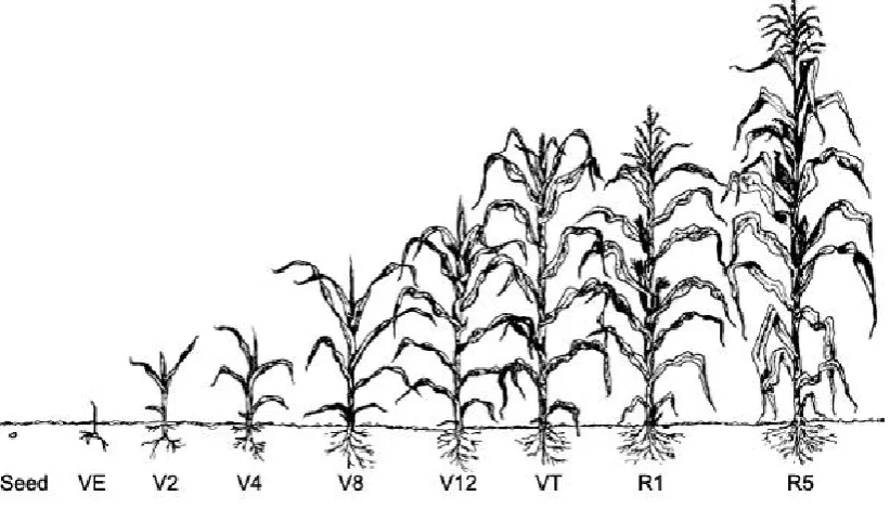
each assay). ... 24 Figure 2.8 Maize plants growing in growth chamber room prior to pathogen application. ... 25 Figure 2.9 Leaves are counted from the base but exclude the growing tip. V4 growth stage
selected for disease application. Diagram courtesy of Purdue University, USA.
https://extension.entm.purdue.edu/fieldcropsipm/corn-stages.php\... 26 Figure 2.10 a) Black pen marks show the area that was treated with plant pathogen. Arrows
indicate treated area. b) Applying the pathogen S. turcica. ... 27 Figure 2.11 Plants after treatment with S. turcica were kept humid for 48 hours to allow
pathogen to germinate and grow. ... 28 Figure 3.1 Example of different morphologies growing on PDA from maize roots. ... 30 Figure 3.2 Microscopy characteristics used in identification of isolates starting from top left; A)
magnification, D) Sporangium of Mucor fragilis (isolate 19) indicated by red arrow. 20X magnification, E) spores of Sordaria fimicola (isolate 36) red arrow indicating ascospores with eight asci present 40X magnification, Fa) the large spores of Curvularia trifolii (isolate 50) with Fb) the spores still attached on conidiophore (red arrow). 40X magnification, G) Chlamydospore forming in Fusarium equiseti (isolate 52). 20X magnification, H) spores of Ascochyta pinodes (isolate 68). 40X
magnification. ... 32 Figure 3.3 Summary of isolates cultured from each genus identified from screening of maize
plants. ... 33 Figure 3.4 Diagram of the number of genera identified from each location as it was sampled
from the maize plant. Source:
https://extension.entm.purdue.edu/fieldcropsipm/corn-stages.php ... 35 Figure 3.5 Number of isolates recovered in each genus from each location of the maize plant 36 Figure 3.6 Epicoccum nigrum morphologies. A) from plant 4 and B) from plant 5 C) top row
with 5 different morphologies of the same isolate subcultured on the same
medium ... 37 Figure 3.7 Isolate 71, microscopy confirmed identification as Fusarium sterilihyphosum. A)
Macroconidia and microconidia spores, Scale bar= 20 µm. B) Coiled hyphae, 40X magnification. ... 38 Figure 3.8 Microscopy confirmed isolate 76 was Fusarium tricinctum with A) distinct napiform
or tear drop microconidia (red arrows) and B) chlamydospore (circled) with spores in a chain. ... 39 Figure 3.9 Dual culture of putative endophytes showing impact the plant disease S. turcica A)
Mucor hiemalis (15) restricting the growth of the S. turcica, B) Trichoderma atroviride (95-8) showing inhibition zones around S. turcica and where the isolate plugs meet. C) Fusarium graminearum (9A) showing definitive inhibition zones-and D) Fusarium avenaceum (60A) also showing distinct inhibition zones. ... 40 Figure 3.10 Bionectria ochroleuca (isolate 13) and B) B. bassiana (isolate J18) increased the
pathogen growth (right) compared to control (left-c1). ... 40 Figure 3.11 Effect of S. turcica on maize leaves pre-treated with selected fungal isolates or
untreated (control). Two cultivars P0021 and 38V12 were scored with a disease severity rating (0-5) and the ratio of disease severity determined compared to control plants. ... 43 Figure 3.12 Growth of Setosphaeria turcica and individual putative endophyte isolates in dual
culture tests. Control = S. turcica alone. Green line represents the average control (S. turcica growth alone) measurement of all assays at 7 days. Maximum possible growth is 8 cm- the width of a Petri dish (Refer to Appendix B.1 for all 79 isolate names). ... 44 Figure 3.13 In planta effects on plant disease severity in assays A- E with different isolates tested
Figure 3.14 A) Cultivar 38V12 showing pathogen treated leaves from plants grown from seed coated with Epicoccum nigrum (24). Score rating for V2 (small leaf) was five. The V3 (large leaf) (right) scored three. The second photo B) shows the left leaf from pot 495. The cultivar used was 38V12, and the seed treated with-Fusarium proliferatum (isolate 51). The treatment applied to the leaf was 0.01 % Triton X 100 (control). The right leaf was from pot 457, cultivar P0021, with the pathogen applied to seed treated of-Penicillium brasilianum (119). The score rating for the right leaf was three. ... 47 Figure 3.15 Maize leaves V2 and V3, from cultivar 38V12, showing visible signs of disease
extending beyond marked lines of pathogen application. Seed was treated using Fusarium equiseti (129B). V2 score =5 (necrosis of whole leaf) and V3 score =4 (leaf tip not showing necrosis but visible lesion within the marked application area). ... 48 Figure 3.16 Helicoverpa armigera feeding in detached leaf assay. A) Left plate where showing
when more food was added and the right plate had been freshly set up. B) H.
armigera caterpillar after 14 days feeding. ... 49 Figure 3.17 Isolates grouped by Abbott’s corrected mortality of Helicoverpa armigera larvae
after being fed on leaves of maize grown from treated seeds. Weights were
Chapter 1
Introduction
1.1
Maize in agriculture
Maize (Zea mays, Poaceae) has been domesticated from teosinte-a wild grass, approximately
7-10,000 years ago and is thought to have originated from Mexico. The Oxford dictionary describes the
word ’maize’ as being derived from mahiz-Taino and maíz-Spanish around the mid-16th Century. The
term maize and corn today are interchangeable and generally have the same meaning. The exception
relates to the geographical location the crop is grown. For example in Scotland and Ireland corn
means oats, in England corn means any cereal crop including wheat, while in the USA and Canada
corn and maize mean the same.
The term maize today is specific to Z. mays but applies to the whole plant; grain (kernel), stem, leaf
and roots. Z. mays is more commonly referred to as maize in the scientific and farming sectors (dairy
sector) in NZ, especially when the plant is used for other commercial agricultural products. The word
corn is more often associated with food products (human consumption), especially when the grains
are used in cereals as in popcorn. For this thesis the term maize will be used and applies to all
growing parts of the plant.
Worldwide maize is an important food crop for both humans and other animals. In NZ maize is grown
for both silage and grain with the end products primarily for stock food (58%) and the remainder for
human consumption and the industrial processing sector (42%) (Booker 2009). The popularity of
maize as a supplementary stock food for the dairy industry has grown rapidly in NZ (Booker 2009;
Millner and Roskruge 2013). Maize grain and silage have a higher food value than more traditional
feed like grass-produced silage or hay with a good cost to dollar/kilogram return (Densley et al.
2003). When making silage, the whole maize plant is shredded and packed into a large stack (bun)
and stored anaerobically until winter feeding. For grain, the maize seed is harvested off the kernel or
cob, stored in silos and is later milled for poultry, pigs and cattle including dairy cows.
Dairy farming is an economically important industry in NZ, providing 37% of total primary industry
export value, which equates to $13.2 billion to the economy (Dairy NZ-Quick stats
http://www.dairynz.co.nz/media/3142896/QuickStats-about-dairying-new-zealand.pdf). NZ provides
3% of the world’s milk products and the industry employs over 48,000 people. Given the importance
of dairy stock to the economy, selected strains of maize are being developed to enhance the feed
quality by DuPont Pioneer® and Advanta seeds (previously known as Pacific seeds), specifically
enhance the plant’s ability to withstand disease, insect and/or climate pressures while retaining good
production such as growth and biomass.
1.2
Important pathogens affecting maize in NZ
There are numerous fungal diseases of maize however only a few diseases commonly occur in NZ.
The diseases covered in this thesis include; 1-Setosphaeria turcica (formerly known as
Helminthosporium turcicum) causing the disease Northern leaf blight (NLB) also known as Northern
corn leaf blight, 2-Aureobasidium zeae (Eyespot) and 3-Diseases caused by Fusarium spp.
1-Setosphaeria turcica
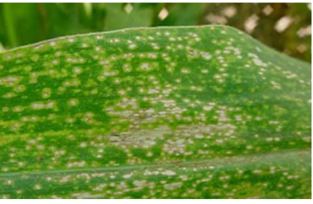
S. turcica causes the disease NLB in maize and is widespread throughout NZ (Perkins 1987). The
fungus overwinters in crop residue and the spores are spread by the wind, capable of travelling long
distances. Symptoms include distinct cigar shaped lesions on the leaf and visible grey shades may be
present on the underside of the leaf. Fungicide treatment is not usually an option as there are few
fungicides registered for use and by the time the disease is noticed the crop is too tall to spray. The
disease NLB decreases the yield and while commercial companies like DuPont Pioneer® continue to
develop resistant strains the disease can still infect the plants resulting in decreased yields (Lipps et
al. 2004).
Figure 1.1 NLB damage in a maize crop. Photo courtesy of DuPont Pioneer®.
2-Aureobasidium zeae
Aureobasidium zeae causes a disease called Eyespot that is spread by wind and moisture (Arny et al.
1971; Munkvold and Martinson 2001). It survives overwintering in crop residue with spores produced
on the underside of the leaves (Lipps and Mills 2005). The disease attacks the maize leaf sheaths and
the leaves covering the ears, causing leaf necrosis and interfering with leaf photosynthesis resulting
in reduced growth, reduced yields and dieback (Arny et al. 1971; Lipps and Mills 2005).
Figure 1.2 Eyespot in maize. Photo courtesy of DuPont Pioneer®.
https://www.pioneer.com/home/site/us/agronomy/crop-management/corn-insect-disease/eyespot/
3-Fusarium spp.
Fusarium spp. affects the quality and yield of maize (Czembor et al. 2015). Species of Fusarium, such
as F. verticillioides and F. proliferatum, produce mycotoxins, which are secondary metabolites. The
main mycotoxins are deoxynivalenol (DON), zearalenone (ZON) and fumonisins which can affect
stock health (Czembor et al. 2015; Fink-Gremmels 2008). The mycotoxins can cause health problems
if consumed by humans and livestock varying from minor upsets through to life threatening
conditions, e.g. liver cancer in humans (Czembor et al. 2015). The extent of the contamination may
mean the plant and/or crop may have to be discarded and is no longer suitable as stock food
Figure 1.3 Fusarium ear rot causes damage to the corn ear and produces mycotoxins. Photo courtesy of DuPont Pioneer®. https://www.pioneer.com/home/site/us/agronomy/crop-management/corn-insect-disease/corn-ear-rots/
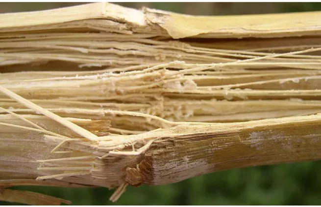
There are numerous species of Fusarium responsible for several diseases in maize targeting the stalk,
root, ear or kernels, e.g. Fusarium ear rot is visible on the tip of the ear or cob (Fig. 1.3) and Fusarium
stalk rot (Fig. 1.4). Fusarium graminearum (telemorph-Giberella zea) can also result in seed rot,
seedling blight, root rot and ear rot (Asran and Buchenauer 2003). Fusarium moniliforme, F.
verticillioides and F. proliferatum can all cause seedling diseases such as seedling blight and seed rot,
root and crown rot, stalk and ear rot (Munkvold 2003; Nelson 1992). More than ten Fusarium spp.
cause seedling blight, wilts, seed and root rots on maize such as F. oxysporum, F. equisetti, F.
culmorum, F. acuminatum, F. graminearum (Asran and Buchenauer 2002; Leslie et al. 2008;
Munkvold 2003).
There are two main diseases affecting the ear and kernels (Munkvold 2003). Giberella stalk rot is
usually caused by F. graminearum but Fusarium culmorum can also be responsible (Czembor et al.
2015). The kernels display a distinct pink colour from the tip towards the base and may encompass a
large area of the ear (Munkvold 2003). The other disease is Fusarium ear rot (Fig. 1.3) caused by
several Fusarium spp.; F. verticillioides (syn. F. moniliforme), F. proliferatum and F. subglutinans, all
Figure 1.4 Fusarium stalk rot breaks down the stem causing death of the plant. Photo courtesy of DuPont Pioneer®.
https://www.pioneer.com/home/site/us/agronomy/crop-management/corn-insect-disease/fusarium-stalk-rot/
1.3
Insect pests of NZ maize
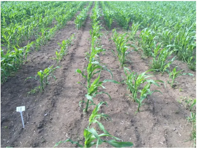
There are four main insect pests of maize in NZ; 1-Argentine stem weevil (ASW) (Listronous
bonariensis) (Coleoptera), 2-Greasy cutworm (Agrotis ipsilon) (Lepidoptera), 3-Corn earworm
(Helicoverpa armigera) (Lepidoptera) and 4-Grass grub (Costelytra zealandica) (Coleoptera).
1-ASW is commonly found throughout NZ (PestWeb 2014; Watson 1981). The adults feed externally
on plant leaves; lay their eggs which hatch and the subsequent larvae then burrow toward the base
of the stem targeting the growing point of the plant (Watson and Hill 1984). The larval stage has the
most economic impact with only one larva at the leaf mining stage capable of killing up to four plants
by eating the centre or growing point of the tiller (Fig. 1.5). Treatment is difficult as spraying the
insects is ineffective at the larval leaf miner stage as chemicals cannot penetrate the leaf surface. The
rate of sowing seeds is denser in forage crops than for seed crops. This makes it easier for pests to
travel between plants in forage crops. To date control has been through seed treatment and pasture
management. The previous season’s plant debris must be removed before planting but this still does
not offer full protection (OEPP/EPPO 1989; PestWeb 2014; Watson and Hill 1985; Watson 1981).
Allowing the sun to dry the crop residue between tractor workings is also a suggested as a method of
Figure 1.5 Argentine Stem Weevil damage to a maize crop. Photo courtesy of DuPont Pioneer®. http://www.pioneer.co.nz/news/2016-08-30/new-research-highlights-seed-treatment-returns.html
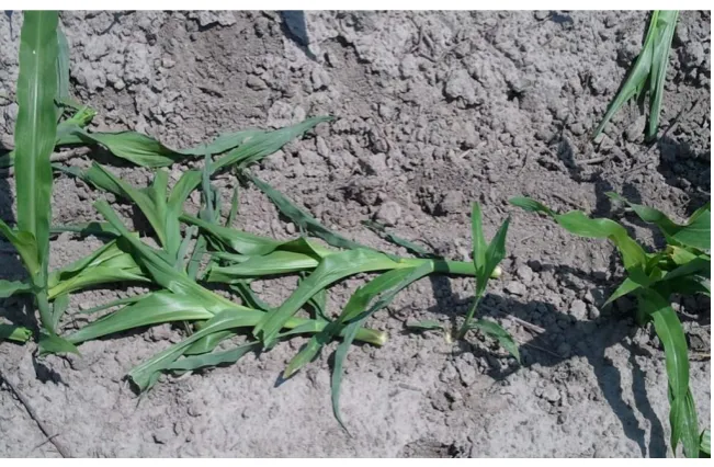
2-The greasy cutworm adult is a nocturnal moth found throughout NZ but the population tends to
fluctuate in densities and may not be seen in one season but prolific in the next (Addison 2007). In
epidemic outbursts the peak time of damage is October through to April and severe damage can
wipe out entire rows of maize. From the 1st to 3rd instar stages the caterpillars feed on the leaves
(Watson 1981). By the 3rd and 4th instar stages, where the larvae can cause the most damage, the
lava may burrow inside the plant then sever the whole plant off at ground level (Fig. 1.6). This makes
it hard for farmers to determine actual levels of infestation in the crop. One caterpillar alone can
destroy large areas in young crops with, in warmer climates, up to three generations completed in a
year. (Addison 2007; Watson 1981). The height of maize plants can make chemical control difficult,
especially when combined with the larval behaviour burrowing underground therefore making
application of pesticides ineffective.
The ASW and greasy cutworm survival into the next season is dependent on overwintering in crop
debris (PestWeb 2014; Watson 1981). Cultural control methods such as reduction of debris left in
paddocks and paddock rotations can be used as a management tool to limit pest numbers but these
Figure 1.6 Greasy cut worm damage seen as plant severed and lying flat on the ground. Photo courtesy of North Carolina State University, https://entomology.ces.ncsu.edu/field-corn-insects/scouting-and-thresholds/scouting-for-seedling-insects/
3-The corn earworm is a polyphagous agricultural pest and is common throughout the world. It is
attracted to the fruiting parts of a wide range of crops but prefers maize when available (Fefelova
and Frolov 2008). Eggs are laid on the upper leaf blade or silk, the caterpillar then travels to the corn
ear and kernel (preferably when at the milk-wax stage of the seed) to feed however it will also eat
the leaves. The corn earworm will normally complete an average of six instars before pupation into
an adult moth. Control of the corn earworm is difficult as the larvae are protected inside the kernel
and resistance has developed to some chemical sprays (Cameron and Walker 2004). Chemical
pesticides are expensive and the crop height of maize limits spraying, especially when the ear
develops. The ear development is usually close to harvest time and spraying may not always be
permitted as the chemical residue may also remain on the crop. The end use of the maize, i.e.
whether for human or animal consumption, will determine if application is acceptable.
4-The grass grub is a native beetle of NZ and a pest of pastures and crops (Young et al. 2009). While
the adult will eat leaves, the majority of the damage done to maize is caused by the larvae (Jackson
et al. 1990; Townsend 2002; Young et al. 2009). The larvae consume the plant roots, causing the
death of the plant, then move on to the next plant. The larvae will eat the roots from seedling stage
right through to mature plants (Cliffe 2011; Jackson 1990; Young et al. 2009). The larvae have three
instar stages with the 2nd and 3rd instar causing the most damage, occurring between March and July
(Cliffe 2011; Jackson 1990; Townsend 2002). The larvae will develop either in a one or a two year
larvae move closer to the surface by the 3rd instar and will be within the top 2 cm by late
summer-early winter (Jackson 1990; Young et al. 2009). Potentially large areas of pasture are affected
depending on the density of grubs. Damage is seen as bare patches in pastures and crops, some
weeks after, making it more difficult to control and prevent.
Control can be difficult, depending on the larval instar stage, as the chemical pesticide does not
penetrate deep enough into the soils (Cliffe 2011; Townsend 2002). Cultural control methods include
cultivation of affected paddocks by ploughing, heavy rolling or stock tramping, or increasing the
number of stock in the mob. These methods can all help reduce numbers but in severe infestations
insecticide sprays may be needed. Insecticides will only be effective when the larvae are towards the
soil surface (Cliffe 2011; Jackson 1990; Townsend 2002). Seeds can be coated with insecticides to
prevent damage while the seedlings establish but this is effective only short-term lasting just one
season. Trials with the bacterium Serratia entomophila as a Biological Control Agent (BCA) had
limited success (Jackson 1990; Popay et al. 2003; Young et al. 2009). Jackson (1990) suggested the
addition of S. entomophila used as a seed coating should be used in conjunction with cultural
practises such as grazing management and using grass resistant seed strains. Young et al. (2009) used
a seed coating technique of two entomopathogenic bacteria, S. entomophila and Yersinia sp. with
the addition of S. entomophila dramatically increasing the seedling establishment of wheat in NZ.
Farrell and Stufkens (1977) noticed while maize was attacked by the grass grub it was to a lesser
degree than grasses and clovers and the grubs themselves did not have the same growth rates. A
different approach by Popay et al. (2003) was with the use of a fungal endophyte in meadow fescue
(Fescue pratensis). They trialled the fescue with and without the endophyte, Neotyphodium
uncinatum, and measured for bioactivity by larvae weight of grass grub. They found with fescue
plants containing endophytes had fewer roots eaten and the larvae had lost weight. This approach
suggests a beneficial endophyte added to a different crop such as maize may be useful for a pest and
disease management approach.
1.4
Endophytes
The term endophyte literally means inside the plant (Greek for endon -inside and phyton -plant) and
has been used broadly to include a wide variety of lifestyle traits (Schulz and Boyle 2005). An
endophyte can be a bacterium or fungus and live within the plant tissues without causing harm or
disease and in fact may give a benefit to the host (Rodriguez et al. 2009; Zakaria et al. 2010).
Endophytes can occur in both above and below ground plant tissues often forming a mutualistic
relationship with plants where the fungus gains protection from the environment with freely
may receive benefits such as enhanced protection from pests and diseases and/or there may be an
increase in stress resistance (Araujo et al. 2000; Raman et al. 2012; Rodriquez et al. 2009; Zakaria et
al. 2012). There is debate over the use of the term ‘endophyte.’ For example mycorrhizal fungi can
be beneficial and can live both externally and internally but are usually not referred to as endophytes
(Hardoim et al. 2015; Hyde and Soytong 2008). Whereas ectomycorrhizal fungi are fungi that live
outside and grow into the rhizosphere but can also colonise within the plant roots but these have
been referred to as endophytes by Rodriguez et al. (2009). The term ‘endophyte’ may also include
different lifestyle traits such as the fungus being a latent pathogen or as part of the reproductive life
cycle the fungus may exit the plant, especially on senescence, to reproduce and sporulate (Hardoim
et al. 2015; Hyde and Soytong 2008; Stone et al. 2017).
In the review by Rodriquez et al. (2009) endophytic fungi were categorised according to taxonomy:
clavicipitaceous (C) containing class 1 endophytes and nonclavicipitaceous (NC) containing classes
2-4. The criteria for each class was characterised by the tissues colonised, the amount of colonisation
within the tissues, transmission-horizontal or vertical, and the host range.
The C group of endophytes include the grass endophytes which have been well studied, such as
Epichloë spp. (anamorph = Neotyphodium). These endophytes are naturally occurring in grasses
including ryegrass (Lolium perenne). The endophytes produce alkaloid toxins, lolitrem B and
ergovaline, causing conditions such as ryegrass staggers and heat stress respectively. Other alkaloids
(loline and peramine) produced by the endophytes have more of a deterrent effect on insects than
on grazing stock (Popay et al. 2012). These endophytes, such as Neotyphodium lolii used in the
development of AR37 by AgResearch NZ, have been selected that show minimum toxicity to sheep,
cattle, dairy cows and horses (Hume et al. 2004; Popay et al. 2012). Since the development of
inoculating endophytes into grasses, specific endophytes have been selected to deter or reduce
insect population specific to the target pasture pests. For example an endophyte (AR37) which
produces more peramine than lolitrem B can deter attack and feeding from ASW, Porina (Wiseana
spp.) and root aphids (Aploneura lentisci) in perennial ryegrass and also potentially increasing the
plants fitness (Charlton and Stewart 1999; Clay 1989; Popay and Hume 2011; Popay et al. 2012).
While the NC fungi are much less studied they are a more diverse group. Both above and below
ground colonisation of plant tissues can occur. The NC group are further divided into three functional
classes; life history, ecological interaction and other traits (Rodriquez et al. 2009). Classification of
the fungi into classes is by traits such as; host range based on which tissues have been colonised,
through the type of reproductive structures, whether the fungus is transmitted vertically and/or
horizontally between plants, or whether the species diversity is low or high (Rodriquez et al. 2009;
fungus-plant interaction can be gained and this knowledge may then be applied to the selection of
endophytes specific to pest and/or diseases in chosen plant species. Some examples like Beauveria
bassiana, Lecanicillium lecanii and Metarhizium anisopliae are already available as commercial
products to control insects by direct chemical applications. The previous fungi are known as
entomopathogenic fungi. As the name suggests they are pathogenic to insects but it has recently
been shown they are also capable of endophytic colonisation (Vidal and Jaber 2015). This is an
important area as both B. bassiana and M. anisopliae, among others, have been and are continuing
to be developed for biocontrol agents (BCA) for agriculture crops.
It has been suggested by Pan and May (2009) that the fungal endophytic community is a
well-structured assembly of different fungi which differ between internal sections of the plant such as the
leaves, stem and the roots. Some endophytes are known to colonise different plant tissues (Hardoim
et al. 2015). This is referred to as multiple habitat levels (Pan and May 2009). For example,
Trichoderma spp. colonise roots while Alternaria spp. have been reported to colonise the stems or
leaves. Others such as Epicoccum and Fusarium species have been found in seeds from fresh cobs of
maize as well as other areas of the plant (Fisher et al. 1992). Rodriquez et al. (2009) note the leaves
in tropical forests contain numerous independent infections and are of high diversity.
Colonisation by endophytes is variable and depends on the host plant, environmental conditions and
fungal species and strain (Carrol 1988: Hardoim et al. 2015; Rodriguez et al. 2009). Vidal and Jaber
(2015) suggested the fungal isolate-host relationship is extremely important to have the right
combination for the endophyte to establish in the host plant. It is suggested that even the strain of
the fungus and the soil mix has an influence on success of endophytic colonisation. The method of
colonisation is just as variable, occurring by entry into the roots, stems or leaves or by vertical
transmission or horizontal transfer (Bais et al. 2006; Hardoim et al. 2015; Rodriguez et al. 2009; Stone
et al. 2017). The endophyte may travel systemically through the plant colonising different tissue
types but where it colonises depends on the specific endophyte and the species of host plant
(Hardoim et al. 2015; Rodriguez et al. 2009).
1.4.1
Maize endophytes
Endophytes are known to occur in maize but there is little information on the diversity of naturally
occurring endophytic species, which plant tissues they colonize or whether they offer the plant
protection from pests and disease (Araújo et al. 2000; Vidal and Jaber 2015). Furthermore, it is
unknown if there is any correlation of endophyte genotypes with geographical location between
The stem and leaf flag of maize were the focus of a study by Fisher et al. (1992) in UK, which looked
at both bacterial and fungal endophytic communities without any visible sign or symptoms of
disease. They found that when the fungal diversity was low there was a high bacterial diversity
present in the cobs of maize plants. Fisher et al. (1992) also examined the vertical transmission and
tissue specificity of certain fungal and bacterial populations. Healthy plants were selected to test if
populations of fungi and bacteria co-exist in maize. Their results suggest the distribution patterns of
these organisms were different between the lower plant, core or pith of the stem and the leaf and
tip areas of the plants studied. They noted that the majority of the bacterial and fungal species show
a high degree of tissue specificity. For example bacteria were found in the core of the stem closer to
the ground while more fungal colonies were recovered towards the lower and middle parts of the
stem. An example was Alternaria alternata which was associated exclusively with the leaves. Pan and
May (2009) studied the internal plant habitat of fungal communities and tested the lower leaf, ear
stalk and upper leaf for whole communities in maize, using both culture dependent and culture
independent methods for interspecific common patterns as well as community assembly. They found
that interspecific interactions affect the endophyte community species composition but this is
influenced by the host’s habitat as well. The fungi found in the previous study contain multiple life
history traits of pathogens, latent pathogens, saprophytes and endophytes. Determining which state
each fungi has presented depends on the site found (internal or external of the plant as well as
leaves or roots) and the time each are found (i.e. close to senescence or if the plant was diseased or
not) (Carroll 1998; Fisher et al. 1992; Schultz and Boyle 2005).
An interesting study by Darvas et al. (2011) looked at the interaction of the caterpillar Helicoverpa
armigera and the disease causing fungus Fusarium verticillioides in genetically modified maize. They
noticed the insect did not grow beyond the third larval instar stage and it would try to move away
from the fungus in the cobs. They also noted that, while the larvae did transfer the fungus to other
places, there was no development of maize pink ear rot disease.
Roots and kernels were examined for endophytes in the study by Seghers et al. (2003). This study
looked at how agricultural practises, such as agrochemical use, can influence endophytic
communities of both bacteria and fungi groups. The authors used plating techniques and counted
colony forming units (CFU’s) todetect communities. DNA was taken from soil and plant (roots and
kernels) samples, followed by denaturing gradient gel electrophoresis (DGGE) to fingerprint the
endophytic community for comparisons between the two groups. The overall results showed
agricultural practises do influence certain populations of the root endophytic communities. The
highest diversity was found in soils with natural organic fertilisers applied while the soils with
Rodriquez et al. (2009) reviewed numerous studies and concluded that endophytes can increase the
plant fitness to abiotic and biotic stresses. The plant’s fitness to cope with insects and disease is more
tolerant if endophytes are present. The review also concluded that endophytes can be specific to
host plant species and the tissue colonised. A study by Singh et al. (2011) also adds to the evidence of
endophytes contributing to the plant’s ability to cope with stress. Their study looked at both class 1
and class 2 endophytes with stress tolerance. They looked at the plants’ ability to cope with drought,
heat, water stress and salinity in conjunction with the mechanisms involved in that response with
different endophytes and hosts plant species. They concluded the effect varied depending on host
species and which endophyte was present, along with which stress was applied.
The studies mentioned above did not sample the ‘whole’ plant but concentrate on specific areas. It is
unknown if these endophytes are the same throughout the plant. For the present study, the plant
was sampled over the entire length of the plant and repeated for each plant sampled. It is hoped a
more conclusive overall picture from the whole plant can be determined from these results and
determine if species are throughout the plant or specific to certain areas.
1.4.2
Endophytes and disease
A study by Zakaria et al. (2010) found 110 fungal isolates from the rice paddy plant (Oryzae sativa)
with the isolates being found from all plant habitat levels. The study looked at healthy plants with no
obvious disease symptoms and to isolate the naturally occurring fungi. Interactions between
endophytes and host plant may change from mutualistic to pathogenic depending on stress factors
or vice versa hence the reason for selecting healthy plants. Fisher et al. (1992) and Schultz and Boyle
(2005) have also cited latent pathogens, found in their studies, becoming pathogenic given the right
conditions such as Fusarium, Curvularia, Penicillium and Aspergillus. Zakaria et al. (2012) suggested
that the latent pathogens found in plants, if put under stress, may become pathogenic. It is therefore
possible that latent pathogens may be found to be naturally occurring in maize plants and would be
hard to distinguish from endophytes without further investigation (Fisher et al. 1992; Zakaria et al.
2010).
The discovery of the endophytes in grasses led to the isolation and selection of strains which produce
higher levels of alkaloids, (secondary metabolites produced by the endophytes), in response to
herbivory (Popay and Hume 2011).One main endophyte genus concerned is Epichloë spp. but the
name ‘Epichloë’ now generally refers to both sexual states (Clay 1989; Kuldau and Bacon 2008).
Inoculating endophytes into grasses can result in the development of specific mutualistic associations
(Kuldau and Bacon 2008). Endophytes are commonly associated with agriculture pasture grasses
(Poaceae) such as ryegrass (Lolium perenne) and fescue species (Festuca spp.). The potential for
development of these endophytes in new host combinations can only add and benefit agriculture
(Easton and Fletcher 2007).
1.4.3
Endophytes and insects
As mentioned above, some endophytes are also referred to as entomopathogenic fungi
(Guesmi-Jouini et al. 2014; Vidal and Jaber 2015). A well-known and studied example is B. bassiana that has
the ability to inhibit plant pathogens as well as insect development (Ownley et al. 2004; Wagner and
Lewis 2000). The addition of an entomopathogenic fungus within a plant has the potential to control
a number of pests. Numerous insects have been studied with the use of entomopathogenic fungi in
different plants and crucially, the plant remains unaffected (Bing and Lewis 1993; Bruck 2010;
Ownley et al. 2004). One such study applied B. bassiana by spraying directly onto maize plants and
found the application to be effective at controlling European corn borer (Ostrinia nubilalis) (Ownley
et al. 2004). The authors also noted B. bassiana was found later to be endophytic in the plant.
Bionectria ochroleuca and B. bassiana have been found as endophytes in artichokes (Cynara
scolymus) and known to have high virulence on the artichoke aphid (Capitophorus elaeagni)
(Hemiptera: Aphididae) (Guesmi-Jouini et al. 2014; Raman et al. 2012). While this study showed the
endophytic capabilities of the fungi it does not mention any effect against insects when the fungus
was added as an endophyte to the plant.
Therefore selection of an endophyte for a BCA will depend on the effect and targeted insect as well
as the fungal strain and host plant compatibility. The chosen endophyte must be capable of
endophytic colonisation in the host plant.
1.5
Research objectives
Little is known of the endophytic community of the entire maize plant rather studies focusing on
particular areas. In this study, putative endophytes were isolated from within the plant tissues from
roots to the tip of the plant. For the purpose of this study the term endophyte refers to any fungus
found within the plant after surface sterilisation with no visible disease symptoms on the plant. This
could include a latent pathogen as well as beneficial endophyte however the latent pathogen can
exist in a plant asymptomatically until a trigger ‘switches on’ the fungus to become disease causing
the study but more important is the location as coming from ‘within’ the plant. This makes the
surface sterilising methods (Chapter 2) extremely important to remove the possibility of anything
other than the isolate being an endophyte. Out of interest, each fungus identified has been reviewed
for the possibility of more than one lifestyle trait (Appendix D1).
In this study it was looked at what part of the plant the endophyte colonised and if there was a
difference between fungal groups. Two geographical areas (Canterbury and Waikato), NZ, were
tested to determine if areas differed in species composition with five sites per area being tested.
Fungi were identified by DNA extraction then sequenced and confirmed where possible by
morphological traits to species level. The sequenced results given from BLAST and UNITE were above
98% and a consensus from the both sites. A further aim of the project was to determine if any of the
identified fungi have potential to act as a BCA. The aim was to find a naturally occurring endophyte
or endophytes that could deter or kill insects and/or prevent disease. The isolate could be taken up
through the seed coating to become endophytic in the plant. This could greatly enhance farmer’s
reliance on chemical sprays (less acceptable in today’s environment) and could be an
environmentally friendly pest management tool (Ownley et al. 2004; Seghers et al. 2004). It was
mentioned earlier (Pests section-pg. 7) that H. armigera have a preference for maize plants (Fefelova
and Frolov 2008). For this reason and ease of supply, H. armigera was used for bioassays. The
pathogen S. turcica was selected to test against plants inoculated with potential endophytes. This
disease has been used previously in another study from our Laboratory with some good results.
Samples of infected plants were tested for evidence of endophytic presence in each insect and
disease trials.
It has been known endophytes are in seeds with a study by Fisher et al. (1992) finding 68% of seeds
used for planting their crop contained endophytes. However young seeds obtained from the mature
cobs only returned 7%. The seed supplied by DuPont Pioneer® were tested to check for the baseline
Chapter 2
General methods
2.1
Introduction
In this section the methods are given for the processing of the plants (2.2) through to identification
(2.2.4), and then application and testing of isolates against plant pathogens (2.4) and insect pests
(2.5). Methods are also provided for the screening for background endophytes (2.6) on the seed
resident endophyte community supplied by DuPont Pioneer®.
2.2
Sampling field collected maize plants for endophytes
Screening of NZ maize plants for endophytes was done over two seasons (2014 and 2015 summer
seasons). Two regions, Waikato and Canterbury, were selected for screening to identify endophytes
naturally occurring within maize plants. Through the Foundation of Arable Research (FAR), maize
crops were sampled, by taking whole plants and processing for isolation of fungi present (Table 2.1).
Plants collected by FAR from the North Island (Waikato) were sent by courier while the Canterbury
plants were collected by myself. Five plants per site, with plants coming from six different farms from
two geographical regions per season (November to May) were collected, for a total of 22 plants in
2014 and 12 plants in 2015. The processing time, by one person, before a plant showed visible
saprophytic growth was less than one week, even with cold storage. This limited the number of
plants that could realistically be sampled at one time within a season.
2.2.1
Recovery of putative endophytes from maize
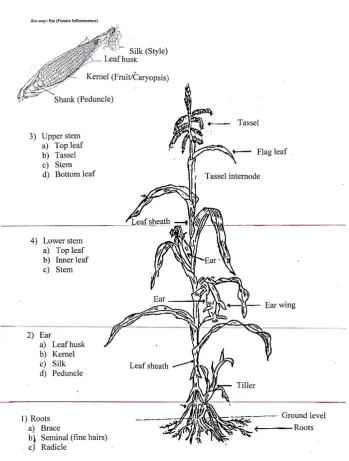
Maize plants were divided into sections (Fig. 2.1); 1-roots, 2-ear, 3-upper stem (which included the
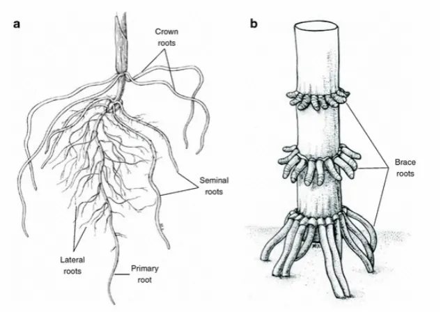
flower if present) and 4-lower stem. Each section was further subdivided. Roots were divided into
three sections; seminal, radicle (also called lateral roots) (Fig. 2.2a) and brace (adventitious roots)
(Fig. 2.2b). The radicle roots contain fine hairs and are found spread throughout the root system (Fig.
2.2a). Samples were taken at random for radicle and seminal root samples. The samples for the brace
roots were taken from the brace node (Fig. 2.2b), as close as possible to the stem just above the
ground, extending to below the soil surface with sufficient length to give multiple tissues samples for
A selection of root samples were taken at random and plated onto agar as a group containing 2-4
roots (Fig. 2.3) with five tissue samples arranged per plate. The three types of root samples were
kept separate with two replicate plates for each root type giving a total of six plates, thirty root tissue
samples in total, sampled from one plant.
The ear was divided into four sites with enough tissue sample taken and plated on agar from each
site. Each of the four sites from the ear (Fig. 2.1-2a-d) had five tissue samples arranged per plate (Fig.
2.4a) and two replicate plates were tested per plant giving a total of 40 tissue samples. The leaf husk
(Fig. 2.1-2a), a group sample of silk (Fig. 2.1-2c) consisting of approximately ten hair thickness,
kernels (Fig. 2.1-2b) and a section of the peduncle (Fig. 2.1-2d) cut in half length wise depending on
the thickness to fit inside a Petri dish were all plated on agar.
Table 2.1 Origin of maize plants for sampling for 2014 and 2015 seasons.
Site
Region
Variety of seed used
Date planted
Sample plant
number
2014
1 Waikato Pioneer P0021 25/10/2013 1-5
2 Waikato
Pioneer 34N41,
Waxy hybrid 10/10/2013 6
3 Canterbury, Ashburton 38V12 9/10/2013 7-11
4 Waikato P0021 hybrid 8/10/2013 12-13
5 Canterbury, Tram Road 39T45 15/10/2013 14-17
38v12 25/10/2013 18-20
6 Canterbury, Oxford
Super sweet NZ yellow
55630, F1 hybrid NA 21-22
2015
1 Waikato P0021 13/10/2014 1-5
2
Canterbury, PGG Wrightsons Farm,
Lincoln NA NA 6-10
3
Canterbury, Lincoln
University (LU) Corson 15/12//2014 11-12
NA- not available
The lower (Fig. 2.1-4a-c) and upper stems (Fig. 2.1-3a-d) contained top and bottom leaf samples from
each section taken close to the stem as well as a section of stem tissue again cut in half to be thin
enough to fit within a Petri dish. The upper stem region (Fig. 2.1-3a-d) had four areas sampled from
the very tip of the plant testing the tassel (Fig. 2.1-3b), then the top leaf (Fig. 2.1-3a), a section of
stem (Fig. 2.1-3c) and a lower leaf (Fig. 2.1-3d) in this area. Five tissue samples per agar plate with
2.1-4a-c) had three tissue samples taken from the top leaf (Fig. 2.1-4a) inner leaf (Fig. 2.1-4b) and the
stem (Fig. 2.1-4c), again five tissue sample per plate (Fig. 2.4.b), with two replicate plates on agar
giving a total of 30 tissues samples from this region. The same process was repeated for each plant
making sure the same area was sampled for every plant from each geographical region in each
season.
2.2.2
Surface sterilisation of maize tissues
The term surface sterilisation for this study refers to all external microbes killed or removed from the
exterior of the maize plant tissue. The internal tissues remain unharmed allowing the endophyte to
exit when plated on potato dextrose agar (PDA) (Difco, NJ). Firstly all soil residues were rinsed off
with tap water and air dried. Each tissue sample was processed following the protocol below. For
surface sterilisation, samples were sequential placed in a deep Petri dish containing: 0.01% Triton
X-100 (BDH) solution for three minutes, agitated gently 2-3 times, removed and placed into the next
Petri dish containing 2% of sodium hypochlorite (bleach; Cyclone-Diversey) for five minutes, then
into 70% EtOH (ethanol) for one minute followed lastly by three rinses in sterile water (dH20) for one
minute each. Samples were placed on sterile paper towel to soak up residual water and allowed to
air dry in laminar flow for 2-3 minutes before cutting and plating. The plant tissue samples were
gently mixed in the solutions to ensure samples were completely submerged. Solutions were
changed after 3-4 samples with all solutions replaced between each plant.
After surface sterilisation, the plant tissue samples were cut into approximately two-three
centimetre segments with the sterile ends cut off and discarded. Five pieces were placed on PDA
after surface sterilisation (Fig. 2.4a & b). More tissue was surface sterilised than needed for plating so
tissue samples were taken at random and placed on the plates without coming into contact with
neighbouring tissue samples. Plates were incubated at 25°C @ 16 hour light, 8 hour dark cycle for 3-4
days or until visible fungal growth was present (Fig 2.4b).
Control plates: A sample of the 0.01% Triton X-100 solution was plated after washing the tissues to
establish the fungal community present in each plant before they had been sterilised. This was
considered the positive control. Three plates were taken at random from the water used to rinse the
plant tissue for fungi and bacteria presence. These plates were expected to have no bacteria or
fungal colonies hence these were called the negative control. Checking was done by pipetting 100 µl
of the rinse water and with a cell spreader (Biologix) spread onto a PDA plate. Two other controls
tested the sterility of the chopping board and the external plant tissue after surface sterilisation. This
compared to an unsterile plant tissue rubbed over a PDA plate. On first use of this method controls
were positive with one fungal colony and some bacterial contamination therefore times were
increased for Triton X-100 from one to three minutes and bleach changed to five minutes. Protocols
were adjusted until plates were clear i.e. no bacterial or fungal growth after 5 days incubation at
25°C.
Figure 2.2 Diagram of maize roots; a) showing lateral or radicle roots with fine hairs and the seminal roots- without hairs. b) The brace roots (also known as adventitious or crown roots) may extend from the first or second node to the ground giving the plant another bracing element as it gets taller Diagram from Hochholdinger (2009).
Figure 2.3 Multiple tissue samples per site of roots showing mycelium
2.2.3
Media
Samples of surface sterilised maize sections as prepared above were plated on PDA. It is known that
some bacteria interferes or inhibits fungal growth (Fisher et al. 1992). This study found it was
necessary to add antibiotics to inhibit bacteria (Fig. 2.4b) from some samples. Antibiotics were added
to PDA were five hundred mg of chlortetracycline hydrochloride (Sigma) and 2500 mg streptomycin
sulphate salt (Sigma) per 100 ml of sterile water. The solution was filtered through a sterile 0.2 µm
filter before adding to the PDA. Antibiotics were used at a rate of 1 ml solution to 100 ml agar to give
whereas F. oxysporum have microconidia produced on false heads on short phialides formed on
hyphae and F. trinctum (Fig. 3.9A) produces a distinct tear drop microconidia spore (Leslie et al.
2008). Microscopy identified conidiation patterns e.g. B. bassiana produces a mass of white ‘cotton
balls’, on a zig zag rachis (spore structure) specific to B. bassiana.
Where multiple samples gave morphological identification as the same species from the same plant,
only one representative culture was taken for identification. Pure cultures were processed for
molecular identification.
2.2.5
DNA extraction
Two to three day old pure fungal cultures were used for DNA extraction. A small amount of hyphae
was placed in 500 μl of Chelex 100 Resin buffer (biotechnology grade 5% or 2 g in 40 ml of deionized
H2O). The cells were ground with a sterile pestle then vortexed thoroughly. The tubes were
incubated by boiling in water for 12 minutes then cooled to room temperature before centrifuging at
13,000 rpm for 20 minutes. The middle and top clear layer of 100-200 μl was transferred to a new 1.7
ml eppendorf tube, avoiding any cell debris or pellet. DNA was stored at minus 20°C.
2.2.6
Polymerase chain reaction
Two μl of the solution was used for polymerase chain reaction (PCR). Each 25 μl PCR reaction
consisted of: 15.75 μl of sterile water, 2.5 μl of buffer (10x) plus MgCl2 (2 mM), 2 μl of
deoxynucleotide (dNTP’s) (2.5 mM) (Roche), 0.25 μl of Fast start polymerase Taq (Roche), 1 μl of
each primer (Integrated DNA technologies (IDT)), 0.5 μl bovine serum albumin (BSA, Bio Labs) and 2
μl of extracted DNA per sample. General primers were used which targeted the internal transcribed
spacer ITS region of the 16s rDNA using the reverse primer ITS4-5’ TCCTCCGCTTATTGATATGC 3’and
forward primer ITS5- 5’ GGAAGTAAAAGTCGTAACAAGG 3’ (White et al. 1990). Thermocycling
conditions were 95°C for 5 min followed by 40 cycles of 95°C for 45 sec, 57°C for 45 sec, 72°C for 2
min and final extension of 72°C for 7 min. PCR products were visualised on a 1% agarose gel and
stained by ethidium bromide or Red safe nucleic acid staining solution for 10 minutes. Samples with
single bands present were then sent for sequencing (Lincoln University (LU) Sequencing Unit) and
analysed using BLASTN through the NCBI website and UNITE (https://unite.ut.ee/analysis.php) to
2.2.7
DNA analysis
Initially 86 fungi from plant tissue samples were sequenced using both reverse and forward primers
(ITS 4 and ITS 5). To keep costs down the remainder were completed with the reverse ITS 4 primer
only. In some cases the quality of the reverse sequence was insufficient, therefore a further 14
samples were re-analysed using both primers to get a consensus. Morphology was also used in
identification but it was also necessary to have isolates confirmed by sequencing for each plant
location (Appendix A. 6) from each plant sampled and for each year.
The top five to eight hits from both UNITE and BLAST were taken to get a consensus. The E-values
returned were 0.0 with a query cover greater than 98%. Ideally the isolate identity was greater than
98% but if the identity returned between 92-98% then a consensus of both BLAST and UNITE was
used where possible. The unidentified (“unknown”) isolates that did not amplify in PCR or gave
multiple names at genus level when compared to databases, or BLAST hits of ‘uncultured fungus’,
were marked as unknown. Where possible with unknowns the DNA extraction and PCR were
repeated to get an identification.
2.3
In vitro bioassay of recovered putative endophytes against a plant
pathogenic fungus
Dual culture assays were chosen as a method for screening putative endophytes isolated in the initial
screening in maize plants (Appendix A. 1 and A. 2). The maize pathogen, S. turcica, causing NLB was
chosen to assess the bioactivity of putative endophytes as it is a major disease in maize and can
result in up to 30% yield loss in susceptible hybrids (Fowler 1985; Perkins and Pederson 1987). S.
turcica was found to have varied growth rates between cultures grown on PDA. Previous research
also indicated S. turcica was slow to form conidia. For this reason an excess number of plates were
cultured and over a longer time frame.
For dual culture assays, isolates were sub-cultured onto fresh PDA. Plates were selected with
sufficient mycelial growth covering the majority of the plate to obtain enough plugs for the isolates
to be tested in each assay. Plugs from both the endophyte isolate and the plant pathogen were
placed onto ¼ strength PDA plates (9.75 g PDA + 10 g of bacteriological agar). A template (Fig. 2.5a)
was used to mark the placement on every plate for the isolate and the pathogen. Two, 5 mm plugs,
were placed opposite each other for both isolate and pathogen (Fig. 2.5b) on each PDA plate with
three replicates per treatment. Where the isolate and pathogen were similar in colour the pathogen
was marked with a ‘P’ (Fig. 2.5b). The control plates consisted of 4 plugs on a plate with three
Figure 2.5 a)-The template used for marking the base of PDA plates and b)-placement of pathogen (P).
Dual culture assays were executed in batches of 7-9 isolates tested at a time. All plates were grown
at a constant 22°C. Isolates were grown in the presence of the plant pathogen, S. turcica, and growth
of the colonies was measured for both pathogen and isolate at seven and 10 days from inoculation. A
total of 79 isolates were tested (Appendix B.1). A number of isolates did not survive the initial
sub-culturing or storage at 4°C.
Figure 2.6 The axis of measurements marked across the widest part of each colony, Alternaria alternata (12) and plant pathogen S. turcica.
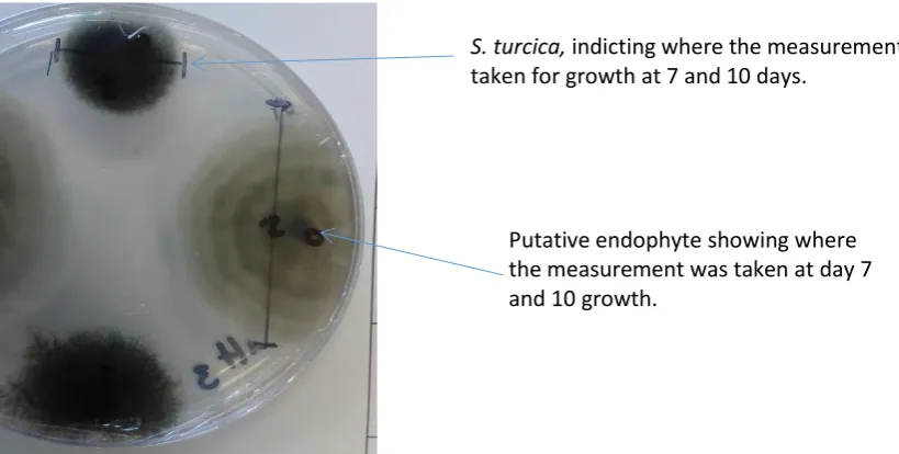
Measurements were recorded across the width of each growing isolate colony (Fig. 2.6). A control
plate of the pathogen only was grown at the same time. Each isolate was compared to the growth of
the pathogen grown on its own (Fig. 2.7).
a b
Site of pathogen placement
S. turcica, indicting where the measurement was taken for growth at 7 and 10 days.
Figure 2.7 Setosphaeria turcica control used to compare each isolate (new control used with each assay).
2.4
In planta maize challenge methods
2.4.1
Seed coating
The aim of seed coating was to establish isolates as endophytes and potentially promote plant
growth and disease and insect resistance.
Spores were harvested in sterile 0.01% Triton X100 solution with haemocytometer counts of
approximately 10⁶-10⁸ spore/ml to treat maize seeds. The seed coating polymer mix (Flo Rite 3330,
BASF) was used, following the protocol of 100 g of seed in 0.56 g polymer + 0.56 g of spore
concentration. Equal volumes of 200 µl polymer and 200 µl of spore suspension were mixed and 35
µl pipetted onto the ten seeds. The seeds were gently agitated until all seeds were evenly coated
with the polymer-spore solution and container walls were free of solution. The seeds were air dried
for approximately 3-5 minutes. For the controls, seeds were coated with a 50:50 mix of 0.01% Triton
X and polymer solution. The seeds were placed in the fridge overnight to encourage all seeds to
germinate at the same time.
2.4.2
Plant disease assay
To determine the effect of endophytes in disease resistance, the known maize pathogen, S. turcica,
was selected as a disease challenge. As a result of the dual culture assays, 21 isolates were selected
for in planta testing (Table 3.4). A preliminary experiment was conducted to ensure that the strain of
S. turcica was able to generate disease symptoms on these maize varieties. After the preliminary
testing it was decided to use both P0021 and 38V12 hybrid seeds supplied by FAR (DuPont Pioneer®)
of disease with one being highly affected by lesions and necrosis of plant tissue and 10 having no
effect. DuPont Pioneer® performance characteristics rated cultivar P0021 at a 7/10 for disease
resistance to NLB and 6/10 for cultivar 38V12.
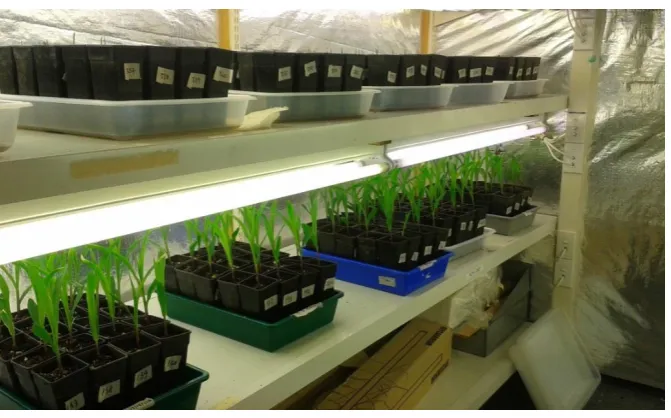
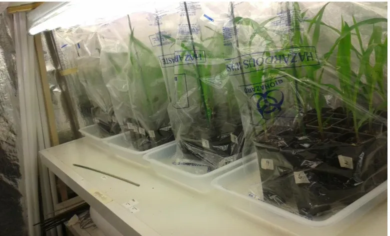
Figure 2.8 Maize plants growing in growth chamber room prior to pathogen application.
After seed coating (method as above) and storage in the fridge overnight, the seeds were removed
from fridge and rested, to acclimatise to room temperature before planting. One seed per pot (0.5
litres) was planted using 3-4 month old potting mix supplied by the LU nursery, Springs Road, Lincoln.
The pots were placed in a growth chamber at 20-25°C and grown to V4 stage (Fig. 2.9). Prior to the
pathogen application plants were kept in separate trays for each seed isolate treatment (Fig. 2.8)
until the pathogen was applied. The experiment was conducted five times with different endophyte