Range of Neurologic Disorders in Patients With Celiac Disease
Nathanel Zelnik, MD; Avi Pacht, MD; Raid Obeid, MD; and Aaron Lerner, MD
ABSTRACT. Objective. During the past 2 decades, ce-liac disease (CD) has been recognized as a multisystem autoimmune disorder. A growing body of distinct neu-rologic conditions such as cerebellar ataxia, epilepsy, myoclonic ataxia, chronic neuropathies, and dementia have been reported, mainly in middle-aged adults. There still are insufficient data on the association of CD with various neurologic disorders in children, adolescents, and young adults, including more common and “soft” neurologic conditions, such as headache, learning disor-ders, attention-deficit/hyperactivity disorder (ADHD), and tic disorders. The aim of the present study is to look for a broader spectrum of neurologic disorders in CD patients, most of them children or young adults.
Methods. Patients with CD were asked to fill in a questionnaire regarding the presence of neurologic dis-orders or symptoms. Their medical charts were reviewed, and those who were reported as having neurologic man-ifestations underwent neurologic examination and brain imaging or electroencephalogram if required. Their neu-rologic data were compared with that of a control group matched for age and gender.
Results. Patients with CD were more prone to de-velop neurologic disorders (51.4%) in comparison with control subjects (19.9%). These disorders include hypo-tonia, developmental delay, learning disorders and ADHD, headache, and cerebellar ataxia. Epileptic disor-ders were only marginally more common in CD. In con-trast, no difference was found in the prevalence of tic disorders in both groups. Therapeutic benefit, with glu-ten-free diet, was demonstrated only in patients with transient infantile hypotonia and migraine headache.
Conclusion. This study suggests that the variability of neurologic disorders that occur in CD is broader than previously reported and includes “softer” and more com-mon neurologic disorders, such as chronic headache, de-velopmental delay, hypotonia, and learning disorders or ADHD. Future longitudinal prospective studies might better define the full range of these neurologic disorders and their clinical response to a gluten-free diet. Pediat-rics 2004;113:1672–1676; celiac disease, neurologic disor-ders, migraine, attention-deficit/hyperactivity disorder, hypotonia.
ABBREVIATIONS. CD, celiac disease; ADHD, attention-deficit/ hyperactivity disorder.
A
lthough in the past celiac disease (CD) wasprimarily considered to be a gluten enterop-athy, during the past 2 decades, its clinical concept has been expanded, and it is now considered a multisystem autoimmune disorder,1with most of the patients being asymptomatic, oligosymptomatic, or present with extraintestinal manifestations.2 Among these extraintestinal manifestations, there is a growing body of publications that report neuro-logic conditions that are associated with CD.3–12 Al-though earlier studies reported neurologic complica-tions in patients with classical gluten enteropathy, some recent studies report neurologic disorders in asymptomatic CD patients.13,14Most of the patients who have CD and were reported as having neuro-logic manifestations were adults, and these manifes-tations were usually chronic and “hard,” such as epilepsy,12 cerebellar ataxia,4,5,13,14chronic neuropa-thies,8,15 myoclonic ataxia,9 progressive leukoen-cephalopathy,7and dementia.16
The aim of this study was to screen for neurologic disorders in children and young adults who have CD and presented with either the classical infantile in-testinal form or the milder late forms, including some asymptomatic patients. We searched for both hard neurologic conditions mentioned above and more common conditions, such as headache, learn-ing disabilities and attention-deficit/hyperactivity disorder (ADHD), developmental delay, hypotonia, and tic disorders.
METHODS
We recruited from the local pediatric gastroenterology clinic all of the patients who had proven CD and were enrolled in our gastroenterology outpatient clinic between 1977 and 2001. Until 1987, our criteria for the diagnosis of CD were based on 3 consec-utive intestinal biopsies demonstrating initially mucosal flattening with typical inflammatory changes, mucosal normalization seen with the second biopsy after strict gluten-free diet, and relapse of pathologic changes with gluten challenge. Since 1988, our diag-nostic approach for CD has been simplified and was based on the demonstration of immunoglobulin A antiendomysial antibodies and 1 intestinal biopsy showing pathologic changes typical of CD. The diagnosis was confirmed when the antiendomysial antibodies disappeared on gluten-free diet.17All patients (or their caregivers) received a questionnaire with a checklist on which they were asked to report on neurologic symptoms or conditions that re-quired medical attention or treatment. Patients with suspected neurologic signs or symptoms underwent a full neurologic eval-uation and laboratory examinations, including brain imaging and electroencephalogram if required. We sent a similar questionnaire for a group of non-CD subjects who were matched for age and gender and underwent a neurologic evaluation if required. In both groups, the diagnosis of the neurobehavioral conditions such as ADHD or specific learning disabilities was based on the diagnostic criteria ofDiagnostic and Statistical Manual of Mental Disorders,18 and the diagnosis of migraine was based on the revised Interna-tional Headache Society classification.19The study was approved
From the Department of Pediatrics, Carmel Medical Center, The Bruce Rappaport Faculty of Medicine, Technion-Israel Institute of Technology, Haifa, Israel.
Received for publication Apr 15, 2003; accepted Aug 7, 2003.
Reprint requests to (N.Z.) Department of Pediatrics, Carmel Medical Center, 7 Michal St, Haifa 34362, Israel. E-mail: nzelnik@netvision.net.il
by the local Helsinki Committee, and informed consent was ob-tained from all subjects (or caregivers).
RESULTS Patients and Control Subjects
Our population consisted of 322 subjects; 111 pa-tients with CD who answered the questionnaires and agreed to take part in this study and 211 subjects, matched for age and gender, who served as controls. The mean ages of the patients with CD and the control subjects were 20.1⫾ 8.9 years (42.3% male, 57.7% female) and 20.1 ⫾ 9.0 years (40.3% male, 59.7% female), respectively. These differences were not significant.
Initial Manifestations of CD
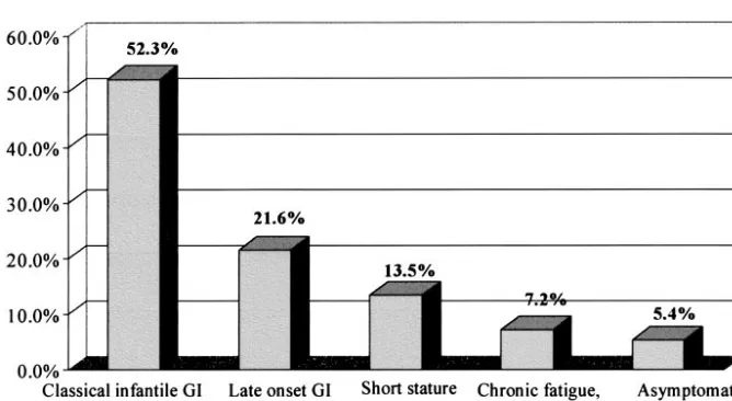
Fifty-eight (52.3%) patients presented with the classical infantile features of CD, such as chronic diarrhea, malabsorption syndrome, failure to thrive, or abdominal pains. The mean age of diagnosis of the infantile form of CD was 1.8⫾0.9 years. CD in the other 53 (47.7%) patients was diagnosed later in life, with a mean age at diagnosis of 14.8⫾8.9 years; 24 (21.6%) had chronic abdominal pain or other gastro-intestinal symptoms without history of infantile di-arrhea or failure to thrive. In 9 patients in this group, the gastrointestinal symptoms were associated with additional findings such as anemia, short stature, and delayed sexual maturation. Fifteen (13.5%) pa-tients received a diagnosis of CD during their eval-uation for short stature. An additional 8 (7.2%) pa-tients had variable manifestations, including isolated anemia (hemoglobin ⬍9 g/dL), chronic fatigue, or irregular menstrual cycles. Six (5.4%) patients were “asymptomatic,” and the search for the diagnosis of CD was made when CD was found in other first-degree relatives (Fig 1).
Neurologic Manifestations
Neurologic disorders or findings were found in 57 (51.4%) patients with CD: 22 with a single manifes-tation, 23 with 2 manifestations, 7 with 3 manifesta-tions, and 5 with 4 manifestations. In contrast, only 42 (19.9%) control subjects reported the presence of neurologic disorder: 26 with a single manifestation,
12 with 2 manifestations, and 4 with 3 manifesta-tions. None had ⬎3 manifestations. Although pa-tients with the late-onset form of CD were somewhat more prone to develop neurologic disorders than patients with the classical infantile CD (54.7% vs 48.3%), this difference was not statistically significant (P⫽.5).
Hypotonia
Hypotonia was recorded from medical files of 16 patients with a history of classical infantile CD. Re-peated examinations revealed that with the excep-tion of 3 patients, 1 of whom had Down syndrome, the hypotonia completely resolved after years of a gluten-free diet. Four patients presented with short stature, and 4 patients had chronic abdominal pains, chronic fatigue, or anemia. Two patients in this group who were still found hypotonic had low se-rum carnitine levels, and with dietary supplements and reinforcement of the gluten-free diet, their symp-toms improved. In the control group, 5 subjects were reported as hypotonic only in infancy and early childhood. Three are still hypotonic, with mental retardation (1 with Down syndrome; Fig 2).
Developmental Delay
Infantile symptoms of CD were present in 12 (70.6%) patients in this group, whereas chronic ab-dominal pain or late-onset gastrointestinal symp-toms, anemia, and short stature were the presenting symptoms of CD in 5 patients. All of the patients in this group had additional neurologic disorders. Ten (58.8%) patients had learning disabilities and/or ADHD (3 with mental retardation), and 2 were ataxic. Five (29.4%) patients were hypotonic during infancy, and their neurologic impairment resolved with a gluten-free diet. One patient had cerebellar ataxia, and another patient had epilepsy. In both patients, the neurologic problems did not seem to respond to dietary management (Fig 2).
Epilepsy and Other Seizure Disorders
Four patients with CD had benign febrile seizures during infancy, and 4 had nonfebrile seizures: 1 with
benign partial epilepsy, another with a single unpro-voked nonfebrile seizure, and 2 others with chronic epilepsy (including 1 patient with intractable epi-lepsy associated with occipital cerebral calcifica-tions). Three control subjects had benign infantile febrile seizures, and 3 had epilepsy (1 with partial complex seizure and 2 with generalized tonic-clonic seizures; Fig 2).
Learning Disabilities and ADHD
In contrast to the usual male preponderance in children with these conditions, among patients with CD, male and female patients were almost evenly affected: 13 (20.3%) of 64 female patients and 10 (21.2%) of 47 male patients. Ten patients presented with the classical infantile form of the disease, and 13 presented with late-onset symptoms (including 1 asymptomatic patient). Three patients had mental retardation, and 3 patients had seizures. Among the patients with mental retardation, 1 had Down syn-drome associated with moderate mental retardation; 1 had epilepsy with occipital calcification; and an-other had mild mental retardation and autism, but no specific cause was found. In the control group, male subjects were predominantly affected: 11 (12.9%) of 85 versus 11 (8.7%) of 126 among female subjects. Two control subjects had mental retarda-tion, 1 with Down syndrome (Fig 2).
Headache
The female/male ratio was 21:10 in the CD group and 14:3 in the control group. Headache was the most commonly found neurologic disorder in our patients with CD. Twenty (64.5%) patients with headache presented with the late-onset symptoms of CD or were asymptomatic, and 11 (35.5%) patients had the classical early infantile form of CD. In 14 (45.1%) patients, the headache filled the criteria for migraine; in 6 (19.4%) patients, the clinical character-istics of the headache were compatible with tension-psychogenic headache; and 11 (35.5%) patients had nonspecific headache. In 16 patients (9 with
mi-graine, 6 with nonspecific headache), the symptoms resolved or significantly improved with the institu-tion of a gluten-free diet. In the control group, 12 subjects had migraineous headache, 3 subjects had tension headache, and 2 subjects had nonspecific headache (Fig 2).
Cerebellar Ataxia
Six patients were ataxic; 3 with ataxia presented with classical infantile CD and 3 others with late-onset symptoms. No correlation was found between the activity of CD and the ataxia. Two patients had cerebellar atrophy; 2 patients had isolated mild ataxia with normal brain imaging; and 2 patients had mental retardation 1 of whom also had autistm. The clinical syndrome consisted of stance and gait ataxia in all patients, limb ataxia in 4 of 6 patients, and nystagmus in 3 of 6 patients. In addition, 4 patients were hypotonic and 2 had sensory neuropathy. None of the 211 control subjects had ataxia (Fig 2).
Tic Disorders
One patient had chronic tic disorder. He also had ADHD and short stature, which led to the diagnosis of CD. One control subject had full-blown Tourette syndrome. Four others had milder forms of simple or chronic tics (Fig 2).
DISCUSSION
The present study clearly demonstrates a strong association between CD and various neurologic manifestations. With the exception of tic disorders and marginally epileptic disorders, all of the other neurologic features were significantly more common in patients with CD (P ⬍.01).
tomatic CD. This finding differs from the trend, which has become widely accepted, to regard CD as gluten sensitivity, found in patients with neurologic disorder and atypical or subclinical CD.13,20
Although hypotonia and developmental delay were more characteristic of the classical infantile-onset CD and in these cases were probably caused by nutritional deficits and toxic effects of severe malab-sorption, most of the other neurologic manifestations were more evenly distributed between early and late onset of CD. In the late-onset forms, they could be related to prolonged exposure to gluten with its mul-tisystem immunologic and inflammatory effects. Clear cause and effect of a gluten-free diet was dem-onstrated mainly in cases of infantile hypotonia, as-sociated with the classical early-onset CD. This tran-sient clinical syndrome could be a nonspecific result of chronic disease and poor nutritional condition or caused by specific nutritional deficiencies, such as lowered levels of vitamin E, vitamin B12, or carnitine, which all were previously reported in patients with CD.21–23 In most of these cases, the hypotonia has been resolved with the improvement of the nutri-tional status of the patients.
Patients who have CD with migraineous or non-specific headache were also markedly improved with the institution of a gluten-free diet. Among these patients, only 11 (35.5%) had the classical early infantile enteropathic CD, whereas 20 (64.5%) pre-sented with late-onset symptoms. Hence, it is clear that malabsorption did not play a significant role in the pathogenesis of headache, and one should look for other causes, including inflammatory or immu-nologic mechanisms. There are only a few reports on the association of CD and headache. Battistella et al24 reported on 2 young patients with migraine-like headache and cerebral calcifications, 1 of whom had CD. Roche-Herrero et al25reported that 39.5% of the children and adolescents with CD manifested head-ache. All were on a gluten-free diet. Recently, Gab-rielli et al26 demonstrated that a significant propor-tion of patients with migraine had subclinical CD and that their symptoms improved with a gluten-free diet.
This is not the case in epileptic disorders or learn-ing disabilities and ADHD. Our epileptic patients were not homogeneous, and strong association with CD probably existed only in the patient with epi-lepsy and occipital calcifications. In all of the other patients, the presence of epilepsy or seizures could be only an incidental finding. Nevertheless, even in this group, one cannot rule out the possibility that CD with gluten toxicity may have played some role in triggering seizures in susceptible subjects. In this respect, Gobbi et al12 has shown that seizures in patients with the syndrome of cerebral calcification, epilepsy, and CD responded to therapy only when the gluten-free diet was initiated shortly after the emergence of epilepsy. In contrast, another study reported of an adequate seizure control even when the diagnosis of epilepsy was made after a few years of exposure to gluten.27
Before this study, the association of ADHD and learning disabilities with CD was not recognized. It
is not clear whether accumulative effects of nutri-tional, immunologic, or inflammatory factors might play some role on learning abilities or attention span in our patients or that the effect is indirect and relates to nonspecific effects of chronic disease. Kieslich et al28reported on the presence of multiple white mat-ter lesions detected by brain magnetic resonance im-aging of patients with CD. One cannot rule out that these lesions might also interfere with high cognitive functions, such as in other white matter diseases.29In regard to mental retardation, both epilepsy with oc-cipital brain calcifications and Down syndrome were previously reported in CD.12,30,31In contrast to these findings, the association between CD and autism is poor and probably does not exist.32
Cerebellar ataxia has been well recognized in pa-tients with CD, primarily in adults.4,5,13,14 Two pa-tients in the study group were also adults, who were diagnosed at 23 and 51 years of age. The first reports emphasized the role of vitamin E deficiency in these patients.33,34 Subsequently, there was a study that reported a patient who had cerebellar syndrome with no evidence of nutritional deficits.6During the last decade, there was a general trend to adopt the concept that in many cases, an immunologic mecha-nism underlies the brain damage, including 1 case in which magnetic resonance imaging studies clearly demonstrated cerebellar (and cerebral) lesions.11 In this context, we suggest that in some patients, chronic immune-mediated inflammation, lympho-cytic infiltration, or vasculitis of the central nervous system might cause irreversible neuronal, glial, or axonal damage, with little clinical improvement even after the institution of a gluten-free diet and cessa-tion of the various autoantibodies in the peripheral blood or the cerebrospinal fluid. However, other studies advocated that the presence of gluten sensi-tivity might contribute to the clinical picture of pa-tients who already have preexisting hereditary atax-ias.35,36
We conclude that the spectrum of neurologic dis-orders in patients with CD is wider than previously appreciated and includes, in addition to previously known entities such as cerebellar ataxia, epilepsy, or neuromuscular diseases, milder and more common problems such as migraine headache and learning disabilities, including ADHD.
REFERENCES
1. Lerner A, Blank M, Shoenfeld Y. Celiac disease and autoimmunity.Isr J Med Sci.1996;32:33–36
2. Branski D, Ashkenazi A, Freier S, et al. Extraintestinal manifestations and associated disorders of celiac disease.Front Gastrointest Res. 1992; 19:110 –121
3. Cooke WT, Smith WT. Neurological disorders associated with adult celiac disease.Brain.1966;89:683–722
4. Finelli PF, McEntee WJ, Ambler M, Kestenbaum D. Adult celiac disease presenting as cerebellar syndrome.Neurology.1980;30:245–249 5. Kinney HC, Burger PC, Hurwitz BJ, Hijmans JC, Grant JP. Degeneration
of the central nervous system associated with celiac disease.J Neurol Sci.
1982;53:9 –22
6. Ward ME, Murphy JT, Greenberg GR. Celiac disease and spinocere-bellar degeneration with normal vitamin E status.Neurology.1985;35: 1199 –1201
7. Kepes JJ, Chou SM, Price LW. Progressive multifocal leucoencephalopa-thy with 10-year survival in a patient with nontropical sprue.Neurology.
8. Kaplan JG, Pack D, Horoupian D, et al. Distal axonopathy associated with chronic gluten enteropathy; a treatable disorder.Neurology.1988; 38:642– 645
9. Bhatia KP, Brown P, Gregory R, et al. Progressive myoclonic ataxia associated with celiac disease. The myoclonus is of cortical origin but the pathology is in the cerebellum.Brain.1995;118:1087–1093 10. Hadjivassiliou M, Chattopadhyay AK, Davies-Jones GAB, Gibson A,
Grunewald RA, Lobo AJ. Neuromuscular disorders as a presenting feature of celiac disease.J Neurol Neurosurg Psychiatry.1997;63:770 –775 11. Ghezzi A, Filippi M, Falini A, Zaffaroni M. Cerebral involvement in celiac disease: a serial MRI study in a patient with brainstem and cerebellar symptoms.Neurology.1997;49:1447–1450
12. Gobbi G, Bouquet F, Greco L, et al. Celiac disease, epilepsy, and cerebral calcifications.Lancet.1992;340:439 – 443
13. Pellecchia MT, Scala R, Perretti A, et al. Cerebellar ataxia associated with subclinical celiac disease responding to gluten-free diet.Neurology.
1999;53:1606 –1608
14. Burk K, Bosch S, Muller CA, et al. Sporadic cerebellar ataxia associated with gluten sensitivity.Brain.2001;124:1013–1019
15. Kelkar P, Ross MA, Murray J. Mononeuropathy multiplex associated with celiac sprue.Muscle Nerve.1996;19:234 –236
16. Collin P, Pirttila T, Nurmikko T, et al. Celiac disease, brain atrophy, and dementia.Neurology.1991;41:372–375
17. Walker-Smith JA, Guandalini S, Schmitz J, et al. Revised criteria for the diagnosis of celiac disease. Report of working group of European Soci-ety of Pediatric Gastroenterology and nutrition.Arch Dis Child.1990;65: 909 –911
18. American Psychiatric Association.Diagnostic and Statistical Manual of Mental Disorders(DSM-IV). 4th ed. Washington, DC: American Psychi-atric Association; 1994
19. Winner P, Martinez W, Mate L, Bello L. Classification of pediatric migraine: proposed revision to the IHS criteria. Headache. 1995;35: 407– 410
20. Wills AJ. The neurology and neuropathology of coeliac disease. Neuro-pathol Appl Neurobiol.2000;26:493– 496
21. Gordon N. Vitamin E deficiency and illness in childhood.Dev Med Child Neurol.1987;29:541–549
22. Dahele A, Ghosh S. Vitamin B12 deficiency in untreated celiac disease.
Am J Gastroenterol.2001;96:745–750
23. Lerner A, Gruener N, Iancu TC. Serum carnitine concentrations in celiac disease.Gut.1993;34:933–935
24. Battistella PA, Mattesi P, Casara GL, et al. Bilateral cerebral occipital calcifications and migraine-like headache.Cephalalgia.1987;7:125–129 25. Roche-Herrero MC, Arcas Martinez J, Martinez-Bermejo A, et al. The
prevalence of headache in a population of patients with celiac disease.
Rev Neurol.2001;32:301–309
26. Gabrielli M, Cremonini F, Fiore G, et al. Association between migraine and celiac disease: results from preliminary case-control and therapeu-tic study.Am J Gastroenterol.2003;98:625– 629
27. Hernandez MA, Colina G, Ortigosa L. Epilepsy, cerebral calcifications and clinical or subclinical coeliac disease. Course and follow up with gluten-free diet.Seizure.1998;7:49 –54
28. Kieslich M, Errazuriz G, Posselt HG, Moeller-Hartmann W, Zanella F, Boehles H. Brain white-matter lesions in celiac disease: a prospective study of 75 diet-treated patients.Pediatrics.2001;108(2). Available at: pediatrics.org/cgi/content/full/108/2/e21
29. Damian MS, Schilling G, Bachmann G, Simon C, Stoppler S, Dorndorf W. White matter lesions and cognitive deficits: relevance of lesion pattern.Acta Neurol Scand.1994;90:430 – 436
30. Book L, Hart A, Black J, Feolo M, Zone JJ, Neuhausen SL. Prevalence and clinical characteristics of celiac disease in Down syndrome in a US study.Am J Med Genet.2001;98:70 –74
31. Zachor DA, Mroczek-Musulman E, Brown P. Prevalence of celiac dis-ease in Down syndrome in the United States.J Pediatr Gastroenterol Nutr.
2000;31:275–279
32. Pavone L, Fiumara A, Bottaro G, Mazzone D, Coleman M. Autism and celiac disease: failure to validate the hypothesis that a link might exist.
Biol Psychiatry.1997;42:72–75
33. Harding AE, Muller DP, Thomas PK, Willison HJ. Spinocerebellar de-generation secondary to chronic intestinal malabsorption: a vitamin E deficiency syndrome.Ann Neurol.1982;12:419 – 424
34. Mauro A, Orsi L, Mortara P, Costa P, Schiffer D. Cerebellar syndrome in adult celiac disease with vitamin E deficiency.Acta Neurol Scand.
1991;84:167–170
35. Pellecchia MT, Scala R, Filla A, De Michele G, Ciacci C, Barone P. Idiopathic cerebellar ataxia associated with celiac disease: lack of dis-tinctive neurological features.J Neurol Neurosurg Psychiatry.1999;66: 32–35
36. Bushara KO, Goebel SU, Shill H, Goldfarb LG, Hallet M. Gluten sensi-tivity in sporadic and hereditary cerebellar ataxia.Ann Neurol.2001;49: 540 –543
AWKWARDNESS IN ELEVATORS
“Riding in elevators is a daily routine of social awkwardness comparable only to urinal conversation or being left at the altar. That is why the most popular button is the one that closes the doors. Elevator engineers are acutely aware of this.”
Brick M.New York Times. April 2, 2003
DOI: 10.1542/peds.113.6.1672
2004;113;1672
Pediatrics
Nathanel Zelnik, Avi Pacht, Raid Obeid and Aaron Lerner
Range of Neurologic Disorders in Patients With Celiac Disease
Services
Updated Information &
http://pediatrics.aappublications.org/content/113/6/1672
including high resolution figures, can be found at:
References
http://pediatrics.aappublications.org/content/113/6/1672#BIBL
This article cites 34 articles, 10 of which you can access for free at:
Subspecialty Collections
sub
http://www.aappublications.org/cgi/collection/neurologic_disorders_
Neurologic Disorders
http://www.aappublications.org/cgi/collection/neurology_sub
Neurology
http://www.aappublications.org/cgi/collection/gastroenterology_sub
Gastroenterology following collection(s):
This article, along with others on similar topics, appears in the
Permissions & Licensing
http://www.aappublications.org/site/misc/Permissions.xhtml
in its entirety can be found online at:
Information about reproducing this article in parts (figures, tables) or
Reprints
http://www.aappublications.org/site/misc/reprints.xhtml
DOI: 10.1542/peds.113.6.1672
2004;113;1672
Pediatrics
Nathanel Zelnik, Avi Pacht, Raid Obeid and Aaron Lerner
Range of Neurologic Disorders in Patients With Celiac Disease
http://pediatrics.aappublications.org/content/113/6/1672
located on the World Wide Web at:
The online version of this article, along with updated information and services, is
by the American Academy of Pediatrics. All rights reserved. Print ISSN: 1073-0397.