R E S E A R C H
Open Access
Neonatal dexamethasone treatment exacerbates
hypoxic-ischemic brain injury
Kan-Hsun Chang
1, Che-Ming Yeh
2, Chia-Yu Yeh
1, Chiung-Chun Huang
1and Kuei-Sen Hsu
1,2*Abstract
Background:The synthetic glucocorticoid dexamethasone (DEX) is commonly used to prevent chronic lung disease in prematurely born infants. Treatment regimens usually consist of high doses of DEX for several weeks, notably during a critical period of brain development. Therefore, there is some concern about adverse effects of this clinical practice on fetal brain development. In this study, using a clinically relevant rat model, we examined the impact of neonatal DEX treatment on subsequent brain injury due to an episode of cerebral hypoxia-ischemia (HI). Results:We found that a 3-day tapering course (0.5, 0.3 and 0.1 mg/kg) of DEX treatment in rat pups on postnatal days 1–3 (P1-3) exacerbated HI-induced brain injury on P7 by a glucocorticoid receptor-mediated mechanism. The
aggravating effect of neonatal DEX treatment on HI-induced brain injury was correlated with decreased glutamate transporter-1 (GLT-1)-mediated glutamate reuptake. The expression levels of mRNA and protein of GLT-1 were significantly reduced by neonatal DEX treatment. We also found that the administration ofβ-lactam antibiotic ceftriaxone increased GLT-1 protein expression and significantly reduced HI-induced brain injury in neonatal DEX-treated rats.
Conclusions:These results suggest that early DEX exposure may lead the neonatal brain to be more vulnerable to subsequent HI injury, which can be ameliorated by administrating ceftriaxone.
Background
Chronic lung disease (CLD) is an important cause of mor-tality and morbidity in preterm infants and inflammation plays a major role in its pathogenesis [1,2]. Due to their strong anti-inflammatory properties, synthetic glucocorti-coids such as dexamethasone (DEX) or betamethasone are frequently used to prevent or lessen the morbidity of CLD in preterm infants. Given that the brain is a major target for glucocorticoids and the developing brain is inherently more susceptible to drug-induced alterations than the adult brain [3], there is concern that neonatal DEX therapy may be associated with increased risk of adverse neurologic outcomes in later life. While there are some controversies about its adverse effects on neurodevelopment, numerous clinical studies have demon-strated that premature infants receiving DEX therapy have a higher incidence of neuromotor dysfunction and an
increased risk of developing cerebral palsy (CP) [4-7]. There is also experimental evidence that DEX exposure in the neonatal rat pups can lead to alterations in hippocam-pal synaptic plasticity and deficits in learning and memory [8-10]. Although these results highlight the adverse conse-quences of neonatal DEX treatment on brain development, little is known about the molecular mechanisms behind these abnormalities.
Neonatal hypoxia-ischemia (HI) is a leading cause of perinatal brain injury, which may ultimately lead to CP, mental retardation, learning disability and epilepsy [11]. Using different neonatal rat models of HI, previous stud-ies have revealed that HI-induced brain injury is associ-ated with excitotoxicity, a type of neuronal death triggered by overstimulation of glutamate receptors and loss of calcium homeostasis [12,13]. Interestingly, there is evidence that pretreatment of neonatal rats with DEX prevents brain injury associated with cerebral HI [14-16]. These findings contrast with clinical observa-tions that early DEX administration in preterm infants may increase the incidence of CP [4-6]. These seemingly discrepant findings are likely related to variations in * Correspondence:richard@mail.ncku.edu.tw
1Department of Pharmacology, College of sMedicine, National Cheng Kung
University, Tainan 701, Taiwan
2Institute of Basic Medical Sciences, College of Medicine, National Cheng
Kung University, Tainan 701, Taiwan
timing and dosage regimens. It was deemed of interest to perform a detailed analysis of the influence of neo-natal DEX treatment on subsequent HI-induced brain injury by using a protocol resembling the one used in clinical practice for preterm infants. In this study, using a well established and clinically relevant 3-day tapering course of DEX treatment in neonatal rat pups on post-natal days 1–3 (P1-3) [9,14,17,18], we asked two main questions: (1) whether neonatal DEX treatment alters the vulnerability of the immature brain to HI-induced brain injury and (2) if so, what is the responsible mo-lecular mechanism(s).
Results
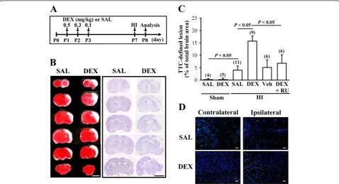
Neonatal DEX treatment enhances HI-induced brain injury To determine the influence of neonatal DEX treatment on HI-induced brain injury, we compared the infarction areas in coronal sections of SAL and DEX groups 24 hours after experimental HI (Figure 1A). We chose this time point because it has been reported to be the peak in expression of neonatal HI-induced cell damage [19].
Figure 1B shows representative images from HI rats stained with 2,3,5-triphenyltetrazolium chloride (TTC) or cresyl violet. Neonatal DEX-treated group exhibited more severe HI-induced brain injury, particularly in the cerebral cortex and the hippocampus, than SAL-treated group. One-way ANOVA revealed a significant main effect of HI treatment on infarct volume (F3,25= 15.3, P< 0.001), and
post hoc analysis showed that infarct volume was signifi-cantly increased (P< 0.05) by neonatal DEX treatment compared with SAL-treated group (Figure 1C). The en-hancement effect of neonatal DEX treatment on HI-induced brain injury was prevented when the animals were given glucocorticoid receptor (GR) antagonist, RU 38486 (40 mg/kg), 1 hour before daily DEX treatment (P< 0.05 vs. DEX). TUNEL analysis was used to determine whether neonatal DEX treatment may sensitize HI-induced cell damage. As shown in Figure 1D, TUNEL-positive apop-totic cells were evident within the frontal cortex ipsilateral to common carotid artery ligation 24 hours after HI. The number of TUNEL-positive cells was significantly greater in DEX-treated group than saline-treated group.
Neonatal DEX treatment decreases GLT-1-mediated glutamate reuptake
Because excitotoxicity due to excessive extracellular glu-tamate is closely associated with HI-induced brain injury [12,13] and transporter-mediated glutamate uptake is es-sential for maintaining low extracellular glutamate con-centrations [20], we therefore examined the influence of neonatal DEX treatment on basal glutamate uptake activ-ity in gliosomes from the frontal cortex on P7. The amounts of total and GLT-1-mediated glutamate uptake in gliosomes were significantly reduced in DEX-treated group compared with SAL-treated group (Total: F1,8=
28.3;P< 0.001; GLT-1: F1,8= 9.8; P= 0.014; Figure 2).
Al-though the amount of non-GLT-1-mediated glutamate up-take tended to be lower in DEX-treated group compared with that from SAL-treated group, the difference did not reach statistical significance (F1,8= 3.7; P= 0.08). In
addition, the observed reduction in basal glutamate uptake activity in DEX-treated group was prevented when rat pups were given RU 38486 (40 mg/kg) 1 hour before daily DEX treatment (F1,14= 21.7;P< 0.001; data not shown).
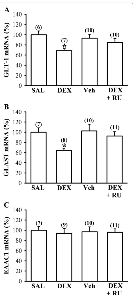
Neonatal DEX treatment decreases basal GLT-1 and GLAST mRNA and protein expression
So far, five distinct mammalian glutamate transporter sub-types, termed EAAT1 (glutamate-aspartate transporter, GLAST), EAAT2 (GLT-1), EAAT3 (excitatory amino acid carrier 1, EAAC1), EAAT4 and EAAT5, have been cloned. GLAST and GLT-1 are found predominantly in glial cells and EAAC1 is expressed in both neurons and glial cells [21]. Because the observed reduction of glutamate uptake
in DEX-treated group could be a result of decreased ex-pression of glutamate transporters, we therefore examined the influence of neonatal DEX treatment on the expres-sion of basal glutamate transporters in the frontal cortex on P7. Quantitative real-time RT-PCR analysis showed that neonatal DEX treatment led to decreased expression of GLT-1 (F1,11= 14.7; P< 0.01) and GLAST mRNAs
(F1,13= 15.5; P< 0.01) compared with SAL-treated group
(Figures 3A and B). The observed reduction in GLT-1 and GLAST mRNA expression in DEX-treated group was prevented when rat pups were given RU 38486 (40 mg/kg) 1 hour before daily DEX treatment (P< 0.05 vs. DEX). However, there was no significant difference between DEX- and SAL-treated group in EAAC1 mRNA expres-sion (F1,14= 0.2;P= 0.63). In parallel, the levels of GLT-1
and GLAST proteins in the frontal cortex were noted to be decreased in DEX-treated group compared with SAL-treated group on P7 (GLT-1: F1,26= 47.3; P< 0.001;
GLAST: F1,11= 13.1;P< 0.01; Figures 4A and B). The
in-hibitory effect of neonatal DEX treatment on GLT-1 and GLAST protein expression was prevented when the ani-mals were given RU 38486 (40 mg/kg) 1 hour before daily DEX treatment (P< 0.05 vs. DEX).
On the basis of the data demonstrating the down-regulation of GLT-1 and GLAST protein levels after neo-natal DEX treatment, it was assumed that reduced GLT-1 and GLAST protein levels may contribute to enhanced HI-induced brain injury by neonatal DEX treatment. To test this possibility, we ran a correlation between them in slices prepared from DEX-treated group. A clear inverse correlation was found between the extent of TTC-defined infarct volume 24 hours after HI and the relative levels of GLT-1 protein (r=−0.55; P= 0.03, n = 15; Figure 4C). In contrast, no such relationship was seen with the relative levels of GLAST protein (r=−0.02; P= 0.96, n = 12; Figure 4D).
DEX treatment decreases GLT-1 mRNA and protein expression in C6 glioma cells
As GLT-1 is expressed predominantly in glial cells [20], further experiments were performed to determine whether DEX treatment may downregulate the expression of GLT-1 in glial cell culturesin vitro. To test this possibility, rat C6 glioma cells were treated with DEX (100 μM) for 24 and 48 hours, respectively. As expected, GLT-1 protein ex-pression in C6 glioma cells was significantly downregulated by DEX treatment, based on Western blotting analysis of whole-cell lysates (24 hours: F1,18= 12.8;P< 0.01; 48 hours:
F1,18= 5.2; P< 0.05; Figure 5A). In contrast, GLAST
protein expression was not altered by DEX treatment (24 hours: F1,18= 0.2; P= 0.67; 48 hours: F1,18= 0.1; P= 0.89;
Figure 5B). In parallel, a significant decrease in GLT-1 mRNA expression was noted in C6 glioma cells of DEX-treated group compared with vehicle-treated group Figure 2Effect of neonatal DEX treatment on glutamate
(24 hours: F1,18= 6.2; P< 0.05; 48 hours: F1,18= 6.4; P<
0.05; Figure 5C), whereas GLAST mRNA level was not al-tered by DEX treatment (24 hours: F1,18= 1.1;P= 0.31; 48
hours: F1,18= 0.1;P= 0.86; Figure 5D).
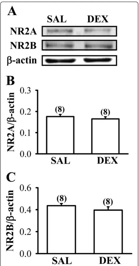
The expression ofN-methyl-D-aspartate receptors is not alter by neonatal DEX treatment
The occurrence of HI-induced excitotoxicity is generally thought to be associated with overstimulation of glutam-ate receptors, particularly the N-methyl-D-aspartate (NMDA) receptor subtype [12,13]. Hence, we determined whether the expression levels of NMDA receptor in the frontal cortex were altered by neonatal DEX treatment. As shown in Figures 6A-C, there were no significant differ-ences between DEX- and SAL-treated group in the ex-pression levels of the two major NMDA receptor subunits, NR2A and NR2B, in the whole tissue lysates of the frontal cortex on P7 (NR2A: F1,14= 0.6; P= 0.45;
NR2B: F1,14= 1.2;P= 0.29).
Ceftriaxone attenuates HI-induced brain injury in neonatal DEX-treated rat pups
The above results clearly indicate the importance of GLT-1 levels in determining the vulnerability of the neonatal brain to HI injury. These findings prompted us to investi-gate whether the elevation of GLT-1 protein expression could attenuate the reinforcing effect of neonatal DEX treatment on HI-induced brain injury. For this, neonatal rat pups were pretreated withβ-lactam antibiotic ceftriax-one (200 mg/kg), that have been shown to effectively exert neuroprotective effect against neonatal HI-induced brain injury through the elevation of GLT-1 expression [22], 1 hour before DEX application (Figure 7A). As expected, we observed that ceftriaxone treatment significantly reduced HI-induced brain injury (F1,26= 5.1; P< 0.05 vs. SAL)
and attenuated the extent of brain damage in DEX-treated rat pups to HI (F1,21= 6.5; P< 0.05 vs. DEX;
Figure 7B). In addition, ceftriaxone treatment substan-tially increased GLT-1 protein expression in the frontal cortex (F1,6= 13.1;P< 0.01 vs. SAL; Figure 7C), the
hippo-campus (F1,6= 6.2; P < 0.05 vs. SAL; Figure 7D) and the
striatum (F1,6= 9.7;P< 0.05 vs. SAL; Figure 7E), and
com-pletely rescued the inhibitory effect of DEX treatment on GLT-1 protein expression in the frontal cortex (F1,8= 9.6; P< 0.05 vs. DEX; Figure 7C) and the hippocampus (F1,6=
7.2; P< 0.05 vs. DEX; Figure 7D). In contrast, GLT-1 protein expression in the striatum was not significantly af-fected by neonatal DEX treatment (F1,6= 0.9; P= 0.39 vs.
SAL; Figure 7E).
Discussion
The concern that neonatal DEX therapy might increase risk of developing neurological dysfunction by altering neural development was an important motivation for Figure 3Effect of neonatal DEX treatment on the expression of
glutamate uptake transporter mRNAs in the frontal cortex.
our study. Using a clinically relevant animal model, our results provide the first evidence that neonatal DEX treatment leads to a sustained downregulation of GLT-1 expression and thereby exacerbates HI-induced brain in-jury. Moreover, we confirm that ceftriaxone can exert a neuroprotective effect against HI-induced brain injury in neonatal rats by increasing expression of GLT-1 [22] and suggest that pretreatment with ceftriaxone in neonatal rats can effectively attenuate DEX-induced augmentation of HI-induced brain injury.
Excitotoxicity related to extracellular accumulation of glutamate plays a critical role in neonatal HI-induced brain injury [23]. The accumulation of extracellular glutamate may result from either decreased uptake or the reversed ac-tion of glutamate transporters [20]. The observaac-tion that DEX-treated group showed a significantly lower GLT-1-me-diated glutamate uptake activity in gliosomes compared with that from SAL-treated group suggests that neonatal DEX treatment may cause decreased glutamate uptake and result in deleterious enhancement of excitotoxic brain in-jury to subsequent HI insults. In addition, we observed that neonatal DEX treatment led to a significant decrease in both mRNA and protein levels of GLT-1 and GLAST, indi-cating that reduced expression of glutamate transporters
may account, at least in part, for the decrease in glutamate uptake observed in P7 rat pups. It is however noteworthy that, although expression levels of both GLT-1 and GLAST were downregulated by neonatal DEX treatment, we observed no significant correlation between the extent of HI-induced brain injury and the expression of GLAST pro-tein. In contrast, our results revealed that GLT-1 protein levels are inversely correlated with the extent of HI-induced brain injury. Therefore, we hypothesize that changes in GLT-1 protein expression underlie the enhancement of neonatal HI-induced brain injury found in DEX-treated rats. This hypothesis is further supported by findings that ceftriaxone treatment attenuates the enhancing effect of neonatal DEX treatment on HI-induced brain injury by in-creasing GLT-1 expression. Moreover, we found no signifi-cant differences in expression of NR2A and NR2B subunits in the frontal cortex between DEX- and SAL-treated groups, suggesting that neonatal DEX treatment does not alter the total amount of NMDA receptors.
how GR activation reduces GLT-1 gene transcription re-mains unclear, it is highly likely that GR acts mainly through interfering with the nuclear transcription factor nuclear factor-κB (NF-κB) signaling to downregulate GLT-1 expression. Using either pharmacological or genetic approaches, the NF-κB signaling has been linked to several drug-induced or neuron-dependent transcriptional activa-tion of GLT-1 [24-26]. Thus, downregulaactiva-tion of NF-κB activity could be associated with a decrease in GLT-1 gene transcription. It has been observed in multiple cell types that DEX can inhibit NF-κB activation by enhancing the cellular levels of IκB-α[27,28] or the protein-protein inter-action between activated GR and the p65 NF-κB subunit [29,30]. Thus, it is reasonable to speculate that DEX may act indirectly by inhibiting NF-κB activity and subse-quently decreasing GLT-1 transcription. However, we could not exclude the possibility that DEX may inhibit transcriptional activation of GLT-1 through a direct DNA binding of activated GR to a specific site in the GLT-1 promoter. Further studies are required to test these possibilities.
We observed that DEX treatment decreases the ex-pression of GLT-1 mRNA and protein in C6 glioma cells
in vitro, consistent with our findingin vivo showing the
reduction in GLT-1 expression in the frontal cortex by neonatal DEX treatment. However, this finding is in con-trast with observations made in a previous study, which reported that DEX provokes an increase of GLT-1 tran-scription and protein levels in cortical astrocytes [31]. The reason for this seemingly contradictory finding is unclear but may be related to differences in experimental design as well as differences in cell types. Zschocke et al. [31] used rat primary cortical astrocytes and examined the extent of GLT-1 induction 72 hours after DEX (100 nM) treatment, whereas the present study examined the expression of GLT-1 in rat C6 glioma cells that were treated with DEX (100 μM) for 24–48 hours. Interestingly, as opposed to GLT-1, expression levels of GLAST mRNA and protein GLAST were not altered by DEX treatment in C6 glioma cells. These observations suggest that these two glial glu-tamate transporter subtypes have different sensitivities to glucocorticoid treatment. Further studies using different dosages of DEX are required to address this issue.
and another way is in combination with other drugs. In the present study, our data revealed the potential clinical benefit of ceftriaxone to ameliorate DEX-induced po-tentiation of HI-induced brain injury. These results are in agreement with recent studies that ceftriaxone can offer neuroprotection in bothin vitroandin vivomodels of ischemic injury and motor neuron degeneration by preventing glutamate excitotoxicity [22,32]. Recent re-ports have also established that ceftriaxone may exert its
neuroprotective effects by inducing GLT-1 transcription through increasing NF-κB binding to the GLT-1 promoter [25,33]. Although ceftriaxone has FDA approved for use in pediatric bacterial meningitis for a long time, there was no evidence of long-term neurodevelopmental sequelae of ceftriaxone treatment in the neonate. Our findings with ceftriaxone suggest that adjunct neuroprotective therapies that elevate GLT-1 activity may minimize glutamate excitotoxicity, thereby allowing a choice of DEX for use in neonates. Further large trials in humans are needed to confirm these results.
Perinatal HI-induced brain injury is one of major causes of CP. There is increasing evidence showing that early DEX administration in preterm infants may increase the incidence of CP [4-6]. In accordance with these clinical findings, the current results show that early DEX exposure is able to increase the vulnerability of the neonatal brain to subsequent HI damage. Although there are some stud-ies indicating that DEX pretreatment can protect neonatal brain against subsequent HI injury [15,16,34], our results do not support a neuroprotective role for neonatal DEX treatment in cerebral HI. One possible explanation of these seemingly discrepant observations is the different doses and regimens of DEX used among studies. These findings reinforce the long-held view that the concentra-tion and duraconcentra-tion of glucocorticoid treatment are major factors determining the beneficial or detrimental effects of glucocorticoids in the brain [35]. In our model, DEX was administered over a long period (P1-3) in tapering doses in an attempt to mimic the longer treatment regimens commonly used in the neonatal intensive care setting [8,9,14,17,18]. While much caution is required when extrapolating data from animal models to the human con-dition, our findings highlight the risk for heightened devel-oping brain vulnerability to HI injury associated with neonatal DEX treatment.
Conclusion
In conclusion, our data provide evidence for a deleteri-ous impact of neonatal DEX treatment on HI injury in the developing brain. Pretreatment wit ceftriaxone, per-haps due to an increase in GLT-1 expression, is able to stimulate glutamate uptake and overcome the excessive HI-induced brain injury resulting from neonatal DEX treatment. Although further investigations are needed to elucidate the molecular mechanisms involved in the GR-mediated downregulation of GLT-1 transcription, our findings demonstrate that supporting GLT-1 expression may exert beneficial effects to ameliorate the lasting en-hancing effect of neonatal DEX treatment on glutamate-mediated excitotoxicity. These findings are of clinical importance because it is now difficult to avoid the use of corticosteroids in neonatology and perinatology to fight the problems of CLD.
Methods Animals
Pregnant Sprague–Dawley rats (body weight 250–280 g) were single-housed under controlled illumination (12/12-hour light–dark cycle) and ambient temperature (24°C), and hadad libitum access to food and water. Pups were born on days 22–23 of gestation. On the day of birth (des-ignated day 0), pups were removed from the nests and eight healthy pups (four males and four females) were ran-domly placed back with each dam. All experimental pro-cedures were performed according to the National Institutes of Health Guide for the Care and Use of Labora-tory Animals and were approved by the Institutional Animal Care and Use Committee of National Cheng Kung University.
Cell culture
Rat C6 glioma cells were obtained from American Type Culture Collection (Manassas, VA) and cultured essentially as described by Amberger et al. [36]. Cells were cultured in 6 cm dishes in Dulbecco’s modified Eagle medium (DMEM; Invitrogen, San Diego, CA) supplemented with 10% fetal bovine serum (Invitrogen, Gaithersburg, MD), 2 mM L-glutamine, and penicillin (100 U/ml)/streptomycin (100μg/ ml) and incubated in 5% CO2-air humidified atmosphere at
Dexamethasone treatmentin vivo
Each litter was assigned to two treatment groups: a SAL-treated and DEX-SAL-treated group. All pups within each litter were removed from their home cage and separated from their mother for injection and body weight measurement (between 11:00 and 13:00) for a period of 5 minutes. Only male offspring were used for experiments. Pups in the DEX group received a daily intraperitoneal injection of DEX (Sigma-Aldrich, St. Louis, MO) from P1 to P3. DEX was given in tapering doses of 0.5 mg/kg on P1, 0.3 mg/kg on P2, and 0.1 mg/kg on P3. Animals in the vehicle group received equivalent volumes of intraperitoneal injection of sterile SAL as the DEX-treated group. In some experi-ments, RU 38486 (40 mg/kg; Tocris Cookson Ltd., Bristol, UK) or ceftriaxone (200 mg/kg; Sigma-Aldrich) was ad-ministered intraperitoneally 1 hour before daily DEX ap-plication. Doses of RU 38486 and ceftriaxone were selected on the basis of previously published and our pilot studies [21,37]. Animals in the vehicle group received equivalent volumes of intraperitoneal injection of propyl-ene glycol.
Production of cerebral hypoxia-ischemia (HI)
Cerebral HI was produced as described previously [34]. Briefly, rat pups at P7 were anesthetized with halothane and underwent the right common carotid artery ligation through a longitudinal midline neck incision. The incision site was infiltrated with 2% lidocaine and the surgery lasted less than 5 minutes. The rat pups were returned to home cage with their dam for 3 hours followed by expos-ure to hypoxia (8% oxygen/92% nitrogen at 37°C) for 2 hours in a temperature-controlled plastic chamber. Sham animals underwent anesthesia and neck incision, the ca-rotid artery was exposed without the ligation and was ex-posed to normoxic condition. The pups were returned to their dam after the hypoxic exposure.
Assessment of infarct volume
Twenty-four hours after HI, rat pups were deeply anesthe-tized with isoflurane. The brains were removed carefully and dissected into coronal 2 mm sections using Leica VT1200S vibrating blade microtome (Leica, Nussloch, Germany). The slices were incubated in 2% TTC solution for 5 minutes in the dark, washed in phosphate buffered saline, and fixed in 10% formaldehyde. The infarct volume was traced and analyzed with Image J Software. The total infarct volume for each brain was calculated by summa-tion of the infarcted area of all brain slices.
Histochemical analysis
Twenty-four hours after HI, rat pups were deeply anesthe-tized with isoflurane and perfused transcardially with 0.1 M phosphate buffered saline (PBS) and 4% paraformalde-hyde. After the perfusion, brains were removed and
continue to fix in 4% paraformaldehyde for 48 hours at 4°C and then transferred to the solution containing 30% su-crose that immersed in 4°C for at least 48 hours before sli-cing. Coronal brain sections (25 μm) were collected, washed with 0.3% Triton X-100, and then incubated for blocking with solution containing 3% goat serum in PBS. The sections were mounted directly on gelatin-coated glass slides and dried. The slides were stained with 1.0% cresyl violet, dehydrated through a series of ethanol, cleared, and coverslipped with permount (Fisher Scientific, Electron Microscopy Sciences, Washington, PA). Stained sections were then examined under a computer-assisted Olympus BX51 microscope and images were taken with an Olympus DP70 microscope digital camera (Olympus, Tokyo, Japan).
TUNEL analysis
Twenty-four hours after HI, rat pups were deeply anesthe-tized with isoflurane and Coronal brain sections (10 μm) were prepared as described above. The presence of apop-totic cells in the frontal cortex was detected by fluorometric detection of DNA fragmentation using an ApopTagW Fluor-escein In Situ Apoptosis Detection Kit (S7110, Millipore, Bedford, MA) according to the manufacturer instructions. Slices were mounted using Vectashield mounting medium containing 40,6-diamidino-2-phenylindole dilactate (DAPI) nuclear stain (Vector Laboratories, Burlingame, CA).
Preparation of gliosomes and glutamate uptake assay The gliosomal fractions of the frontal cortex were prepared as previously described with some modifications [38]. In brief, the microdissected tissue samples were homogenized in 0.32 M sucrose, 1 mM EDTA, 4 mM Tris and 10 mM glucose, pH 7.4, using a glass-Teflon homogenizer. Homog-enates were centrifuged at 1,000 × g for 5 minutes, 4°C. The resultant pellet was discarded, and the supernatant was spun at 14,000 × g for 10 minutes in a microcentrifuge, 4°C. The pellets constituted the crude gliosomal fractions. The crude gliosomal fractions were resuspended in Krebs-Ringer buffer (in mM: 120 NaCl, 4.7 KCl, 2.2 CaCl2, 1.2
MgCl2, 25 HEPES, 1.2 MgSO4, 1.2 KH2PO4and 10 glucose,
was determined with sodium-free solution that was pre-pared by replacing NaCl with choline chloride. The non-glutamate transporter-1 (non-GLT-1)-mediated non-glutamate uptake was calculated in the presence of GLT-1 inhibitor dihydrokainate (100μM; Sigma-Aldrich).
Western blotting
The microdissected tissue samples from the frontal cortex, the hippocampus or the striatum were transferred into ice-cold Tris–HCl buffer solution (TBS; pH 7.4) con-taining a cocktail of protein phosphatase and proteinase inhibitors (50 mM Tris–HCl, 100 mM NaCl, 15 mM dium pyrophosphate, 50 mM sodium fluoride, 1 mM so-dium orthovanadate, 5 mM EGTA, 5 mM EDTA, 1 mM phenylmethylsulfonyl fluoride, 1μM microcystin-LR, 1μM okadaic acid, 0.5% Triton X-100, 2 mM benzamidine, 60 μg/ml aprotinin, and 60μg/ml leupeptin) to avoid dephos-phorylation and degradation of proteins, and ground with a pellet pestle (Kontes glassware, Vineland, NJ). In some experiments, cultured C6 glioma cells were dissolved in ice-cold TBS containing a cocktail of protein phosphatase and proteinase inhibitors and collected by cell scraper. Samples were sonicated and spun down at 15,000 × g at 4°C for 10 minutes. The supernatant was then assayed for total protein concentration using Bio-Rad Bradford Protein Assay Kit (Hercules, CA). Each sample from tissue hom-ogenate was separated using 8-10% SDS-PAGE gel. Follow-ing the transfer on nitrocellulose or polyvinylidene fluoride membranes, blots were blocked in buffer solution containing 5% milk and 0.1% Tween-20 in PBS (in mM: 124 NaCl, 4 KCl, 10 Na2HPO4and 10 KH2PO4; pH 7.2)
for 1 hour and then blotted for 2 hours at room tem-perature with antibodies that recognize GLT-1 (1:1000; Abcam Cambridge, MA), GLAST (1:1000; Abcam). NR2A (1:1000; Santa Cruz Biotechnology, Santa Cruz, CA), NR2B (1:1000; Santa Cruz Biotechnology) or β-actin (1:4000; Sigma-Aldrich, St Louis, MO). It was then probed with HRP-conjugated secondary antibody for 1 hour and developed using the ECL immunoblotting detection system (Amersham Biosciences, Buckinghamshire, UK), according to manufacturer’s instructions. Immunoblots were analyzed by densitometry using Bio-profil BioLight PC software (Vulber Lourmat, France). Only film exposures that were not saturated were used for quantification analysis. Expres-sion of GLT-1, GLAST, NR2A or NR2B was evaluated rela-tive to that forβ-actin. Background correction values were subtracted from each lane to minimize the variability across membranes.
Quantitative real-time RT-PCR
Total RNA was isolated from rACC tissue samples using a Tri Reagent kit (Molecular Research Center, Cincinnati, OH) and treated with RNase-free DNase (RQ1; Promega, Madison, WI) to remove potential contamination by
genomic DNA. Total RNA (1μg) from samples was reverse transcribed using a SuperScript cDNA synthesis kit (Invitrogen, Carlsbad, CA). Real-time PCR was performed on the Roche LightCycler instrument (Roche Diagnostics, Indianapolis, IN) using the FastStart DNA Master SYBR Green I kit (Roche Applied Science) according to the man-ufacturer’s instructions. The following primers were used: GLT-1 (1618–1780), 50-ATTGACTCCCAACACCG-30(for
ward) and 50-CATTGGCCGCCAGAGTTA-30 (reverse);
GLAST, 50-TATACAGTGACAGTCATCGTC-30 (forward)
and 50-ACAAATCTGGTGATGCGT-30 (reverse); excita-tory amino acid carrier 1 (EAAC1), 50- GTCATTCTGC CACTGATTAT-30(forward) and 50-GATGCCGTCTG.
AGTACAG-30 (reverse); β-actin, 50-TTCTACAATGA
GCTGCGTGTGGC-30(forward) and 50-CTCATAGCTCT
TCTCCAGGGAGGA-30(reverse). PCR cycles consisted of an initial denaturation step at 95°C for 10 minutes, followed by 45 cycles of 10 seconds at 95°C, 10 seconds at 65°C, and 20 seconds at 72°C. After amplification, equal volumes of PCR products were subjected to electrophoresis on 1.5% (w/v) agarose gels and visualized with ethidium bromide. A melting curve was created at the end of the PCR cycle to confirm that a single product had been amplified. Data were analyzed by LightCycler quantification software to de-termine the threshold cycle above background for each re-action. The relative transcript amount of the gene of interest, which was calculated using standard curves of ser-ial RNA dilutions, was normalized to that ofβ-actin of the same RNA.
Data analysis
All data are expressed as means ± S.E.M. Number of ani-mals used is indicated by n. The significance of the differ-ence between the groups was calculated by one-way analysis of variance followed by Fisher’s least significant difference post hoc test. Probability values (P) of less than 0.05 were considered to represent significant differences.
Abbreviations
CEF:Ceftriaxone; CP: Cerebral palsy; CLD: Chronic lung disease; DAPI: 40 ,6-diamidino-2-phenylindole dilactate; DEX: Dexamethasone; GR: Glucocorticoid receptor; GLAST: Glutamate-aspartate transporter; GLT-1: Glutamate transporter-1; HI: Hypoxia-ischemia; NMDA:N-methyl-D-aspartate; P1-3: Postnatal days 1-3; PBS: Phosphate buffered saline; SAL: Saline; TTC: 2,3,5-triphenyltetrazolium chloride; Veh: Vehicle.
Competing interests
The authors declare no competing financial interests.
Authors’contributions
KHC, CMY and CYY performed the experiments and the statistical analysis. KHC, CCH and KSH designed the study and wrote the manuscript. All authors read and approved the final manuscript.
Acknowledgements
Received: 16 November 2012 Accepted: 27 March 2013 Published: 18 April 2013
References
1. Lee SK, McMillan DD, Ohlsson A, Pendray M, Synnes A, Whyte R, Chien LY, Sale J:Variations in practice and outcomes in the Canadian NICU network: 1996–1997.Pediatrics2000,106(5):1070–1079.
2. Lemons JA, Bauer CR, Oh W, Korones SB, Papile LA, Stoll BJ, Verter J, Temprosa M, Wright LL, Ehrenkranz RA, Fanaroff AA, Stark A, Carlo W, Tyson JE, Donovan EF, Shankaran S, Stevenson DK:Very low birth weight outcomes of the National Institute of Child health and human development neonatal research network, January 1995 through December 1996. NICHD Neonatal Research Network.Pediatrics2001,
107(1):E1.
3. Harris A, Seckl J:Glucocorticoids, prenatal stress and the programming of disease.Horm Behav2011,59(3):279–289.
4. O’Shea TM, Kothadia JM, Klinepeter KL, Goldstein DJ, Jackson BG, Weaver RG 3rd, Dillard RG:Randomized placebo-controlled trial of a 42-day tapering course of dexamethasone to reduce the duration of ventilator dependency in very low birth weight infants: outcome of study participants at 1-year adjusted age.Pediatrics1999,104(1):15–21. 5. Shinwell ES, Karplus M, Reich D, Weintraub Z, Blazer S, Bader D, Yurman S,
Dolfin T, Kogan A, Dollberg S, Arbel E, Goldberg M, Gur I, Naor N, Sirota L, Mogilner S, Zaritsky A, Barak M, Gottfried E:Early postnatal dexamethasone treatment and increased incidence of cerebral palsy.Arch Dis Child Fetal Neonatal Ed2000,83(3):F177–F181.
6. Barrington KJ:The adverse neuro-developmental effects of postnatal steroids in the preterm infant: a systematic review of RCTs.BMC Pediatr 2001,1:1.
7. Yeh TF, Lin YJ, Lin HC, Huang CC, Hsieh WS, Lin CH, Tsai CH:Outcomes at school age after postnatal dexamethasone therapy for lung disease of prematurity.N Engl J Med2004,350(13):1304–1313.
8. Kamphuis PJ, Gardoni F, Kamal A, Croiset G, Bakker JM, Cattabeni F, Gispen WH, Van Bel F, Di Luca M, Wiegant VM:Long-lasting effects of neonatal dexamethasone treatment on spatial learning and hippocampal synaptic plasticity: involvement of the NMDA receptor complex.FASEB J2003,
17(8):911–913.
9. Lin HJ, Huang CC, Hsu KS:Effects of neonatal dexamethasone treatment on hippocampal synaptic function.Ann Neurol2006,59(6):939–951. 10. Wang YC, Huang CC, Hsu KS:The role of growth retardation in lasting
effects of neonatal dexamethasone treatment on hippocampal synaptic function.PLoS One2010,5(9):e12806.
11. Vannucci RC:Hypoxic-ischemic encephalopathy.Am J Perinatol2000,
17(3):113–120.
12. McDonald JW, Silverstein FS, Johnston MV:MK-801 protects the neonatal brain from hypoxic-ischemic damage.Eur J Pharmacol1987,140(3):359–361. 13. Vexler ZS, Ferriero DM:Molecular and biochemical mechanisms of
perinatal brain injury.Semin Neonatol2001,6(2):99–108.
14. Bakker JM, Schmidt ED, Kroes H, Kavelaars A, Heijnen CJ, Tilders FJ, Van Rees EP:Effects of short-term dexamethasone treatment during pregnancy on the development of the immune system and the hypothalamo-pituitary adrenal axis in the rat.J Neuroimmunol1995,63(2):183–191.
15. Feng Y, Rhodes PG, Bhatt AJ:Dexamethasone pre-treatment protects brain against hypoxic-ischemic injury partially through up-regulation of vascular endothelial growth factor A in neonatal rats.Neuroscience2011,
179:223–232.
16. Tuor UI, Simone CS, Arellano R, Tanswell K, Post M:Glucocorticoid prevention of neonatal hypoxic-ischemic damage: role of hyperglycemia and antioxidant enzymes.Brain Res1993,604(1–2):165–172.
17. Cummings JJ, D’Eugenio DB, Gross SJ:A controlled trial of dexamethasone in preterm infants at high risk for bronchopulmonary dysplasia.N Engl J Med1989,320(23):1505–1510.
18. Dobbing J, Sands J:Comparative aspects of the brain growth spurt.
Early Hum Dev1979,39(1):79–83.
19. Cheng Y, Deshmukh M, D’Costa A, Demaro JA, Gidday JM, Shah A, Sun Y, Jacquin MF, Johnson EM, Holtzman DM:Caspase inhibitor affords neuroprotection with delayed administration in a rat model of neonatal hypoxic-ischemic brain injury.J Clin Invest1998,101(9):1992–1999. 20. Camacho A, Massieu L:Role of glutamate transporters in the clearance
and release of glutamate during ischemia and its relation to neuronal death.Arch Med Res2006,37(1):11–18.
21. Anderson CM, Swanson RA:Astrocyte glutamate transport: review of properties, regulation, and physiological functions.Glia2000,32(1):1–14. 22. Lai PC, Huang YT, Wu CC, Lai CJ, Wang PJ, Chiu TH:Ceftriaxone attenuates
hypoxic-ischemic brain injury in neonatal rats.J Biomed Sci2011,18:69. 23. Silverstein FS, Buchanan K, Johnston MV:Perinatal hypoxia-ischemia
disrupts striatal high-affinity [3H]glutamate uptake into synaptosomes.
J Neurochem1986,47(5):1614–1619.
24. Ghosh M, Yang Y, Rothstein JD, Robinson MB:NF-κB contributes to neuron-dependent induction of glutamate transporter-1 expression in astrocytes.J Neurosci2011,31(50):9159–9169.
25. Lee SG, Su ZZ, Emdad L, Gupta P, Sarkar D, Borjabad A, Volsky DJ, Fisher PB:
Mechanism of ceftriaxone induction of excitatory amino acid transporter-2 expression and glutamate uptake in primary human astrocytes.J Biol Chem2008,283(19):13116–13123.
26. Rodriguez-Kern A, Gegelashvili M, Schousboe A, Zhang J, Sung L, Gegelashvili G:β-amyloid and brain-derived neurotrophic factor, BDNF, up-regulate the expression of glutamate transporter GLT-1/EAAT2 via different signaling pathways utilizing transcription factor NF-κB.
Neurochem Int2003,43(4–5):363–370.
27. Auphan N, DiDonato JA, Rosette C, Helmberg A, Karin M:
Immunosuppression by glucocorticoids: inhibition of NF-κB activity through induction of IκB synthesis.Science1995,270(5234):286–290. 28. Scheinman RI, Cogswell PC, Lofquist AK, Baldwin AS Jr:Role of
transcriptional activation of IκB-αin mediation of immunosuppression by glucocorticoids.Science1995,270(5234):283–286.
29. Heck S, Bender K, Kullmann M, Göttlicher M, Herrlich P, Cato AC:IκB-α -independent downregulation of NF-κB activity by glucocorticoid receptor.EMBO J1997,16(15):4698–4707.
30. Ray A, Prefontaine KE:Physical association and functional antagonism between the p65 subunit of transcription factor NF-κB and the glucocorticoid receptor.Proc Natl Acad Sci USA1994,91(2):752–756. 31. Zschocke J, Bayatti N, Clement AM, Witan H, Figiel M, Engele J, Behl C:
Differential promotion of glutamate transporter expression and function by glucocorticoids in astrocytes from various brain regions.J Biol Chem 2005,280(41):34924–34932.
32. Rothstein JD, Patel S, Regan MR, Haenggeli C, Huang YH, Bergles DE, Jin L, Dykes Hoberg M, Vidensky S, Chung DS, Toan SV, Bruijn LI, Su ZZ, Gupta P, Fisher PB:β-Lactam antibiotics offer neuroprotection by increasing glutamate transporter expression.Nature2005,433(7021):73–77. 33. Kim K, Lee SG, Kegelman TP, Su ZZ, Das SK, Dash R, Dasgupta S, Barral PM,
Hedvat M, Diaz P, Reed JC, Stebbins JL, Pellecchia M, Sarkar D, Fisher PB:
Role of excitatory amino acid transporter-2 (EAAT2) and glutamate in neurodegeneration: opportunities for developing novel therapeutics.
J Cell Physiol2011,226(10):2484–2493.
34. Barks JD, Post M, Tuor UI:Dexamethasone prevents hypoxic-ischemic brain damage in the neonatal rat.Pediatr Res1991,29(6):558–563. 35. Li Y, Gonzalez P, Zhang L:Fetal stress and programming of hypoxic/
ischemic-sensitive phenotype in the neonatal brain: mechanisms and possible interventions.Prog Neurobiol2012,98(2):145–165.
36. Amberger VR, Hensel T, Ogata N, Schwab ME:Spreading and migration of human glioma and rat C6 cells on central nervous system myelinin vitro is correlated with tumor malignancy and involves a metalloproteolytic activity.Cancer Res1998,58(1):149–158.
37. Saal D, Dong Y, Bonci A, Malenka RC:Drugs of abuse and stress trigger a common synaptic adaptation in dopamine neurons.Neuron2003,
37(4):577–582.
38. Nakamura Y, Iga K, Shibata T, Shudo M, Kataoka K:Glial plasmalemmal vesicles: a subcellular fraction from rat hippocampal homogenate distinct from synaptosomes.Glia1993,9(1):48–56.
39. Chen CC, Yang CH, Huang CC, Hsu KS:Acute stress impairs hippocampal mossy fiber-CA3 long-term potentiation by enhancing cAMP-specific phosphodiesterase 4 activity.Neuropsychopharmacol2010,35(7):1605–1617.
doi:10.1186/1756-6606-6-18