Open Access
Research article
ApoE isoform-dependent changes in hippocampal synaptic function
Kimberly M Korwek
1, Justin H Trotter
4, Mary Jo LaDu
2, Patrick M Sullivan
3and Edwin J Weeber*
4Address: 1Neuroscience Graduate Program, Vanderbilt University, Nashville, Tennessee 37232, USA, 2Department of Anatomy and Cell Biology, University of Illinois at Chicago, Chicago Illinois 60612, USA, 3Department of Medicine, Centers for Aging/Geriatric Research Education and Clinical Center, Durham Veteran Affairs Medical Center, Duke University Medical Center, Durham, North Carolina 27710, USA and 4Department of Molecular Pharmacology and Physiology, Johnnie B Byrd Sr Alzheimer's Center & Research Institute, University of South Florida Tampa, Florida 33612, USA
Email: Kimberly M Korwek - [email protected]; Justin H Trotter - [email protected]; Mary Jo LaDu - [email protected]; Patrick M Sullivan - [email protected]; Edwin J Weeber* - [email protected]
* Corresponding author
Abstract
The lipoprotein receptor system in the hippocampus is intimately involved in the modulation of synaptic transmission and plasticity. The association of specific apoE isoform expression with human neurodegenerative disorders has focused attention on the role of these apoE isoforms in lipoprotein receptor-dependent synaptic modulation. In the present study, we used the apoE2, apoE3 and apoE4 targeted replacement (TR) mice along with recombinant human apoE isoforms to determine the role of apoE isoforms in hippocampus area CA1 synaptic function. While synaptic transmission is unaffected by apoE isoform, long-term potentiation (LTP) is significantly enhanced in apoE4 TR mice versus apoE2 TR mice. ApoE isoform-dependent differences in LTP induction require NMDA-receptor function, and apoE isoform expression alters activation of both ERK and JNK signal transduction. Acute application of specific apoE isoforms also alters LTP induction while decreasing NMDA-receptor mediated field potentials. Furthermore, acute apoE isoform application does not have the same effects on ERK and JNK activation. These findings demonstrate specific, isoform-dependent effects of human apoE isoforms on adult hippocampus synaptic plasticity and highlight mechanistic differences between chronic apoE isoform expression and acute apoE isoform exposure.
Introduction
More than a decade ago, the allelic variation of apolipo-protein E (apoE) was associated with an altered risk of Alzheimer's disease (AD) development [1,2]. The human population maintains three commonly occurring apoE isoforms that differ at two amino acid positions: apoE2 (Cys112, Cys158), apoE3 (Cys112, Arg158), and apoE4
(Arg112, Arg158). The apoE3 allele is maintained at an
allele frequency of approximately 78% in populations of
European descent and is described as having no effect on AD risk. ApoE4, with an allele frequency of 14% in these populations, is linked to an increased risk of developing sporadic AD as well as a decreased age of onset compared to inherited apoE3. In contrast, apoE2 expression decreases disease risk compared to apoE3 [1,3].
In the CNS, apoE binds to the seven identified mamma-lian members of the highly conserved low-density lipo-Published: 27 May 2009
Molecular Neurodegeneration 2009, 4:21 doi:10.1186/1750-1326-4-21
Received: 6 January 2009 Accepted: 27 May 2009
This article is available from: http://www.molecularneurodegeneration.com/content/4/1/21
© 2009 Korwek et al; licensee BioMed Central Ltd.
protein receptor (LDLR) family [4]. Research in the last decade has established that the LDLR family is intimately involved in neuronal signal transduction, modulation of ligand-gated ion channels, and control of neurite out-growth, synapse formation and neuronal migration (for review see [5]). Of particular interest is the association of apoE with two highly expressed members of the LDLR family: apolipoprotein E receptor 2 (apoER2) and the very low density lipoprotein receptor (VLDLR). Through the experimental use of reelin, an apoER2 and VLDLR ligand, these receptors have been linked to several signal trans-duction pathways that play a role in synaptic maturation and NMDA receptor modulation in the adult hippocam-pus. Furthermore, disruption of reelin binding or apoER2 and VLDLR function through deletion or mutation results in learning and memory defects [6-10]. While apoE also associates with these important signaling receptors, rela-tively little is known about apoE signaling in the CNS in general, or how apoE isoforms may specifically affect syn-aptic function.
The targeted replacement apoE isoform-expressing mice (apoE TR) are a valuable tool for understanding the role of apoE in memory formation and synaptic function. These mice express one of the three human apoE isoforms under the control of the endogenous murine promoter [11]. The high degree of conservation between murine and human apoE receptors allows for these mice to be used as a general mammalian model for direct compari-son of the actions of apoE isoforms [12,13]. Previous work has shown that apoE4 TR mice have impaired spatial memory retention during tests that apoE3 TR, apoE-defi-cient, and murine apoE-expressing animals were able to perform [14]. Studies of the electrophysiologic response to hippocampal perforant path stimulation revealed tha LTP induction in apoE3 TR mice was equivalent to the wild-type controls, but both apoE2 TR and apoE-deficient animals show significant reduction in LTP induction with further reduction observed in apoE4 TR mice [15]. An investigation of a similar line of apoE targeted replace-ment mice reveals an age-dependent enhancereplace-ment of CA1 LTP in young apoE4 TR animals compared to wild-type controls [16]. While these data suggest that the actions of apoE isoforms can alter synaptic plasticity, an in-depth study of hippocampus area CA1 synaptic function using all of the available apoE TR mice is unfortunately absent.
Work from our group and others suggest that apoE has similar signaling capabilities as the extracellular matrix protein reelin and can modulate synaptic function in an isoform-dependent manner. The present study deter-mines apoE necessity and isoform-dependent changes of synaptic transmission and plasticity in area CA1 of the adult hippocampus in apoE TR mice. We reveal differ-ences in the effects of apoE-isoforms under chronic or
acute exposure conditions and also explore the possible mechanisms of hippocampal apoE isoform-dependent signaling using both electrophysiological and biochemi-cal techniques.
Results
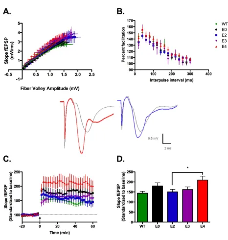
LTP induction in apoE TR animals is isoform-dependent The extracellular matrix protein reelin signals through both apoER2 and VLDLR; these receptors also bind apoE. However, unlike reelin-deficient mice, apoE TR and apoE-deficient animals develop normally without gross patho-logical changes to brain organization suggesting that apoE signaling or apoE isoform expression are not involved in neuronal migration during development. This has clear advantages when examining parameters of synaptic func-tion in apoE TR and apoE deficient mice. We find that hip-pocampal slices from 3–5 month-old animals are equivalent across genotypes and are identical to wild-type hippocampi on a gross anatomical level.
Supporting the lack of structural changes, hippocampus area CA1 synaptic transmission does not vary significantly with apoE isoform expression (figure 1A). These data ensure an important parameter in plasticity studies: that specific electrical 'input' elicits an equivalent synaptic response 'output' regardless of genotype. Thus, subse-quent variation in measured plasticity can be attributed to apoE isoform rather than changes in CA1 synaptic con-nectivity. Determination of short-term plasticity using paired-pulse facilitation revealed typical percent facilita-tion between apoE TR, apoE-deficient and wild-type mice (C57BL/6J) (figure 1B).
ApoE4 TR animals show increased LTP induction without changes in synaptic transmission
Figure 1
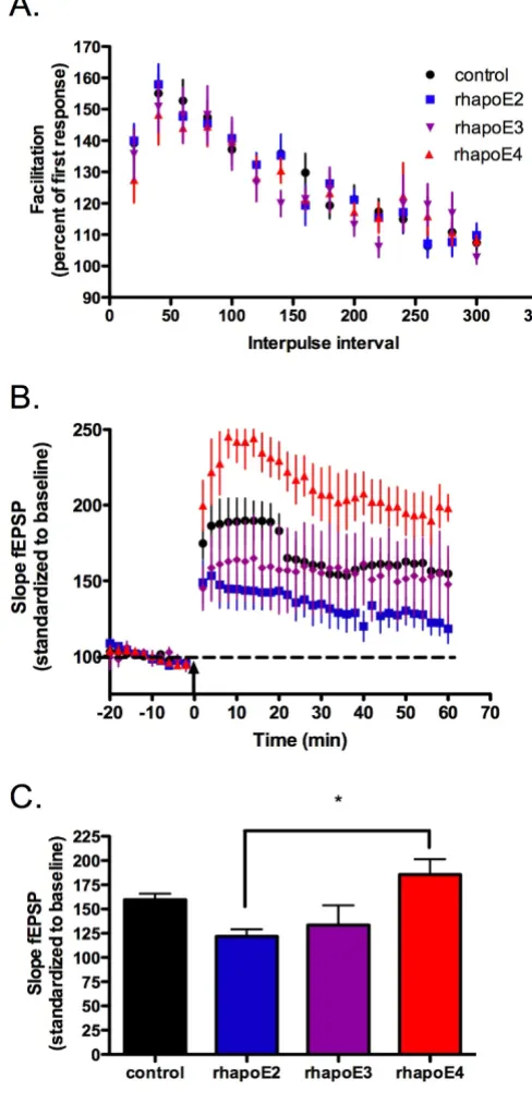
We hypothesize that apoE isoforms differentially act as ligands to a set of lipoprotein receptors that have the abil-ity to modulate synaptic activabil-ity. However, the changes in the amount of plasticity between specific apoE isoforms may be due to intrinsic properties of the apoE TR mouse or the production of compensatory protein expression during chronic apoE isoform expression. To test for iso-form-dependent signaling differences that could account for our electrophysiology results in the TR mice, we pre-pared hippocampal slices from apoE-deficient mice and perfused with 100 nM of human recombinant E2, E3 and E4 (rhapoE) prior to theta burst stimulation. ApoE-defi-cient mice were chosen to insure that the presence of endogenous murine apoE did not interfere with the applied rhapoE. Perfusion of rhapoE2, rhapoE3 or rhapoE4 has no effect on overall baseline synaptic trans-mission (data not shown) or short-term plasticity (figure 2A). However, there is an isoform-dependent change in synaptic plasticity following theta burst stimulation (fig-ure 2B,C). Interestingly, the change in plasticity with spe-cific isoform perfusion follows a trend similar to that measured in the corresponding apoE isoform TR mouse; both apoE4 TR animals and slices treated with rhapoE4 demonstrate significantly increased LTP induction over apoE2 TR mice or treatment with rhapoE2, respectively (rhapoE: ANOVA, p = 0.0112). This suggests that differ-ences in synaptic plasticity are not developmentally-dependent and that apoE acts as an isoform-specific sign-aling molecule in the adult hippocampus.
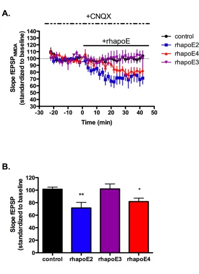
Effect of apoE isoform on LTP is NMDAR dependent The NMDA receptor is intimately associated with many forms of synaptic plasticity and long-term potentiation, including theta-burst-induced LTP [18,20]. In addition, signal transduction via apoE receptors is linked to NMDA receptor maturation [21,22], increased NMDA receptor currents [23], and activation of signaling pathways that involve NMDA receptor function [9,23,24]. These results lead us to hypothesize that the observed alterations in LTP may be due to apoE isoform-specific changes in NMDA receptor function. Thus, we induced NMDA-receptor independent LTP by delivering two one-second trains of 200 Hz stimulation concurrent with application of the NMDA receptor antagonist APV (100 μM). Long-lasting potentiation was induced in wild-type, apoE-deficient, and apoE TR animals; however, we eliminated apoE iso-form-dependent alterations of LTP induction (figure 3A– B).
The normalization of NMDAR-independent LTP induc-tion suggests that NMDARs are being modified by both the presence of specific apoE isoforms in the TR mice and by exogenous application of apoE isoforms. This can occur through direct changes in NMDAR function indi-cated by changes in phosphorylation and subsequent
modulation of NMDAR conductance. We took advantage of the acute apoE-isoform application strategy to investi-gate the ability of apoE isoforms to change CA1 NMDA receptor function over time.
NMDA receptor field potentials were isolated by applica-tion of 20 μM of the AMPA receptor antagonist CNQX fol-lowed by application of 100 nM of rhapoE2, rhapoE3 or rhapoE4. While we hypothesized that the enhanced LTP seen in the presence of apoE4 was related to increased NMDA receptor currents similar to the effect of reelin application, we found that application of either rhapoE2 or rhapoE4 significantly reduced NMDA field potentials from control levels (figure 4A–B). Interestingly, there was no effect on NMDAR field potentials with the application of rhapoE3. Although these results suggest a specific change to NMDA receptors, there is also the possibility that apoE isoforms can alter apoE receptors known to influence NMDA receptors in the hippocampus, such as apoER2.
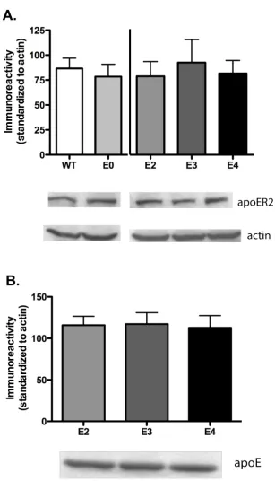
ApoE isoforms do not alter apoER2 expression levels Alterations in apoER2 expression and decreased reelin expression can adversely affect spatial memory [9,25]. Furthermore, changes in reelin concentration and expres-sion can alter LTP strength and induction as well as NMDAR conductance [21,23]. This suggests that differ-ences in receptor expression and concentration as well as changes to lipoprotein receptor ligands may affect synap-tic transmission or learning and memory. Therefore, we probed for changes in overall expression levels of the main apoE receptor in the brain, apoER2. Isolated hippoc-ampus tissue was probed for apoER2 expression using western blot analysis. Figure 5A indicates that apoE2, apoE3 and apoE4-expressing animals exhibit no altera-tions in total apoER2 expression in the hippocampus (ANOVA, p = 0.8269); apoE deficient and murine apoE-expressing animals are also equivalent in receptor expres-sion (t-test, p = 0.6175). Recent studies by Riddell et al. [26] suggest a differential apoE isoform expression in whole hippocampus or frontal cortex homogenates. How-ever, our results obtained with slightly different condi-tions show that targeted replacement did not alter overall apoE expression levels in the hippocampus (figure 5B, ANOVA, p = 0.6912).
Acute recombinant apoE isoform application recapitulates alterations in LTP induction
Figure 2
NMDA receptor-independent LTP is not affected by apoE isoform
Figure 3
ApoE isoforms alter NMDA receptor-dependent field potentials
Figure 4
ApoE isoform expression does not affect apoER2 expression levels
Figure 5
state of NMDA-receptor subunit tyrosine phosphoryla-tion.
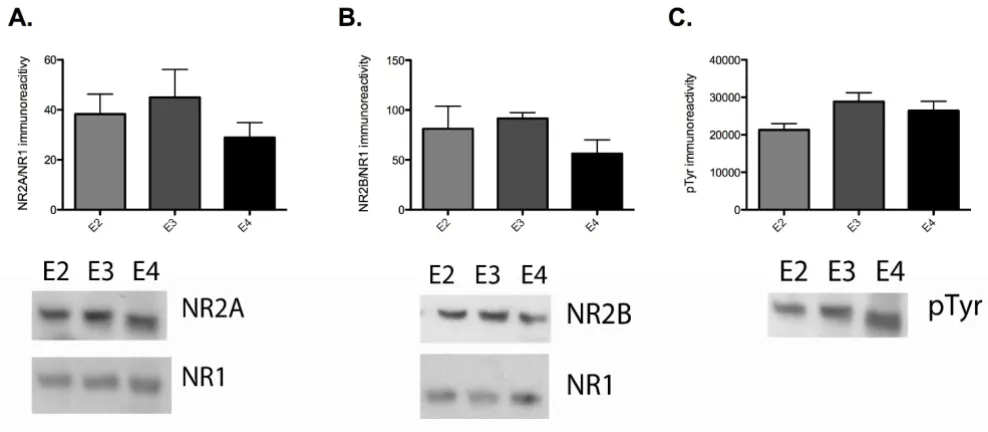
To test this possibility, we first attempted to immunopre-cipitate NR2A and NR2B from hippocampus area CA1 samples from apoE TR animals. We found that there were significant difficulties in our ability to immunoprecipitate equivalent amounts either NR2A or NR2B from the differ-ent apoE TR animals (data not shown). While this indi-cates that there may be structural differences or unidentified protein interactions with NMDA receptors that prevent complete immunoprecipitation due to the presence of apoE isoforms, it also hindered our ability to determine potential isoform-specific modulation of NR2A and NR2B phosphorylation. Therefore, we chose to probe for NR1, NR2A and NR2B in homogenates of hip-pocampus area CA1. We found that there were no signifi-cant differences in total NR2A or NR2B levels, standardized to NR1, between apoE2, apoE3 and apoE4 TR animals (figure 6A, B). We also probed for total tyro-sine phosphorylation at the corresponding to NR2A and NR2B protein band. Tyrosine phosphorylation at this molecular weight was also unchanged between apoE TR animals (figure 6C).
We were able to immunoprecipitate NR2A and NR2B from area CA1 of the hippocampus that had been treated with rhapoE isoforms. ApoE-deficient hippocampus slices
were isolated and incubated with 100 nM recombinant apoE for 40 minutes before flash freezing on dry ice and rapidly dissecting out CA1. We found that there were no significant differences in the ratio of pNR2A to NR2A between any of the conditions, but there was a trend towards a reduced ratio in the rhapoE3 and rhapoE4 treated groups (figure 7A). Similarly, there were no signif-icant differences in pNR2B to NR2B ratios (figure 7B).
ApoE isoform alters signal transduction
ApoE has been previously shown in neuronal cell culture to have isoform specific effects on signal transduction pathways crucial to synaptic plasticity such as ERK and JNK [31]. As those effects were blocked with LDL receptor inhibitors and the NMDA receptor antagonist MK-801, we hypothesized that similar mechanisms may be underlying the isoform-specific alterations in CA1 LTP.
Area CA1 dissected from acute hippocampal slices of apoE TR, wild-type, and apoE-deficient animals were homoge-nized and subjected to SDS-PAGE analysis. Membranes were probed for pERK1/2, ERK1/2, pJNK1/2 or JNK1/2. This revealed that pERK1/2 was significantly increased in apoE4 TR animals versus apoE2 TR or apoE3 TR (figure 8Ai, ANOVA p = 0.0005). There were no accompanying significant changes in total ERK (figure 8Aii). The ratio of pERK to total ERK showed a significant increase in the
Effect of chronic apoE isoform expression on NR2A, NR2B levels and tyrosine phosphorylation
Figure 6
Effect of acute apoE exposure on NR2A and NR2B tyrosine phosphorylation
Figure 7
Chronic apoE isoform expression alters activation of ERK1/2 and JNK1/2
Figure 8
apoE4 TR over apoE2 TR and apoE3 TR (figure 8Aiii, ANOVA p = 0.003).
When pJNK levels were analyzed, we found a significant reduction in apoE-deficient, versus wild-type animals (fig-ure 8Bi, t-test p = 0.0003) and apoE2 and apoE3 TR versus apoE4 TR animals (figure 8Bi, ANOVA p = 0.0075). This was not accompanied by any significant changes in total JNK. The pJNK/JNK ratio revealed a significant reduction in apoE-deficient from wild-type (figure 8Bii, t-test 0.0015), and apoE2 TR and apoE3 TR animals from apoE4 TR (figure 8Biii, ANOVA p = 0.0025).
In light of the differences in specific signal transduction proteins in the apoE TR mice, we wanted to test the effects of acute apoE exposure on signal transduction. ApoE-defi-cient hippocampus slices were isolated and incubated with 100 nM recombinant apoE for 40 minutes before flash freezing on dry ice and rapidly dissecting out CA1. Tissue samples were probed by western analysis for pERK, pJNK, ERK and JNK. Unlike comparable apoE TR mice, apoE isoform application did not significantly alter total pERK levels (data not shown). There were no significant differences in the pERK/ERK ratio (figure 9A). Further-more, in contrast to what was seen with chronic apoE iso-form expression, acute apoE isoiso-form application did not significantly alter either JNK or pJNK levels (data not shown) or pJNK/JNK ratio (figure 9B). Taken together, these data suggest that the effects on signal transduction pathways and the potential compensatory changes of pro-longed apoE isoform expression result in similar electro-physiology results, but may be more representative of different mechanisms underlying those physiologic effects.
Discussion
Essential to defining how apoE influences the memory disruption and etiology of neurodegenerative disorders such as Alzheimer's disease is the establishment of the role of apoE in synaptic function. However, the physio-logic actions of apoE as a signaling molecule in the adult CNS are unclear. In the present study we investigated both the necessity for murine apoE expression in adult hippoc-ampus area CA1 synaptic plasticity as well as potential dif-ferences due to specific human apoE isoform expression. We found a lack of an overt physiologic defect in apoE-deficient animals with a C57Bl/6J background. This lack of phenotype was especially valuable in comparing differ-ences with specific isoform expression in apoE TR mice. The apoE TR mice are particularly useful for these investi-gations as they allow for the direct in vivo comparison of the effects of apoE isoforms and reduce the potential cave-ats associated with apoE isoform over-expression. Impor-tantly, in the absence of other genetic manipulations commonly used in mouse models of AD, these animals
do not develop the potentially confounding pathological abnormalities know to affect memory formation and syn-aptic plasticity. Moreover, we were able to utilize the lack of a strong physiologic phenotype in apoE-deficient mice in conjunction with acute application of human recom-binant apoE isoforms to preclude the potential confound-ing results of interactions or competition with endogenous murine apoE.
Previous electrophysiological studies of apoE TR mice have revealed that both perforant path LTP induction and the vulnerability of this potentiation by oligomeric beta amyloid (Aβ) vary with apoE isoform expression [15,19]. These studies show a decrease in LTP induction in apoE-deficient, apoE2 TR and apoE4 TR animals, with the great-est decrease in potentiation observed in apoE4 TR [15]. Our current study focuses on the well-characterized Schaf-fer collateral synapses, as does the previous work on reelin and its effects through the lipoprotein receptors apoER2 and VLDLR [9]. Although we predicted that the LTP induc-tion profile would mirror that in the dentate gyrus, it was surprising to see that apoE4 TR mice demonstrated selec-tive enhancement of CA1 LTP and apoE2 TR mice show the least amount of LTP induction.
ApoE isoforms acting as signaling ligands can change hip-pocampal physiology and alter LTP induction in a number of ways. One possibility is alteration of NMDA receptors through changes in subunit composition and/or phosphorylation. This possibility is already established as an action of reelin through apoER2 and VLDLR [21,22,32,33]. Our attempts to monitor changes in NMDAR subunit composition and phosphorylation through immunoprecipation revealed differences in our ability to pull down either NR2A or NR2B consistently between the apoE TR mice. Interestingly, total NR1 levels in these assays did not vary with NR2A or NR2B levels. We hypothesize that our ability to immunoprecipitate NR2A and NR2B is being hindered by differential treatment of NMDA receptors within neurons due to apoE signaling and our experimental conditions are not accurately con-trolling for these differences.
Effects of acute apoE isoform exposure on ERK1/2 and JNK1/2 activation
Figure 9
chronic changes in signaling resulting in slight alterations to NMDAR subunit composition, localization or function that may cumulatively exert a physiologic effect on LTP. As shown by figure 10, it is likely that alterations in signal transduction via apoER2 that impact NMDA receptor function plus alterations in other signal transduction cas-cades combine to produce apoE isoform-specific changes in synaptic plasticity. Ongoing research in our laboratory is investigating these potential possibilities.
The changes in LTP induction with acute apoE isoform application are more difficult to explain. In this instance changes in NMDAR subunit composition are unlikely,
and there were no significant changes in the ERK or JNK signal transduction pathways. Interestingly, rhapoE4 did not increase isolated NMDA receptor field potentials as expected, but rather caused a significant decrease. Yet there were no significant differences in the pNR2A/NR2A or pNR2B/NR2B ratios with treatment of any rhapoE iso-form. While the observed trend towards decreased NR2A and NR2B phosphorylation with rhapoE4 treatment may actually contribute to the apoE-induced decreases in NMDAR field potentials, further studies will be necessary to determine if there are other mechanisms such as desen-sitization or internalization of NMDARs at work. Another possibility is that application of a bolus of apoE isoform
Model of chronic apoE signaling in adult hippocampus
Figure 10
selectively blocks lipoprotein receptor activity. This situa-tion occurs with an antibody that binds to the reelin asso-ciation site of apoER2 (CR-50), which results in a decrease in NMDA receptor whole cell currents [34]. Thus, the decrease in NMDA receptor currents with rhapoE2 and rhapoE4 may reflect an isoform-specific affinity for apoER2 when using non-cholesterol associated human recombinant protein. However, the ability for rhapoE4 to increase LTP induction despite these decreases in NMDAR field potentials suggests that apoE isoforms may also be acting differentially as signaling molecules to alter other components of the LTP machinery.
The idea of apoE as a signaling molecule is not new. In pri-mary neuronal cell culture, application of either full length apoE or a tandem repeat peptide of the receptor binding domain of apoE significantly enhances phospho-rylation of ERK1/2 and Dab-1 [32]. This pathway is well validated as essential to the underlying mechanisms of LTP [35,36], as well as learning and memory [37-39]. Modulation of these pathways by either pharmacological or genetic manipulation has dramatic effects on both syn-aptic plasticity and memory formation. With this in mind, an important aspect of these studies that should not be overlooked is the lack of an LTP phenotype in the apoE knockout mouse. This raises an interesting question: if apoE is acting as a signaling molecule important for neu-ronal function, then why is there no overt change in phys-iology in its absence? Our results suggest a unique role for specific apoE isoforms in the modulation of hippocampal CA1 synaptic plasticity. In addition, both apoE isoforms and reelin can bind to the same family of lipoprotein receptors. The role of reelin in synaptic function and memory formation is now well established, and specific isoform expression can have a direct effect on reelin sign-aling and modulation of synaptic function. Thus, apoE isoforms may function by changing reelin's ability to modulate NMDA receptors and activate specific signal transduction pathways by as yet unknown mechanisms. Furthermore, changes in apoE availability, due to spatial changes or associations with other proteins, may have a profound affect on synaptic function in the different sub-fields of the hippocampus.
With renewed focus on the role of apoE in neurodegener-ative disorders and as a potentially rich area for therapeu-tic intervention comes the necessity to better understand how apoE can affect synaptic function. Evolution appears to have co-opted apoE to perform duties encompassing far more than cholesterol transport. Acting directly through lipoprotein receptor activation, or indirectly by altering reelin action, apoE isoforms can have a significant effect on NMDA receptor function. How might changes in apoE during normal and pathological states of neurode-generation affect synaptic plasticity and memory
forma-tion? While this question is the foundation for future studies, our results here highlight the previously unappre-ciated role of apoE as a modulator of hippocampus syn-aptic plasticity.
Experimental procedures
Animal maintenance
ApoE2, apoE3 and apoE4 targeted replacement animals were obtained from a colony maintained at Taconic (Hudson, New York USA). ApoE-deficient and C57BL/6J animals were obtained from Jackson Laboratories (Bar Harbor, Maine, USA). Animals were housed in a standard 12 hour light cycle and bred and maintained in accord-ance with the Vanderbilt University Institutional Animal Care and Use Committee protocol.
Electrophysiology
Hippocampus slices were prepared from 3- to 5-month old mice as previously reported [9]. The brain was rapidly removed and placed in oxygenated ice-cold high sucrose cutting saline solution containing (in mM) 110 sucrose, 60 NaCl, 3 KCl, 28 NaHCO3, 1.25 NaH2PO4, 5 glucose, 0.6 ascorbate, 7 MgCl2, and 0.5 CaCl2. Horizontal 400 μm
sections were cut in high sucrose cutting solution using a vibratome. Slices were maintained in cold, oxygenated cutting solution until dissection. After dissection, the hip-pocampus slices were transferred to room temperature cutting solution diluted 1:1 with artificial cerebral spinal fluid (ACSF). ACSF contains, in mM, 125 NaCl, 2.5 KCl, 26 NaHCO3, 1.25 NaH2PO4, 25 glucose, 1 MgCl2, and 2 CaCl2. Slices were maintained in this solution with
con-stant 95% O2/5% CO2 perfusion for 10 min before trans-ferring to the brain slice recording chamber (Fine Science Tools, San Francisco, California, USA).
elicited 40–50% of the maximum fEPSP response as determined from the input-output curve.
Long-term potentiation (LTP) was induced by theta-burst protocol. Theta-burst LTP protocol consisted of five trains of 10 bursts at a 5 Hz frequency with each burst consisting of 4 stimulations delivered at 100 Hz and an inter-train interval of 20 seconds. NMDAR-independent LTP was induced by 2 one-second trains of stimulation delivered at 200 Hz; concurrent application of 100 μM 2-amino-5-phosphonovaleric acid (APV) confirmed NMDAR inde-pendence. NMDAR field potentials were isolated by incu-bating slices on the rig in ACSF containing no Mg2+ for 15
minutes followed by addition of 20 μM 6-cyano-7-nitro-quinoxaline-2,3-dione (CNQX) and 25 μM picrotoxin. After 15 minutes of stabilization, slices were treated with 100 nM of recombinant human apoE isoforms (Calbio-chem).
Statistics
Electrophysiological data was also analyzed using one-way ANOVA with Bonferroni's post hoc tests. Significance was set at p < 0.05 for all tests.
Biochemistry
ApoE/apoER2
Bilateral dissections of the whole hippocampus from aged (1 year old) animals were performed. Brain tissue was rap-idly dissected and flash frozen on dry ice. Tissue was homogenized in NP-40 lysis buffer containing (in mM) 50 Tris-HCL ph 8.0, 150 NaCl, 1 EDTA, 1 PMSF, 1 Na3VO4, 1 NaF, 1 μg/mL each of aprotinin, leupeptin and pepstatin, and 1% NP-40. Protein concentration was determined by Bradford Assay (Bio-rad). For western blot analysis, 10 μg of protein was resolved by 4–20% gradient SDS-PAGE. The proteins were then transferred to PVDF membranes. Membranes were probed with goat anti-human apoE (Academy Bio-medical, Houston Texas, USA), rabbit anti-apoER2 (a gift of Drs. Gary Olson and Ray Burk, Vanderbilt University [40]), and rabbit anti-actin (Sigma) diluted in 0.24% I-block. Membranes were developed using HRP-conjugated secondary antibodies and enhanced chemiluminescence. Optical density of immunoreactivity was quantified by densitometry using Image J software (NIH).
For investigations of chronic apoE exposure, slices were obtained from apoE2 TR, apoE3 TR, apoE4 TR, apoE-defi-cient and C57BL/6J (wild-type) mice in an identical fash-ion as for electrophysiology (see above). The slices were frozen on dry ice and CA1 dissected unless otherwise stated. For investigations of acute apoE exposure, slices were obtained from apoE-deficient mice in an identical fashion as for electrophysiology (see above). Slices (n = 3– 4 per treatment) were incubated in ACSF at 30°C for 1
hour prior to the addition of 100 nM recombinant human apoE2, apoE3 or apoE4 (Calbiochem) for 40 minutes. Slices were then flash frozen on dry ice and CA1 dissected.
ERK/JNK activation
Pooled tissue was homogenized in NP-40 lysis buffer con-taining (in mM) 50 Tris-HCL ph 8.0, 150 NaCl, 1 EDTA, 1 PMSF, 1 Na3VO4, 1 NaF, 1 μg/mL each of aprotinin, leu-peptin and pepstatin, and 1% NP-40. Protein concentra-tion was determined by Bradford Assay (Bio-Rad). Ten μg of protein was resolved by SDS-PAGE on 4–15% Tris-HCL gradient gels (Bio-Rad) and transferred to PVDF mem-brane. Membranes were probed with rabbit anti-ERK1/2, anti-ERK1/2 pTpY185/187, anti-JNK1/2, anti-JNK1/2
pTpY183/185 (Invitrogen) diluted in 0.24% I-block.
Mem-branes were developed using HRP-conjugated secondary antibodies and enhanced chemiluminescence.
NMDAR subunit phosphorylation
Pooled tissue was sonicated in modified RIPA buffer (Tris/ HCl pH 7.4, 2 mM EDTA, 150 mM NaCl, 0.1% SDS, 0.5% sodium deoxycholate, 1% triton X100, 1× phosphatase inhibitors I and II (Sigma), and 1× complete protease inhibitors (Sigma). Protein concentrations were deter-mined by BCA Protein Assay (Bio-Rad). Ten μg of protein was resolved by SDS-PAGE on 4–15% Tris-HCL gradient gels (Bio-Rad) and transferred to PVDF membrane. Mem-branes were probed with mouse anti-phosphotyrosine, clone 4G10 (Millipore), rabbit NR2A, rabbit anti-NR2B (Upstate), and mouse anti-NR1 (Millipore) diluted in 0.24% I-block. Membranes were developed using HRP-conjugated secondary antibodies and enhanced chemilu-minescence.
For immunoprecipitation assays, a total of 400 μg of pro-tein lysate was used to immunoprecipitate either rabbit anti-NR2A or rabbit anti-NR2B (Upstate) overnight at 4°C with agitation. Protein A/G magnetic beads (New England BioLabs) were added to each reaction (25 μl bead slurry/reaction) and samples were incubated for 2 hours at 4°C with agitation. Following three wash cycles, the protein was eluted with 1× Laemmli sample buffer sepa-rated by SDS-PAGE on 4–15% Tris-HCl gradient gels (Bio-Rad) and transferred to PVDF membranes. Membranes were probed with mouse anti-pTyr, clone 4G10 (Milli-pore), rabbit anti-NR2A, and rabbit anti-NR2B in 2% BSA-TBST. Membranes were developed using HRP-conju-gated secondary antibodies and enhanced chemilumines-cence.
Optical density of immunoreactivity was quantified by densitometry using Image J software (NIH).
Competing interests
Authors' contributions
KK performed the electrophysiological and biochemical studies. JT performed the immunoprecipitation assays and aided in the biochemical studies. ML provided the apoE TR mice and aided in the conceptual analysis. PS developed the apoE TR mice and approved their use in this study. EW directed the design and coordination of this study. All authors read and approved the final manu-script.
References
1. Corder EH, Saunders AM, Strittmatter WJ, Schmechel DE, Gaskell PC, Small GW, Roses AD, Haines JL, Pericak-Vance MA: Gene dose of apolipoprotein E type 4 allele and the risk of Alzheimer's disease in late onset families. Science 1993, 261:921-923. 2. Rebeck GW, Reiter JS, Strickland DK, Hyman BT: Apolipoprotein
E in sporadic Alzheimer's disease: allelic variation and recep-tor interactions. Neuron 1993, 11:575-580.
3. Strittmatter WJ, Saunders AM, Schmechel D, Pericak-Vance M, Eng-hild J, Salvesen GS, Roses AD: Apolipoprotein E: high-avidity binding to beta-amyloid and increased frequency of type 4 allele in late-onset familial Alzheimer disease. Proc Natl Acad Sci USA 1993, 90:1977-1981.
4. Beffert U, Stolt PC, Herz J: Functions of lipoprotein receptors in neurons. J Lipid Res 2004, 45:403-409.
5. Qiu S, Korwek KM, Weeber EJ: A fresh look at an ancient tor family: emerging roles for low density lipoprotein recep-tors in synaptic plasticity and memory formation. Neurobiol Learn Mem 2006, 85:16-29.
6. Koch S, Strasser V, Hauser C, Fasching D, Brandes C, Bajari TM, Sch-neider WJ, Nimpf J: A secreted soluble form of ApoE receptor 2 acts as a dominant-negative receptor and inhibits Reelin signaling. Embo J 2002, 21:5996-6004.
7. Nakajima K, Mikoshiba K, Miyata T, Kudo C, Ogawa M: Disruption of hippocampal development in vivo by CR-50 mAb against reelin. Proc Natl Acad Sci USA 1997, 94:8196-8201.
8. Qiu S, Korwek KM, Pratt-Davis AR, Peters M, Bergman MY, Weeber EJ: Cognitive disruption and altered hippocampus synaptic function in Reelin haploinsufficient mice. Neurobiol Learn Mem
2006, 85:228-242.
9. Weeber EJ, Beffert U, Jones C, Christian JM, Forster E, Sweatt JD, Herz J: Reelin and ApoE receptors cooperate to enhance hip-pocampal synaptic plasticity and learning. J Biol Chem 2002,
277:39944-39952.
10. Trommsdorff M, Gotthardt M, Hiesberger T, Shelton J, Stockinger W, Nimpf J, Hammer RE, Richardson JA, Herz J: Reeler/Disabled-like disruption of neuronal migration in knockout mice lacking the VLDL receptor and ApoE receptor 2. Cell 1999,
97:689-701.
11. Sullivan PM, Mezdour H, Aratani Y, Knouff C, Najib J, Reddick RL, Quarfordt SH, Maeda N: Targeted replacement of the mouse apolipoprotein E gene with the common human APOE3 allele enhances diet-induced hypercholesterolemia and atherosclerosis. J Biol Chem 1997, 272:17972-17980.
12. Kim DH, Iijima H, Goto K, Sakai J, Ishii H, Kim HJ, Suzuki H, Kondo H, Saeki S, Yamamoto T: Human apolipoprotein E receptor 2. A novel lipoprotein receptor of the low density lipoprotein receptor family predominantly expressed in brain. J Biol Chem
1996, 271:8373-8380.
13. Nimpf J, Schneider WJ: The VLDL receptor: an LDL receptor relative with eight ligand binding repeats, LR8. Atherosclerosis
1998, 141:191-202.
14. Grootendorst J, Bour A, Vogel E, Kelche C, Sullivan PM, Dodart JC, Bales K, Mathis C: Human apoE targeted replacement mouse lines: h-apoE4 and h-apoE3 mice differ on spatial memory performance and avoidance behavior. Behav Brain Res 2005,
159:1-14.
15. Trommer BL, Shah C, Yun SH, Gamkrelidze G, Pasternak ES, Ye GL, Sotak M, Sullivan PM, Pasternak JF, LaDu MJ: ApoE isoform affects LTP in human targeted replacement mice. Neuroreport 2004,
15:2655-2658.
16. Kitamura HW, Hamanaka H, Watanabe M, Wada K, Yamazaki C, Fujita SC, Manabe T, Nukina N: Age-dependent enhancement of hippocampal long-term potentiation in knock-in mice expressing human apolipoprotein E4 instead of mouse apol-ipoprotein E. Neurosci Lett 2004, 369:173-178.
17. Larson J, Wong D, Lynch G: Patterned stimulation at the theta frequency is optimal for the induction of hippocampal long-term potentiation. Brain Res 1986, 368:347-350.
18. Larson J, Lynch G: Role of N-methyl-D-aspartate receptors in the induction of synaptic potentiation by burst stimulation patterned after the hippocampal theta-rhythm. Brain Res
1988, 441:111-118.
19. Trommer BL, Shah C, Yun SH, Gamkrelidze G, Pasternak ES, Blaine Stine W, Manelli A, Sullivan P, Pasternak JF, LaDu MJ: ApoE isoform-specific effects on LTP: blockade by oligomeric amyloid-beta1-42. Neurobiol Dis 2005, 18:75-82.
20. Abraham WC, Huggett A: Induction and reversal of long-term potentiation by repeated high-frequency stimulation in rat hippocampal slices. Hippocampus 1997, 7:137-145.
21. Qiu S, Weeber EJ: Reelin signaling facilitates maturation of CA1 glutamatergic synapses. J Neurophysiol 2007, 97:2312-2321. 22. Sinagra M, Verrier D, Frankova D, Korwek KM, Blahos J, Weeber EJ, Manzoni OJ, Chavis P: Reelin, very-low-density lipoprotein receptor, and apolipoprotein E receptor 2 control somatic NMDA receptor composition during hippocampal matura-tion in vitro. J Neurosci 2005, 25:6127-6136.
23. Qiu S, Zhao LF, Korwek KM, Weeber EJ: Differential reelin-induced enhancement of NMDA and AMPA receptor activ-ity in the adult hippocampus. J Neurosci 2006, 26:12943-12955. 24. Rebeck GW, Ladu MJ, Estus S, Bu G, Weeber EJ: The generation
and function of soluble apoE receptors in the CNS. Mol Neu-rodegener 2006, 1:15.
25. Beffert U, Weeber EJ, Durudas A, Qiu S, Masiulis I, Sweatt JD, Li WP, Adelmann G, Frotscher M, Hammer RE, Herz J: Modulation of syn-aptic plasticity and memory by reelin involves differential splicing of the lipoprotein receptor apoER2. Neuron 2005,
47:567-579.
26. Riddell DR, Zhou H, Atchison K, Warwick HK, Atkinson PJ, Jefferson J, Xu L, Aschmies S, Kirksey Y, Hu Y, et al.: Impact of apolipopro-tein E (ApoE) polymorphism on brain ApoE levels. J Neurosci
2008, 28:11445-11453.
27. Ferrani-Kile K, Leslie SW: Modulation of protein tyrosine phos-phatase activity alters the subunit assembly in native N-methyl-D-aspartate receptor complex. J Pharmacol Exp Ther
2005, 314:86-93.
28. Goebel SM, Alvestad RM, Coultrap SJ, Browning MD: Tyrosine phosphorylation of the N-methyl-D-aspartate receptor is enhanced in synaptic membrane fractions of the adult rat hippocampus. Brain Res Mol Brain Res 2005, 142:65-79.
29. Lu YM, Roder JC, Davidow J, Salter MW: Src activation in the induction of long-term potentiation in CA1 hippocampal neurons. Science 1998, 279:1363-1367.
30. Tezuka T, Umemori H, Akiyama T, Nakanishi S, Yamamoto T: PSD-95 promotes Fyn-mediated tyrosine phosphorylation of the N-methyl-D-aspartate receptor subunit NR2A. Proc Natl Acad Sci USA 1999, 96:435-440.
31. Hoe HS, Harris DC, Rebeck GW: Multiple pathways of apolipo-protein E signaling in primary neurons. J Neurochem 2005,
93:145-155.
32. Bock HH, Herz J: Reelin activates SRC family tyrosine kinases in neurons. Curr Biol 2003, 13:18-26.
33. Chen Y, Beffert U, Ertunc M, Tang TS, Kavalali ET, Bezprozvanny I, Herz J: Reelin modulates NMDA receptor activity in cortical neurons. J Neurosci 2005, 25:8209-8216.
34. Groc L, Choquet D, Stephenson FA, Verrier D, Manzoni OJ, Chavis P: NMDA receptor surface trafficking and synaptic subunit composition are developmentally regulated by the extracel-lular matrix protein Reelin. J Neurosci 2007, 27:10165-10175. 35. Hugues S, Chessel A, Lena I, Marsault R, Garcia R: Prefrontal
infu-sion of PD098059 immediately after fear extinction training blocks extinction-associated prefrontal synaptic plasticity and decreases prefrontal ERK2 phosphorylation. Synapse
2006, 60:280-287.
ele-Publish with BioMed Central and every scientist can read your work free of charge "BioMed Central will be the most significant development for disseminating the results of biomedical researc h in our lifetime."
Sir Paul Nurse, Cancer Research UK
Your research papers will be:
available free of charge to the entire biomedical community
peer reviewed and published immediately upon acceptance
cited in PubMed and archived on PubMed Central
yours — you keep the copyright
Submit your manuscript here:
http://www.biomedcentral.com/info/publishing_adv.asp
BioMedcentral
ment-binding protein phosphorylation is required for the long-term facilitation process of aversive olfactory learning in young rats. Neuroscience 2003, 121:9-16.
37. Cestari V, Costanzi M, Castellano C, Rossi-Arnaud C: A role for ERK2 in reconsolidation of fear memories in mice. Neurobiol Learn Mem 2006, 86:133-143.
38. Fischer A, Radulovic M, Schrick C, Sananbenesi F, Godovac-Zimmer-mann J, Radulovic J: Hippocampal Mek/Erk signaling mediates extinction of contextual freezing behavior. Neurobiol Learn Mem 2007, 87:149-158.
39. Giovannini MG: The role of the extracellular signal-regulated kinase pathway in memory encoding. Rev Neurosci 2006,
17:619-634.
40. Olson GE, Winfrey VP, Nagdas SK, Hill KE, Burk RF: Apolipopro-tein E receptor-2 (apoER2) mediates selenium uptake from selenoprotein P by the mouse testis. J Biol Chem 2007,