Open Access
Research
Differential effects of age on circulating and splenic leukocyte
populations in C57BL/6 and BALB/c male mice
Lesya M Pinchuk
1and Nikolay M Filipov*
1,2Address: 1Department of Basic Sciences, College of Veterinary Medicine, Mississippi State University, Mississippi State, MS, USA and 2Center for Environmental Health Sciences, Department of Basic Sciences, College of Veterinary Medicine, Mississippi State University, Mississippi State, MS, USA
Email: Lesya M Pinchuk - pinchuk@cvm.msstate.edu; Nikolay M Filipov* - filipov@cvm.msstate.edu * Corresponding author
Abstract
Background: Despite several reports on age-related phenotypic changes of the immune system's cells, studies that use a multipoint age comparison between the specific and innate immune cell populations of prototypical Th1- and Th2-type polarized mouse strains are still lacking.
Results: Using a multipoint age comparison approach, cells from the two major immune system compartments, peripheral blood and spleen, and flow cytometry analysis, we found several principal differences in T cell and professional antigen presenting cell (APC) populations originating from a prototypical T helper (Th) 1 mouse strain, C57BL/6, and a prototypical Th2 strain, BALB/c. For example, regardless of age, there were strain differences in both peripheral blood mononuclear cells (PBMC) and spleens in the proportion of CD4+ (higher in the BALB/c strain), CD8+ T cells and CD11b+/CD11c+ APC (greater in C57BL/6 mice). Other differences were present only in PBMC (MHC class II + and CD19+ were greater in C57BL/6 mice) or differences were evident in the spleens but not in circulation (CD3+ T cells were greater in C57BL/6 mice). There were populations of cells that increased with age in PBMC and spleens of both strains (MHC class II+), decreased in the periphery and spleens of both strains (CD11b+) or did not change in the PBMC and spleens of both strains (CD8+). We also found strain and age differences in the distribution of naïve and memory/activated splenic T cells, e.g., BALB/c mice had more memory/activated and less naive CD8+ and CD4+ T cells and the C57BL/6 mice.
Conclusion: Our data provide important information on the principal differences, within the context of age, in T cell and professional APC populations between the prototypical Th1 mouse strain C57BL/6 and the prototypical Th2 strain BALB/c. Although the age-related changes that occur may be rather subtle, they may be very relevant in conditions of disease and stress. Importantly, our data indicate that age and strain should be considered in concert in the selection of appropriate mouse models for immunological research.
Background
Recent studies indicate that the immune system under-goes gradual age-related shifts in cell populations, which
lead to functional changes of the immune responses. The compensatory modulations, including lymphocyte altera-tions, were recently defined as immunosenescence. This is
Published: 11 February 2008
Immunity & Ageing 2008, 5:1 doi:10.1186/1742-4933-5-1
Received: 12 October 2007 Accepted: 11 February 2008
This article is available from: http://www.immunityageing.com/content/5/1/1
© 2008 Pinchuk and Filipov; licensee BioMed Central Ltd.
a complex process of multiple reorganizational and devel-opmentally regulated changes rather than a simple unidi-rectional decline in all immune functions [1,2]. Nevertheless, for the most part, the activity of the immune system declines with age, with the most pronounced alter-ations found in cell-mediated immunity (CMI), especially in the T cell functions, which are related to thymic involu-tion [3-8]. Although decline in adaptive immunity repre-sents a major problem for the aged, evidence accumulated within the last decade indicates that aging also has a pro-found impact on innate immunity [9].
Despite the maintenance of normal CD3+ cell numbers with age, there is a considerable decrease in CD4- and CD8-mediated responses [10,11]. One major reason for CMI decreases with age is the substantial reduction in the representation of naïve T lymphocytes with a concomitant increase in memory T cells. This is a consequence of com-pensatory homeostatic proliferation in response to the reduced numbers of naïve cells and the influence of cumulative exposure to pathogens and environmental antigens [12,13]. A second key age-related change is the alteration of the activation potential of memory T cells [14,15], leading to hyporesponsivity [16]. Also, there is an increased oligoclonal expansion of nontransformed T cell populations [17,18].
Additional shifts have also been documented in other cells of the ageing immune system, such as changes in the levels of CD4+ cells and proportion of CD4+/CD8+ pop-ulations in peripheral tissues [19,20]. The most consistent finding associated with a repressed immune response has been a decrease in the proportion of CD4+ T cells [21,8]. The appearance of multiple CD8+ T cell clonal expansions is one of the most dramatic qualitative changes in the memory cell population during ageing [22].
There is an agreement that ageing results in perturbation of peripheral blood B cells in two important ways. First, the number of newly made B cells that migrate to the spleen from the bone marrow is reduced [23,24]. Second, there is an accumulation of B lineage cells in the splenic compartments [23,24]. Many of these effects may be a consequence of functional defects intrinsic to the B cells [25,26], but others may be secondary to age-related changes in CD4+ T cells. Indeed, aged CD4+ T cells are less efficient at inducing germinal center formation and promoting somatic hypermutation [27,25]. This possibly reflects a shift from T helper 1 cell (Th1) to Th2-type cytokine patterns associated with age in mice and humans [28]. The factors that determine whether a proliferating CD4+ T cell in mice and humans will differentiate into a Th1 or Th2 cell are not fully understood. However, the consequences of inducing Th1 versus Th2 profiles are pro-found: the selective production of Th1 cells leads to CMI,
whereas the production of predominantly Th2 cells pro-vides humoral immunity. Recent studies have shown that the interaction of the most powerful APC, dendritic cells (DC), directly with pathogens through toll-like receptor (TLR)-dependent mechanisms or with innate lym-phocytes represents a major control mechanism for adap-tive immunity, including Th polarization [29-31].
Age-related shifts in cell population profiles may lead to a different humoral or cellular immune response bias in mice. In addition to age, genetics play a major role in the shaping of the immune response. Thus, the CD4/CD8 ratio, B cell apoptosis, and pre-B cell expansion are under genetic control in mice and humans [32-34]. Significant strain differences have been found in hematopoietic pro-genitor cell functions between B6, BALB and D2 mice [33-35]. Multiple reports suggest a preferential bias for the C57BL/6 mouse strain to develop Th1-type response, whereas the BALB/c strain is biased towards a Th2-type cytokine polarization to some infectious agents, including
Leishmania major, Pseudomonas aeruginosa, and Porphyro-manas gingivalis [36-40].
Previous studies have focused on the genetic basis of strain differences in peripheral blood cell populations [41]. However, the other major component of the immune system, the spleen, has been overlooked. Moreo-ver, effects of ageing on APC have not received much attention. Despite several reports on age-related changes in the cells of the immune system (discussed in [2]), com-prehensive studies that use a multipoint age comparison between the leukocyte populations of mouse strains that develop different immune responses, are still lacking. Hence, our objective was to perform a detailed side-by-side comparison of the age-related changes in peripheral blood and splenic T cell and professional APC popula-tions in prototypical Th1 and Th2 mouse strains, C57BL/ 6 and BALB/c, respectively.
Results
Body and spleen weight changes with age in C57BL/6 and BALB/c mouse strains
Spleens of BALB/c were consistently heavier than the C57BL/6 mice's spleens. For example, 3- and 5-month-old mice's spleens weighted 3.6 + 0.14 vs. 2.2 + 0.09 and 3.0 + 0.17 vs. 1.8 + 0.17 g/kg BW; BALB/c vs. C57BL/6, 3- and 5-month olds, respectively. With age, relative spleen weight decreased in both strains with the most prominent decline occurring in the C57BL/6 mice from 1 to 3 months (27% decrease: from 3.0 + 0.21 to 2.2 + 0.09 g/kg BW).
Changes on the number of PBMC and splenocytes in C57BL/6 and BALB/c mice at selected ages
In addition to all phenotypic differences due to strain and age (described below), we also evaluated the effect of strain on the number of PBMC and splenocytes in 3- and 5-month old mice. In accord with the spleen weight data, BALB/c mice had more splenocytes than C57BL/6 mice. In both strains, a moderate decrease in the number of splen-ocytes was observed as the animals aged from 3 to 5 months (1.8 × 10⁷ vs. 0.9 × 10⁷ and 1.6 × 10⁷ vs. 0.8 × 10⁷; cells/spleen, BALB/c vs. C57BL/6, 3- and 5-month old mice, respectively). In circulation, the difference between the two strains was present in both 3- and 5-month old mice. In addition, a moderate increase in the number of circulating PBMC was observed in both strains from 3 to 5 months of age (5.1 × 10⁶ vs. 3.5 × 10⁶ and 6.0 × 10⁶ vs. 4.2 × 10⁶; cells/ml, BALB/c vs. C57BL/6, 3- and 5-month old mice, respectively).
Effects of age and strain on the cells of adaptive and innate immunity
B cell-specific molecules expression
Percentage of peripheral blood B cells expressing CD19+ cells increased with age in both strains, peaking in 18-month-old animals (Fig. 1A). There was a decline at 3 and 5 months of age, which was significant only in the BALB/ c strain PBMC (Fig. 1A). Overall, the PBMC of C57BL/6 mice had more CD19+ B cells than did BALB/c PBMC (Fig. 1A). However, in the spleens, due to differences in the kinetics of CD19 alterations with age, the % of CD19+ B cells in the BALB/c strain was significantly higher than in the C57BL/6 strain at 3 months of age (Fig. 1B).
T cell phenotypes
We did not find any significant age- or strain-related dif-ferences in the levels of CD3-bearing T cells in the periph-eral blood. Although the percentage of CD3+ cells fluctuated with age in both strains, the fluctuations were non-significant and usually occurred in opposite direc-tions (data not shown). The kinetics of CD3 expression in spleen T cells was different from CD3 expression in peripheral blood T cells in both strains, revealing some strain differences. Thus, the C57BL/6 mice had higher % of circulating CD3+ T cells than the BALB/c mice (data not
shown). CD3+ T cells declined only in 5 and 18 months old C57BL/6 mice (data not shown).
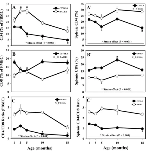
As expected, BALB/c mice had significantly higher propor-tions of circulating CD4+ cells than C57BL/6 mice at all ages (Fig. 2A). There were some age-related differences in CD4 expression in BALB/c mice T cells. At the ages of 3 and 5 months, the % of CD4+ cells was significantly higher than the % of CD4+ cells in 1-month old BALB/c mice (Fig. 2A). In both strains, the CD4+ T cells were the lowest in 18-month old mice. However, the decreases in CD4+ T cells occurred earlier and were more pronounced
Effects of age and strain on circulating (A) and splenic (B) CD19+ lymphocytes collected from male C57BL/6 and BALB/c mice up to 18 months of age
Figure 1
Effects of age and strain on circulating (A) and splenic (B) CD19+ lymphocytes collected from male C57BL/6 and BALB/c mice up to 18 months of age. * Indicates strain differences within a particular age category (P < 0.05). a,b Indicates age differences within a particular strain
Effects of age and strain on CD4+ and CD8+ lymphocytes, as well as on the CD4/CD8 ratio in circulation (A, B, and C, respec-tively) and in the spleen (A', B', and C', respecrespec-tively) of male C57BL/6 and BALB/c mice up to 18 months of age
Figure 2
Effects of age and strain on CD4+ and CD8+ lymphocytes, as well as on the CD4/CD8 ratio in circulation (A, B, and C, respectively) and in the spleen (A', B', and C', respectively) of male C57BL/6 and BALB/c mice up to 18 months of age. * Indicates strain differences within a particular age category (P < 0.05, or as indicated). a,b Indicates age
in the C57BL/6 mice (5 months of age) than in the BALB/ c strain (10 months of age, Fig. 2A). Similar to PBMC, the % of CD4+ splenocytes was significantly higher in the BALB/c strain than in C57BL/6 mice at all ages (Fig. 2A'). Both strains followed a similar pattern, different from the one in PBMC: moderate decrease up to 5 months of age (significant differences in 5-month-old animals), fol-lowed by increase at the age of 10 months and, finally, decline in CD4+ cell numbers in 18-month-old animals that was more pronounced in the C57BL/6 mice (Fig. 2A').
The C57BL/6 mice had higher % of peripheral blood CD8+ T cells than the BALB/c animals at all ages except 3-and 5-month old mice (Fig. 2B). At these ages, CD8+ T cells decreased in the C57BL/6 strain (Fig. 2B). The
expres-sion pattern diverged afterwards such that 10- and 18-month-old C57BL/6 mice had more CD8+ cells than BALB/c mice (Fig. 2B). Again, similar to peripheral blood, the C57BL/6 strain had higher % of CD8+ T cells in the spleen at all age groups (Fig. 2B'). There were no striking age-dependent changes in CD8 expression, but there was a significant increase of CD8+ cells in 10-month-old C57BL/6 mice and a significant decrease in the % of cyto-toxic T cells in 5-month-old BALB/c mice (Fig. 2B').
Mainly due to the greater % of CD4+ PBMC, the prototyp-ical Th2 mouse strain, BALB/c, had significantly greater CD4/CD8 ratio at all ages (Fig. 2C). Overall, the kinetics of the CD4/CD8 ratio were similar to the kinetics of CD4+ T cells in both strains. Significant age-related drops in the CD4/CD8 ratio occurred at the age of 10 months in the
Effects of age and strain on CD4+/CD44med/high (A), CD4+/CD44neg/low (B), CD8+/CD44med/high (C), and CD8+/CD44neg/low (D)
splenocytes isolated from the spleens of male C57BL/6 and BALB/c mice up to 18 months of age
Figure 3
Effects of age and strain on CD4+/CD44med/high (A), CD4+/CD44neg/low (B), CD8+/CD44med/high (C), and CD8+/
CD44neg/low (D) splenocytes isolated from the spleens of male C57BL/6 and BALB/c mice up to 18 months of
age. * Indicates strain differences within a particular age category (P < 0.001). a,b,c,d Indicates age differences within a particular
C57BL/6 mice PBMC and at the age of 18 months in both strains (Fig. 2C). As expected, and similar to PBMC, the CD4/CD8 ratio was significantly greater in the spleen of the BALB/c strain throughout ageing (Fig. 2C). The CD4/ CD8 ratio was relatively constant at all ages in BALB/c strain having some insignificant fluctuations, whereas the CD4/CD8 ratio decreased beginning at the age of 5 months in the C57BL/6 animals, with the decrease being significant at 18 months of age (Fig. 2C').
In addition to the CD4+ and CD8+ T cells, in the spleens, we investigated the proportion of naïve (CD44neg/low) and activated and/or memory (CD44med/high) among helper (CD4+) and cytotoxic (CD8+) T cell populations [42-44].
The % of CD4+/CD44med/high T cells increased with age in both strains (Fig. 3A), with the increase being more prom-inent in the BALB/c mice. There were no strain differences in the % of activated/memory CD4+ T cells in one-month-old mice; beginning at three months of age, BALB/c mice had more CD4+/CD44med/high T cells (Fig. 3A).
The percentage of naïve CD4+ T cells (CD44neg/low) declined with age in both strains, although the decline in the BALB/c mice was more precipitous (Fig. 3B). C57BL/6 mice had significantly more CD4+/CD44neg/low T cells at all ages (Fig. 3B).
The % of CD8+/CD44med/high cytotoxic T cells was greater in the BALB/c mice up until 5 months of age (Fig. 3C). In both strains, the % of CD8+/CD44med/high cytotoxic T cells increased with age (Fig 3C).
CD8+/CD44neg/low T cells declined with age in both mouse strains (Fig. 3D). The decrease in the BALB/c mice continued at 18 months or age, whereas the decrease in 18-month-old C57BL/6 mice was similar to the one observed in 10-month old C57BL/6 mice (Fig. 3D).
Molecules related to professional antigen presentation
The % of MHC class II+ cells in peripheral blood increased with age in the animals of both strains, peaking at 18 months (Fig. 4A). There were several age-related differ-ences in the MHC class II+ cells % within each strain. First, in the C57BL/6 strain, the levels of the MHC class II-expressing APC were fairly stable up to 10 months of age (Fig. 4A). Second, there was a significant decrease in MHC class II+ cells up to 5 months of age in the BALB/c strain (Fig. 4A). Due to the decreases of MHC class II molecules at the ages of 3 and 5 months and the more moderate increases at the age of 18 months in the BALB/c strain, the % of MHC II+ cells at these ages was lower in the BALB/c strain than in the C57BL/6 strain (Fig. 5A).
The increases in the % of MHC class II-bearing cells in the spleen were similar to increases in PBMC populations in both strains but were less pronounced (Fig. 4A'). Increases in the level of MHC class II+ cells in 5- and 18-month-old BALB/c mice approached significance (P < 0.07), while 18-month-old C57BL/6 mice had significant increases. The % of splenocytes expressing MHC class II was signifi-cantly greater in 1- and 18-month-old C57BL/6 mice than in their BALB/c counterparts, revealing a difference between the two strains (Fig. 4A').
The proportion of CD11b+ PBMC decreased in both strains, with 18-month-old animals having the lowest amounts of CD11b+ lymphocytes (Fig. 4B). Some age-related fluctuations in the % of CD11b+ were apparent in both strains. Thus, the expression of CD11b increased in both strains at the age of 3 months, and only in the C57BL/6 strain at the age of 10 months (Fig. 4B). There were age-related decreases in the % of splenocytes express-ing CD11b molecules in both strains, with the lowest % of CD11b+ APC being in 18-month-old mice (Fig. 4B'). There was a strain difference at this age: C57BL/6 mice had significantly more CD11b-bearing APC than BALB/c mice (Fig. 4B').
Variations in the % of APC-expressing CD11c molecules were similar to the variations in CD11b in both circula-tion and spleen (Figs. 4B, 4C, 4B' and 4C'). The % of dou-ble-positive CD11b/c PBMC overall was significantly greater in C57BL/6 mice than in BALB/c mice at all ages except 5 and 18 months (Fig. 4D). Age-related fluctua-tions in the % of CD11b/c+ cells were more prominent in the C57BL/6 strain than in BALB/c animals (Fig. 4D). In the spleen, the fluctuations in the % of CD11b/c cells fol-lowed similar pattern to the fluctuations of CD11b/c cells in the circulation, except that they declined with age in both mouse strains, with the effect being more prominent in the BALB/c mice (Fig. 4D'). To demonstrate that CD11c+ and/or CD11b+/CD11c+ cells in mouse periph-eral blood and spleens are primarily DC and their mye-loid progenitors, we assessed the adhesion molecule expression in CD3+ T cells and CD19+ B cells in periph-eral blood and spleens of 3-month-old mice by using three-color flow cytometry analysis. Both, CD11c and CD11b markers were expressed insignificantly in CD3+ T cells and CD19+ B cells compared to the expression levels in total PBMC or splenic populations (Fig. 5C).
Discussion
Effects of age and strain on MHCII+, CD11b+, CD11c+, and CD11b+/CD11c+ positive lymphocytes in circulation (A, B, C, and D, respectively) and in the spleen (A', B', C' and D', respectively) of male C57BL/6 and BALB/c mice up to 18 months of age
Figure 4
Effects of age and strain on MHCII+, CD11b+, CD11c+, and CD11b+/CD11c+ positive lymphocytes in circula-tion (A, B, C, and D, respectively) and in the spleen (A', B', C' and D', respectively) of male C57BL/6 and BALB/ c mice up to 18 months of age. * Indicates strain differences within a particular age category (P < 0.05). a,b Indicates age
Flow Cytometry analysis of cell-specific surface molecule expression on peripheral blood and spleen mononuclear cells in C57BL/6 and BALB/c male mice
Figure 5
Flow Cytometry analysis of cell-specific surface molecule expression on peripheral blood and spleen mononu-clear cells in C57BL/6 and BALB/c male mice. Shown on the figure are PBMC (A) and spleen (B) mononuclear cells from randomly chosen mice regardless of age. Cells were separated, stained with directly conjugated mAbs to several cell-specific markers and isotype-matching controls, and gated as low FSC and SSC populations. The MHC class II, CD19 and CD3 staining was analyzed by using single histogram statistics (columns 1, 2, 3, respectively). Two-color analysis for the CD4/CD8, CD11b/ CD11c staining was performed by using dot plots with quadrant statistics (columns 4 and 5, respectively). Analysis of the CD4/ CD44 and CD8/CD44 staining in the spleen was performed by using dot plots with multiple gate statistics (columns 6 and 7, respectively). The numbers (1, 2, 3, 4 on the dot plots in columns 6 and 7 represent the indicated CD4+ (column 6) and CD8+ (column 7) CD44high, CD44med, CD44low, CD44neg T cell sub-populations, respectively. For statistical analysis, as indicated on
(B), the CD44high and CD44med sub-populations were combined into CD44med/high (activated/memory) and, similarly, the
CD44low and CD44neg sub-populations were combined into CD44neg/low (naïve) T cell populations. (C) To eliminate the
about DC changes with age, although their number in the epidermis decreases with age [48,49]. Studies of age-related effects on B cell-mediated immunity are also not as advanced as those of the T cell immune response [2].
Similar to [50], we observed that the expression of MHC antigens on splenic lymphocytes in C57BL/6 mice increases with age. Our data indicated that C57BL/6 ani-mals have higher proportion of B cells at all ages than BALB/c mice. Similarly, previous reports suggests that the relative proportion of B220+ cells is high in C57BL/6 and intermediate in BALB/c mouse strains [8,51]. We also found that the % of CD19+ B cells increases with age in peripheral blood of both strains, while the increases in splenic B cells are prominent in C57BL/6 strain only. This finding, at least for PBMC, differs from earlier reports sug-gesting that the number of peripheral blood B lym-phocytes (B220+) in C57BL/6 and BALB/c mice does not change with age [8,52]. This difference may be due to the different markers used to identify B cells (CD19 versus B220) or to the different sex of the animals (all males used in our study; females in [52]; males and females in [8]).
Our comparison of the kinetics of MHC class II, CD19, and the adhesion molecules CD11b and CD11c expressed on professional APC indicated that, except at the age of one month, C57BL/6 mice had higher % of CD19+ cells. At the same time, we did not observe any strain-related differences in the proportion of CD11b+ or CD11c+ APC in the same age groups. Our data that peripheral blood T and B cells virtually do not express CD11b or/and CD11c are in agreement with the report that the population of peripheral blood CD11b+/CD11c+ APC are predomi-nantly DC [53]. The proportion of CD11b+/CD11c+ APC was significantly greater in 1, 3, and 10 months old C57BL/6 mice than in the BALB/c mice. As previously reported, in general, the proportion of CD11b+/CD11c+ DC in peripheral blood is relatively small [53]. Therefore, increases in the % of DC could not contribute dramati-cally to the increases in the % of cells expressing MHC class II molecules. Based on the kinetics of MHC class II, CD19, CD11b and CD11c markers in aged animals, we suggest that strain-related differences in MHC class II+ cells were most likely due to the differences in the number of CD11b+/CD11c+ DC in 1 month old mice, CD19+ B cells and DC at the age of 3 months, and CD19+ B cells in 5- and 18-month-old animals.
Unlike in PBMC populations, the spleen cell expression of MHC class II, but not of CD19, was significantly higher in 1-month old C57BL/6 mice. At the same time we did not find any strain-related differences in the % of CD11b+, CD11c+ or CD11b+/CD11c+ APC at this age. Most likely, the CD11b-/CD11c- DC populations contribute to the strain differences at this age. Our data suggest that
18-month-old mice from both strains did not differ in the % of CD19+ B cells. Interestingly, we found significant strain differences in the % of CD11b+, CD11c+ and CD11b+/ CD11c+ APC in 18 months old animals. This suggests that the strain difference in the % of MHC class II + APC at the age of 18 months was due to the differences in the % of monocytes and DC [53,54].
In contrast to previously reported data that T cell number increases dramatically with age in mice bred by a cross between CB6F1 mothers and C3D2F1 fathers [55], we conclude that age does not affect the % of total T cells in mouse PBMC from the strains used here. However, differ-ences were evident in splenic T cell populations. C57BL/6 mice had a greater CD3+ T cell % than the BALB/c strain up to the age of 10 months, followed by a substantial decline up to the age of 18 months, resulting in signifi-cantly lower % of T cells than their BALB/c counterparts. These results might be explained by the age-related differ-ential accumulation of T cell clones in the spleens of mice from different strains.
Our data are in accord with earlier reports that the proto-typical Th2-type strain BALB/c has a greater % of CD4-bearing cells. We observed that at all age groups BALB/c mice have higher % of CD4+ cells than C57BL/6 strain in both PBMC and spleen [8,51]. Similarly, our finding that the % of CD4+ cells decreases with age in peripheral blood regardless of strain agrees with previous reports [17,52,55].
The decline of CD4+ cells with age that occurs in circula-tion was not observed in the spleens of BALB/c mice. Sim-ilar, other groups have found little no or change with age in CD4+ cell proportions in spleen of several inbred mouse strains, including BALB/c [50,56-58]. Yet, in other mouse strains, including C57BL/6, the splenic CD4+ T cells decreased with age [17,52,59], which is what we observed here, albeit of smaller magnitude than in the blood. Thus, CD4+ T cell clonal expansion, which has been described in previous reports apparently occurs with age only in the spleens of the prototypical Th2-type strain BALB/c [17,18].
In general, our results support a gradual, age-dependent shift from naïve CD44neg/low cells towards an increase in CD44med/high cell populations, representing activated or memory phenotypes in mice and humans [8,28,52,55,60,61]. The data presented here are signifi-cant regarding these earlier observations in two major ways. First, we found strain differences in the distribution of naïve and activated and/or memory cells in splenic CD4+ and CD8+ T cell populations; BALB/c mice had more activated/memory T-cells and less naïve T cells than C57BL/6 mice. Second, the overall kinetics of CD4+ naïve and activated/memory phenotypes were similar between the two strains and resembled the kinetics of their CD8+ naïve and activated/memory T cells.
Overall, the strain-related phenotypic changes in the splenic APC and T cell populations did not always corre-late with the changes in the APC and T cells residing in peripheral blood. There were prominent strain differences in both PBMC and spleen populations in the % of CD4+ (higher in the BALB/c strain), CD8+ T cells (higher in C57BL/6 mice) and CD11b+/CD11c+ APC (higher in C57BL/6 mice). Other strain differences however, were present only in PBMC. Namely, the differences in the % of MHC class II + and CD19+, which were greater overall in the C57BL/6 strain than in BALB/c mice. Of note, the strain difference in the % of CD3+ T cells was only evident in the spleens but not in the peripheral blood. The C57BL/ 6 strain had greater % of T cells than BALB/c mice. Because of the differences in the composition of the peripheral blood cells and splenocytes [28] spleen cells could not always be used as surrogates for peripheral blood [52].
Age as a factor, influenced phenotypic changes in both strains. There were populations of cells that increased with age in the PBMC and spleens of both strains (i.e., MHC class II+), decreased in the periphery and spleens of both strains (CD11b+) or did not change in the PBMC and spleens of both strains (CD8+). However, in many cases the age-related differences were genetically determined, strongly supporting the evidence of intrinsic connection between genetic background and ageing.
Conclusion
Taken together, our data provide important information on the principal differences in T cell and professional APC populations between the prototypical Th1 mouse strain C57BL/6 and the prototypical Th2 strain BALB/c. Although many of the age-related changes that occur may be rather subtle and not of much consequence to animals in normal condition, they may become very relevant in conditions of disease and stress. This information might foster development of new strategies to enhance the abil-ity of the immune system to cope with infection at
differ-ent ages within the context of a particular genetic background.
Methods
Animals
Male BALB/c and C57BL/6 mice, 1, 3, 5, 10 (purchased from Harlan, Indianapolis, IN), and 18 months of age (purchased from the National Institute of Aging, NIH, Bethesda, MD) were used in this study. Animals were housed (up to 3/cage) on a 12 h light/dark cycle, with water and food available ad libitum. All animal procedures were in accordance with the Animal Welfare Act and the Guide for the Care and Use of Laboratory Animals (NIH publication No. 86-23) and were approved in advance by the Institutional Animal Care and Use Committee (IACUC) of Mississippi State University. At the designated times, animals were sacrificed via CO2 asphyxiation, blood was obtained by a cardiac puncture and then imme-diately transferred into 2 ml vacutainers containing citric buffer (BD Biosciences Pharmingen, San Diego, CA). The tubes were maintained on a rocker platform until periph-eral blood mononuclear cells (PBMC) isolation and sub-sequent analysis. In addition, body weights were recorded; spleens were collected, weighed, placed in 3 ml saline, and maintained on ice until further processing and analysis.
Cell preparation PBMC
Blood samples were diluted with PBS (1:15), and plasma was removed by centrifugation. To remove red blood cells, samples were incubated with ACK lysing buffer (Bio-Wittaker, Walkersville, MD) for 7 min on ice. Then PBMC were washed twice in PBS and stained with directly conju-gated mAbs to several cell-specific markers. PBMC were gated as low forward scatter (FSC) and low side scatter (SSC) populations using Flow Cytometer FACS Calibur (Becton Dickinson, San Jose, CA).
Splenocytes
Cell dissociation sieves (Sigma, St. Louis, MO) were used to isolate spleen mononuclear cells. Following dissocia-tion, splenocytes were incubated with ACK lysing buffer for 7 min on ice, washed twice in PBS, and stained with mAbs to different cell-specific markers. Spleen mononu-clear cells were gated as described for PBMC.
Cell counting
Antibodies and Flow Cytometry
Fluorescein-conjugated mAbs to CD4 (H129.19), CD19 (ID3), MHC class II (28-16-85), CD11b (M1/70), phyco-erythrin-conjugated mAbs to CD3 (17A2), CD8 (53-6.7), CD11c (HL3), CD44 (IM7), CD4 (H129.19), Per-CP-con-jugated mAbs to CD3 (145-2C11), CD19 (eBIOID3) and isotype-matched controls were used. All conjugated mAbs were purchased from PharMingen/BD Biosciences (San Diego, CA). Isotype-matched controls were purchased from ID Labs (Ontario, Canada). Immunofluorescent staining was analyzed using Cell Quest Version 3.3 Soft-ware (Becton Dickinson). The CD19, MHC class II, and CD3 staining was analyzed by using single histogram sta-tistics (Fig. 5A and 5B). Two-color analysis for the CD4/ CD8, CD11b/CD11c, staining was performed by using dot plots with quadrant statistics (Fig. 5A, 5B). In the spleen, analysis of the CD4/CD44 and CD8/CD44 stain-ing was performed by usstain-ing dot plots with multiple gates statistics (Fig. 5B). To eliminate the contribution of B and T cells to the % of CD11b/c cells, a three-color analysis (CD19, CD11b and CD11c; CD3, CD11b, CD11c; for B-and T-cell, respectively) was performed by gating on CD3+ T cells and CD19+ B cells and analyzed by dot plots with quadrant statistics (Fig 5C).
Statistical Analysis
All cell marker-specific lymphocyte sub-populations were expressed as a percentage of the total PBMC and spleno-cytes. Then, data was subjected to a two-way (age, strain) analysis of variance (ANOVA). When ANOVA P-value was < 0.05 for a main effect or an interaction, group means were separated by Student-Newman-Keul's multiple com-parison post hoc test.
Competing interests
The author(s) declare that they have no competing inter-ests.
Authors' contributions
LP was responsible for cell separation, immunophenotyp-ing, flow cytometry analysis, manuscript preparation and review. NF was responsible for animal sacrifice, blood and spleen separation, statistical analysis, manuscript prepara-tion and review. Both LP and NF read and approved the final version of the manuscript.
Acknowledgements
This research was funded by Competitive Research Initiation Grant Pro-gram (Mississippi State University) and in part by grant ES011654 from the National Institutes of Health NIH (NIEHS, NIH). The technical assistance of B. Boyd, T. Lee, T. Hurt, S. Sistrunk, M. Stewart, P. Crittenden, and M. Pinchuk is greatly appreciated.
References
1. Globerson A, Effros RB: Ageing of lymphocytes and lym-phocytes in the aged. Immunol Today 2000, 21:515-21.
2. Linton PJ, Dorshkind K: Age-related changes in lymphocyte development and function. Nat Immunol 2004, 5:133-9. 3. Solana R, Villanueva JL, Pena J, De la Fuente M: Cell mediated
immunity in ageing. Comp Biochem Physiol A 1991, 99:1-4. 4. Kay RA: TCR gene polymorphisms and autoimmune disease.
Eur J Immunogenet 1996, 23:161-77.
5. Pawelec G, Solana R: Immunosenescence. Immunol Today 1997, 18:514-6.
6. Hirokawa K: Age-related changes of signal transduction in T cells. Exp Gerontol 1999, 34:7-18.
7. Pawelec G, Effros RB, Caruso C, Remarque E, Barnett Y, Solana R: T cells and aging (update february 1999). Front Biosci 1999, 4:D216-69.
8. Chen J, Flurkey K, Harrison DE: A reduced peripheral blood CD4(+) lymphocyte proportion is a consistent ageing pheno-type. Mech Ageing Dev 2002, 123:145-53.
9. Solana R, Pawelec G, Tarazona R: Aging and innate immunity.
Immunity 2006, 24:491-4.
10. Effros RB, Cai Z, Linton PJ: CD8 T cells and aging. Crit Rev Immunol
2003, 23:45-64.
11. Grubeck-Loebenstein B, Wick G: The aging of the immune sys-tem. Adv Immunol 2002, 80:243-84.
12. Timm JA, Thoman ML: Maturation of CD4+ lymphocytes in the aged microenvironment results in a memory-enriched pop-ulation. J Immunol 1999, 162:711-7.
13. Kapasi ZF, Murali-Krishna K, McRae ML, Ahmed R: Defective gen-eration but normal maintenance of memory T cells in old mice. Eur J Immunol 2002, 32:1567-73.
14. Miller JF, Heath WR, Allison J, Morahan G, Hoffmann M, Kurts C, Kosaka H: T cell tolerance and autoimmunity. Ciba Found Symp
1997, 204:159-68. discussion 168–71
15. Nel AE, Slaughter N: T-cell activation through the antigen receptor. Part 2: role of signaling cascades in T-cell differen-tiation, anergy, immune senescence, and development of immunotherapy. J Allergy Clin Immunol 2002, 109:901-15. 16. Lerner A, Yamada T, Miller RA: Pgp-1hi T lymphocytes
accumu-late with age in mice and respond poorly to concanavalin A.
Eur J Immunol 1989, 19:977-82.
17. Callahan JE, Kappler JW, Marrack P: Unexpected expansions of CD8-bearing cells in old mice. J Immunol 1993, 151:6657-69. 18. Schwab R, Szabo P, Manavalan JS, Weksler ME, Posnett DN, Pannetier
C, Kourilsky P, Even J: Expanded CD4+ and CD8+ T cell clones in elderly humans. J Immunol 1997, 158:4493-9.
19. Hayashi Y, Takemura T, Akashi T, Esaki Y, Kurashima C, Hirokawa K: Immunopathological analysis of interstitial renal lesions in elderly people. Pathobiology 1990, 58:230-5.
20. Ben-Yedidia T, Abel L, Arnon R, Globerson A: Efficacy of anti-influ-enza peptide vaccine in aged mice. Mech Ageing Dev 1998, 104:11-23.
21. Flurkey K, Miller RA, Harrison DE: Cellular determinants of age-related decrements in the T-cell mitogen response of B6CBAF1 mice. J Gerontol 1992, 47:B115-20.
22. Posnett DN, Sinha R, Kabak S, Russo C: Clonal populations of T cells in normal elderly humans: the T cell equivalent to "benign monoclonal gammapathy". J Exp Med 1994, 179:609-18.
23. Johnson SA, Rozzo SJ, Cambier JC: Aging-dependent exclusion of antigen-inexperienced cells from the peripheral B cell reper-toire. J Immunol 2002, 168:5014-23.
24. Kline GH, Hayden TA, Klinman NR: B cell maintenance in aged mice reflects both increased B cell longevity and decreased B cell generation. J Immunol 1999, 162:3342-9.
25. Zheng B, Han S, Takahashi Y, Kelsoe G: Immunosenescence and germinal center reaction. Immunol Rev 1997, 160:63-77. 26. Whisler RL, Grants IS: Age-related alterations in the activation
and expression of phosphotyrosine kinases and protein kinase C (PKC) among human B cells. Mech Ageing Dev 1993, 71:31-46.
27. Yang X, Stedra J, Cerny J: Relative contribution of T and B cells to hypermutation and selection of the antibody repertoire in germinal centers of aged mice. J Exp Med 1996, 183:959-70. 28. Effros RB: Long-term immunological memory against viruses.
Mech Ageing Dev 2000, 121:161-71.
Publish with BioMed Central and every scientist can read your work free of charge "BioMed Central will be the most significant development for disseminating the results of biomedical researc h in our lifetime."
Sir Paul Nurse, Cancer Research UK
Your research papers will be:
available free of charge to the entire biomedical community
peer reviewed and published immediately upon acceptance
cited in PubMed and archived on PubMed Central
yours — you keep the copyright
Submit your manuscript here:
http://www.biomedcentral.com/info/publishing_adv.asp
BioMedcentral 30. Fujii S, Shimizu K, Hemmi H, Steinman RM: Innate Valpha14(+)
natural killer T cells mature dendritic cells, leading to strong adaptive immunity. Immunol Rev 2007, 220:183-98.
31. Steinman RM: Dendritic cells: understanding immunogenicity.
Eur J Immunol 2007, 37(Suppl 1):S53-60.
32. Hoag KA, Clise-Dwyer K, Lim YH, Nashold FE, Gestwicki J, Cancro MP, Hayes CE: A quantitative-trait locus controlling periph-eral B-cell deficiency maps to mouse Chromosome 15. Immu-nogenetics 2000, 51:924-9.
33. Clementi M, Forabosco P, Amadori A, Zamarchi R, De Silvestro G, Di Gianantonio E, Chieco-Bianchi L, Tenconi R: CD4 and CD8 T lym-phocyte inheritance. Evidence for major autosomal reces-sive genes. Hum Genet 1999, 105:337-42.
34. Kraal G, Weissman IL, Butcher EC: Genetic control of T-cell sub-set representation in inbred mice. Immunogenetics 1983, 18:585-92.
35. Amadori A, Zamarchi R, De Silvestro G, Forza G, Cavatton G, Danieli GA, Clementi M, Chieco-Bianchi L: Genetic control of the CD4/ CD8 T-cell ratio in humans. Nat Med 1995, 1:1279-83. 36. von Stebut E, Belkaid Y, Nguyen BV, Cushing M, Sacks DL, Udey MC:
Leishmania major-infected murine langerhans cell-like den-dritic cells from susceptible mice release IL-12 after infec-tion and vaccinate against experimental cutaneous Leishmaniasis. Eur J Immunol 2000, 30:3498-506.
37. Misslitz AC, Bonhagen K, Harbecke D, Lippuner C, Kamradt T, Aeb-ischer T: Two waves of antigen-containing dendritic cells in vivo in experimental Leishmania major infection. Eur J Immu-nol 2004, 34:715-25.
38. Pinto EF, de Mello Cortezia M, Rossi-Bergmann B: Interferon-gamma-inducing oral vaccination with Leishmania amazon-ensis antigens protects BALB/c and C57BL/6 mice against cutaneous leishmaniasis. Vaccine 2003, 21:3534-41.
39. Ritchie AJ, Yam AO, Tanabe KM, Rice SA, Cooley MA: Modification of in vivo and in vitro T- and B-cell-mediated immune responses by the Pseudomonas aeruginosa quorum-sensing molecule N-(3-oxododecanoyl)-L-homoserine lactone. Infect Immun 2003, 71:4421-31.
40. Gemmell E, Winning TA, Carter CL, Ford PJ, Bird PS, Ashman RB, Grieco DA, Seymour GJ: Differences in mouse strain influence leukocyte and immunoglobulin phenotype response to Por-phyromonas gingivalis. Oral Microbiol Immunol 2003, 18:364-70. 41. Chen J, Astle CM, Harrison DE: Development and aging of
prim-itive hematopoietic stem cells in BALB/cBy mice. Exp Hema-tol 1999, 27:928-35.
42. Swain SL, Bradley LM: Helper T cell memory: more questions than answers. Semin Immunol 1992, 4:59-68.
43. Griffin JP, Orme IM: Evolution of CD4 T-cell subsets following infection of naive and memory immune mice with Mycobac-terium tuberculosis. Infect Immun 1994, 62:1683-90.
44. Mobley JL, Rigby SM, Dailey MO: Regulation of adhesion mole-cule expression by CD8 T cells in vivo. II. Expression of L-selectin (CD62L) by memory cytolytic T cells responding to minor histocompatibility antigens. J Immunol 1994, 153:5443-52.
45. Chen J, Astle CM, Harrison DE: Genetic regulation of primitive hematopoietic stem cell senescence. Exp Hematol 2000, 28:442-50.
46. Ortega E, Garcia JJ, De La Fuente M: Ageing modulates some aspects of the non-specific immune response of murine mac-rophages and lymphocytes. Exp Physiol 2000, 85:519-25. 47. Varas A, Sacedon R, Hernandez-Lopez C, Jimenez E, Garcia-Ceca J,
Arias-Diaz J, Zapata AG, Vicente A: Age-dependent changes in thymic macrophages and dendritic cells. Microsc Res Tech 2003, 62:501-7.
48. Uyemura K, Castle SC, Makinodan T: The frail elderly: role of dendritic cells in the susceptibility of infection. Mech Ageing Dev 2002, 123:955-62.
49. Sprecher E, David D, Yadin H, Peleg BA, Becker Y: Mouse footpad Langerhans cells as an indicator for safety of foot and mouth disease virus vaccines. J Virol Methods 1990, 29:189-96.
50. Sidman CL, Luther EA, Marshall JD, Nguyen KA, Roopenian DC, Worthen SM: Increased expression of major histocompatibil-ity complex antigens on lymphocytes from aged mice. Proc Natl Acad Sci USA 1987, 84:7624-8.
51. Chen J, Harrison DE: Quantitative trait loci regulating relative lymphocyte proportions in mouse peripheral blood. Blood
2002, 99:561-6.
52. Oughton JA, Pereira CB, DeKrey GK, Collier JM, Frank AA, Kerkvliet NI: Phenotypic analysis of spleen, thymus, and peripheral blood cells in aged C57B1/6 mice following long-term expo-sure to 2,3,7,8-tetrachlorodibenzo-p-dioxin. Fundam Appl Toxi-col 1995, 25:60-9.
53. Manfra DJ, Chen SC, Jensen KK, Fine JS, Wiekowski MT, Lira SA: Conditional expression of murine Flt3 ligand leads to expan-sion of multiple dendritic cell subsets in peripheral blood and tissues of transgenic mice. J Immunol 2003, 170:2843-52. 54. Maraskovsky E, Brasel K, Teepe M, Roux ER, Lyman SD, Shortman K,
McKenna HJ: Dramatic increase in the numbers of functionally mature dendritic cells in Flt3 ligand-treated mice: multiple dendritic cell subpopulations identified. J Exp Med 1996, 184:1953-62.
55. Miller RA: Age-related changes in T cell surface markers: a longitudinal analysis in genetically heterogeneous mice.
Mech Ageing Dev 1997, 96:181-96.
56. Kirschmann DA, Murasko DM: Splenic and inguinal lymph node T cells of aged mice respond differently to polyclonal and antigen-specific stimuli. Cell Immunol 1992, 139:426-37. 57. Komuro T, Sano K, Asano Y, Tada T: Analysis of age-related
degeneracy of T-cell repertoire: localized functional failure in CD8+ T cells. Scand J Immunol 1990, 32:545-53.
58. Gonzalez-Quintial R, Theofilopoulos AN: V beta gene repertoires in aging mice. J Immunol 1992, 149:230-6.
59. Dubiski S, Ponnappan U, Cinader B: Strain polymorphism in pro-gression of aging: changes in CD4, CD8 bearing subpopula-tions. Immunol Lett 1989, 23:1-7.
60. Globerson A: T lymphocytes and aging. Int Arch Allergy Immunol
1995, 107:491-7.
61. Lynch F, Ceredig R: Mouse strain variation in Ly-24 (Pgp-1) expression by peripheral T cells and thymocytes: implica-tions for T cell differentiation. Eur J Immunol 1989, 19:223-9. 62. Filipov NM, Pinchuk LM, Boyd BL, Crittenden PL: Immunotoxic