King’s Research Portal
DOI:
10.1016/j.jns.2018.02.032
Document Version
Peer reviewed version
Link to publication record in King's Research Portal
Citation for published version (APA):
Yousaf, T., Pagano, G., Niccolini, F., & Politis, M. (2018). Excessive daytime sleepiness may be associated with caudate denervation in Parkinson disease. Journal of the Neurological Sciences, 387, 220-227.
https://doi.org/10.1016/j.jns.2018.02.032
Citing this paper
Please note that where the full-text provided on King's Research Portal is the Author Accepted Manuscript or Post-Print version this may differ from the final Published version. If citing, it is advised that you check and use the publisher's definitive version for pagination, volume/issue, and date of publication details. And where the final published version is provided on the Research Portal, if citing you are again advised to check the publisher's website for any subsequent corrections.
General rights
Copyright and moral rights for the publications made accessible in the Research Portal are retained by the authors and/or other copyright owners and it is a condition of accessing publications that users recognize and abide by the legal requirements associated with these rights. •Users may download and print one copy of any publication from the Research Portal for the purpose of private study or research.
•You may not further distribute the material or use it for any profit-making activity or commercial gain •You may freely distribute the URL identifying the publication in the Research Portal
Take down policy
If you believe that this document breaches copyright please contact librarypure@kcl.ac.uk providing details, and we will remove access to the work immediately and investigate your claim.
Excessive daytime sleepiness may be associated with caudate denervation in Parkinson disease
Tayyabah Yousaf, Gennaro Pagano, Flavia Niccolini, Marios Politis
PII: S0022-510X(18)30090-X
DOI: doi:10.1016/j.jns.2018.02.032
Reference: JNS 15800
To appear in: Journal of the Neurological Sciences
Received date: 10 August 2017
Revised date: 19 February 2018
Accepted date: 20 February 2018
Please cite this article as: Tayyabah Yousaf, Gennaro Pagano, Flavia Niccolini, Marios Politis , Excessive daytime sleepiness may be associated with caudate denervation in Parkinson disease. The address for the corresponding author was captured as affiliation for all authors. Please check if appropriate. Jns(2018), doi:10.1016/j.jns.2018.02.032
This is a PDF file of an unedited manuscript that has been accepted for publication. As a service to our customers we are providing this early version of the manuscript. The manuscript will undergo copyediting, typesetting, and review of the resulting proof before it is published in its final form. Please note that during the production process errors may be discovered which could affect the content, and all legal disclaimers that apply to the journal pertain.
ACCEPTED MANUSCRIPT
Excessive daytime sleepiness may be associated with caudate denervation in Parkinson disease
Tayyabah Yousaf1, MSc, Gennaro Pagano1, MD, MSc, Flavia Niccolini1, MD, MSc and Marios Politis1, MD, PhD, FRCP, FEAN
1
Neurodegeneration Imaging Group, Institute of Psychiatry, Psychology and Neuroscience (IoPPN), King’s College London, London, UK.
Corresponding author:
Professor Marios Politis, MD, MSc, PhD, FRCP, FEAN Neurodegeneration Imaging Group
Maurice Wohl Clinical Neuroscience Institute
Institute of Psychiatry, Psychology & Neuroscience (IoPPN) 125 Coldharbour Lane, Camberwell, London, SE5 9NU Telephone: +44-207-8485682
email: marios.politis@kcl.ac.uk website: http://nig-politis.com/
Tayyabah Yousaf: tayyabah.yousaf@kcl.ac.uk Gennaro Pagano: gennaro.pagano@kcl.ac.uk Flavia Niccolini: flavia.niccolini@kcl.ac.uk
Abstract word count: 271
ACCEPTED MANUSCRIPT
Author contributions
Miss Yousaf - Study concept and design, statistical analysis and interpretation of data and drafting of the manuscript
Dr. Pagano - Study concept and design, study supervision, interpretation of data and drafting of the manuscript
Dr. Niccolini - critical revision of the manuscript for important intellectua l content
Dr. Politis - Study concept and design, study supervision, critical revision of the manuscript for important intellectual content and final approval of the manuscript
Financial Disclosure Statement
Data used in the preparation of this article were obtained from the Parkinson’s Progression Markers Initiative (PPMI) database (www.ppmi-info.org/data). For up-to-date information on the study, visit www.ppmi-info.org. PPMI – a public-private partnership - is sponsored by the Michael J. Fox Foundation for Parkinson's Research (MJFF) and is co-funded by MJFF, Abbvie, Avid Radiopharmaceuticals, Biogen Idec, Bristol-Myers Squibb, Covance, Eli Lilly & Co., F. Hoffman-La Roche, Ltd., GE Healthcare, Genentech, GlaxoSmithKline, Lundbeck, Merck, MesoScale, Piramal, Pfizer and UCB.PPMI. Industry partners are contributing to PPMI through financial and in-kind donations and are playing a lead role in providing feedback on study parameters through the Industry Scientific Advisory Board (ISAB). Through close interaction with the study, the ISAB is positioned to inform the selection and review of potential progression markers that could be used in clinical testing.
Miss Yousaf, Dr. Pagano, Dr. Niccolini and Dr. Politis report no disclosures.
Declaration of interests
ACCEPTED MANUSCRIPT
ABSTRACT
Excessive daytime sleepiness (EDS) is one of the earliest and most common non-motor symptoms of PD, substantially impacting on patient’s quality of life. Using the Parkinson's Progression Markers Initiative database, we performed a case-control study to investigate whether dopaminergic deficit is associated with the development of EDS using dopaminergic specific single photon emission computed tomography (SPECT) molecular imaging of dopamine transporters (DAT). We enrolled 84 early de novo PD patients with EDS and 84 without EDS, who were matched for age, gender, age of diagnosis, years of education and disease duration. We assessed and compared semi-quantified [123I]FP-CIT SPECT, and motor and non-motor features amongst these two groups, alongside exploring the clinical and imaging correlates of EDS and the predictive significance of these markers in the development of EDS. PD patients with EDS had worse non-motor (MDS-UPDRS Part-I,
P<0.001) and motor (MDS-UPRDS Part-II, P=0.005) experiences of daily living, as well as worse autonomic (SCOPA-AUT, P<0.0001) and cognitive (MoCA P=0.05) function, depression (GDS, P=0.002), and reduced caudate DAT ([123I]FP-CIT, P=0.024) compared to PD patients without EDS. Lower caudate [123I]FP-CIT values correlated with higher EDS scores (r=-0.192, P=0.013). Among patients without EDS, 47 PD patients (56%) developed EDS over a median follow-up of 36 months. Cox multivariate analysis including all clinical and imaging data available, revealed that abnormal caudate [123I]FP-CIT uptake (P=0.030) and disease duration (P=0.018) were predictors for the development of EDS. Although our findings indicate that dopaminergic deficits in the caudate may be associated to EDS in patients with PD, the pathophysiological causality is debateable, given that dopamine caudate denervation may covary with dopaminergic involvement at other targets and with non-dopaminergic involvement.
ACCEPTED MANUSCRIPT
1.0 INTRODUCTION
Excessive daytime sleepiness (EDS) is one of the most common and troublesome non-motor symptoms (NMS) in both early and advanced Parkinson’s disease (PD) [1]. Although a range of NMS are primarily reported to be associated with non-dopaminergic deficits, such as depression [2], BMI changes [3] and fatigue [4], the dopaminergic system has also been identified as a pivotal contributor to non-motor symptoms in PD [5], reflecting the multisystem nature of the disorder. Politis and colleagues reported in vivo evidence of dopamine dysfunction in the hypothalamus of PD patients, suggesting a dopaminergic contribution to several non-motor symptoms, including sleep disorders, neuro-endocrinal problems and autonomic dysfunction [6]. NMS are often poorly identified and inadequately treated, highlighting the challenge attached with NMS management. Therefore, gaining an insight into the neurobiological mechanisms underlying these symptoms could improve management and the development of novel treatments.
The clinical heterogeneity of PD has led to the discovery of phenotypic subtypes of motor and non-motor symptoms [7]. Patients with daytime sleepiness, along with cognitive impairment, autonomic dysfunction and depression were recognised as a specific subtype of PD [7], instigating an interest in the clinical associations of EDS, alongside phenotypic and neurobiological risk factors of EDS development.
An amalgamation of dopaminergic denervation, nocturnal sleep disruption and dopaminergic medication is likely to be causative [8, 9], though several studies have suggested that EDS may be a primary feature of PD, unrelated to dopaminergic therapies or nocturnal sleep disturbances [10]. A magnetic resonance imaging (MRI) brain morphometry revealed that EDS in PD patients was related to atrophy of the medial cerebellar peduncle, suggesting that degeneration of the pontomedullary respiratory centres may underlie EDS development in PD [11]. Further, patients experiencing daytime sleepiness demonstrated a prominent impairment in circadian melatonin secretion, suggesting that circadian dysfunction may underlie excessive sleepiness [12]. A genetic component contributing to the pathogenesis of EDS has also been reported [13]. Furthermore, Saper and colleagues proposed that a ‘flip-flop switch’ is responsible for the sleep-wake cycle in primates, whereby the suprachiasmatic nucleus regulates the internal rhythm between two switches, with hypocretin potentially playing a regulatory role [14]. The quantity of hypocretin-producing neurons and the CSF level of hypocretin-1 have been shown to be reduced in PD [15-17], with consistent loss of
ACCEPTED MANUSCRIPT
hypocretin-producing neurons demonstrated by a reduction in the number of post-mortem hypothalamic hypocretin neurons, acknowledged as the ‘gold standard’ [16, 17].
Disease variables found to be associated with EDS include longer disease duration, older age, severity of motor manifestations, depression, cognitive impairment and, non-tremor dominant motor phenotype [8, 18], though not all epidemiological studies of PD have discovered the same picture or disease variables to be associated with EDS [19].
Studies investigating EDS and dopaminergic pathways discovered that subjective daytime sleepiness is associated with striatonigral degeneration [20], though others have proposed that it may be associated with extrastriatal dopaminergic loss in specific brain structures involved in alertness [21]. Thus, the role of the striatum in the development of EDS remains unclear. We aimed to explore the relationship between EDS and striatal dopamine terminals, using [123I]FP-CIT single photon emission computed tomography (SPECT), alongside determining the clinical correlates and risk factors in the development of EDS, in de novo (drug-naïve) PD patients. We hypothesised that EDS would be associated with striatal dopaminergic denervation.
2.0 METHODS
The Parkinson’s Progression Markers Initiative (PPMI) is a five-year observational, international, multi-centre study designed to provide insight into disease aetiology by identifying PD progression biomarkers. The present study was written according to the STROBE guidelines. This study is registered with ClinicalTrials.gov, number NCT01141023.
2.1 Cohort selection
We included PD patients aged >30 years old, who had a disease duration ≤ 2 years and were not on dopamine replacement therapy. Institutional review boards approved the study and written informed consent was obtained from all participants. From a total of 412 patients with PD, 84 had EDS according to the Movement Disorder Society Unified PD Rating Scale (MDS-UPDRS) Part-I, item 1.8 ‘daytime sleepiness’. This self-reported, self-completed instrument consists of 5 statements, rating from 0: normal to 4: severe, with a higher rating corresponding to a higher degree of EDS. The presence of EDS was defined as daytime
ACCEPTED MANUSCRIPT
sleepiness interfering with daily activities and social interactions (cut-off >1). All clinical and imaging assessments were performed in subjects who were not receiving any dopaminergic medication, at baseline. The cut-off values considered abnormal for all clinical and imaging variables were calculated as 2 S.D. from the mean of HCs, as previously stated [22].
Using propensity scores, PD patients with EDS were matched 1:1 for age, gender, age at disease diagnosis, duration of disease and years of education with 84 PD patients without EDS. Characteristics of patients included are summarised in Table 1.
2.2 Dopaminergic imaging
SPECT images were obtained 4 ± 0.5 h after administrating an injection of approximately 185 MBq [123I]FP-CIT. [123I]FP-CIT SPECT scans were analysed following the imaging technical operations manual (http://ppmi-info.org/). In brief, SPECT image volumes were spatially normalised to an Ioflupane template. The eight most prominent axial slices containing the striatum were summed and a standardised volume of interest (VOI) template was then applied to this image. VOI analyses were performed on the right and left caudate and putamen, employing the occipital region as the reference tissue. Specific binding ratios (SBR) were calculated as the ratio of the putamen or caudate VOI count density divided by the occipital cortex count density minus one. This measure approximates the binding potential, BPnd, when the radioligand is in equilibrium at the target site and has previously been reported with Ioflupane SPECT [23].
2.3 Structural imaging
T1-weighted Magnetic Resonance Imaging (MRI) scans were acquired in the saital plane on 3T Siemens (TIM Trio and Verio) scanners (Erlangen, Germany), employing a magnetisation-prepared rapid-acquisition gradient echo sequence. The parameters of the MRI sequence were as follows: repetition time: 2,300/1,900 milliseconds; echo time: 2.98/2.96/2.27/2.48/2.52 milliseconds; inversion time: 900 milliseconds; flip angle: 9°; matrix: 256 × 256; and 1mm3 isotropic voxel.
Surface-based analysis was carried out using the freely available software package FreeSurfer (version 5.3, http://surfer.nmr.mgh.harvard.edu). In brief, the FreeSurfer preprocessing
pipeline includes (1) removal of non-brain tissue; (2) automated Talairach transformation; (3) segmentation of subcortical white matter and deep grey matter structures (4) intensity
ACCEPTED MANUSCRIPT
normalisation (5) tessekkation of the gray matter/white matter boundary; (6) automated topology correction; (7) surface deformation; and (8) registration of the subjects’ brains to the common spherical atlas. The results implemented in FreeSurfer were visually inspected and manually edited, if required.
Regions-of- interest (ROIs) included subcortical structures such as caudate, putamen, accumbens, pallidum, thalamus, hippocampus and amygdala, as well as cortical structures such as precentral and postcentral gyrus, anterior and posterior cingulate, entorhinal cortex, fusiform gyrus, inferior and superior parietal cortex, inferior, middle and superior frontal cortex, insula cortex, parahippocampal cortex, inferior, middle and superior temporal cortex, lateral occipital and lateral orbitofrontal cortex.
2.4 Assessment of outcomes and follow-up
PD patients attended a follow-up visit once every year, where the physician reviewed the patient’s condition. This included reassessing the patient for EDS development. Follow-up period was terminated either at the patient’s last visit or if the patient developed EDS.
2.5 Dopaminergic therapy
Study participants could start dopamine replacement therapy (DRT) at any point after baseline as part of routine clinical care. A participant was considered to have received DRT (dopamine agonists, levodopa, monoamine oxidase type B (MAO-B) inhibitors) from the first time it was recorded at an annual study visit. Information about medication dosages was not readily ascertained in the database and is not included.
2.6 Statistical methods
Statistical analyses were performed using Statistical Package for the Social Sciences (SPSS), version 22 and graphical illustration in GraphPad Prism 6. For all variables, Gaussianity was tested with Kolmogorov-Smirnov test. Multivariate analysis of variance (MANOVA) was used to assess the main effects of all clinical and imaging variables between the group of PD patients with EDS and PD patients without EDS. If the overall multivariate test was significant, P values for each variable were calculated following Bonferroni correction. Categorical variables were compared using a χ2 test. A one-way ANOVA and Dunnett’s post-hoc test was carried out to determine differences between the means of the three groups (patients who scored 0, 1 or 2 on UPDRS-Part 1, Item 1.8). Cohen’s d was calculated for
ACCEPTED MANUSCRIPT
each variable by dividing the mean difference by the pooled standard deviation. Pearson and Spearman's correlation tests were carried out to assess the relationship between [123I]FP-CIT SPECT and EDS. To determine the independent predictors of EDS, multivariate Cox proportional hazards analyses (backward conditional) were used including variables achieving P<0.10 on univariate analysis. Only the time to occurrence of the first event in a category for a given subject was used. To increase the stability of our findings, we confirm that the outcome present at one visit was still present at subsequent visits. Statistical significance was set at P<0.05, Bonferroni corrected.
3.0 RESULTS
3.1 Dopaminergic imaging
Caudate [123I]FP-CIT binding was reduced in PD patients with EDS compared to PD patients without EDS (1.87±0.54 vs 2.06±0.53, P=0.025; Figure 1 and 2A). Caudate [123I]FP-CIT uptake did not correlate with insomnia (MDS-UPDRS, Part 1, item 1.7, P=0.288, ρ=0.052) or RBD (RBDSQ, P=0.091, ρ=-0.082). Putamen [123
I]FP-CIT binding was not significantly different between the two groups (0.77 ±0.30 vs 0.83 ±0.27, P=0.164).
3.2 Structural imaging
There were no differences in cortical thickness or subcortical volumes between PD patients with EDS compared to those without (P>0.1).
3.3 Clinical phenotype analysis
PD patients with EDS had an overall worse clinical picture (MDS-UPDRS Total, P<0.001), worse non-motor (MDS-UPDRS Part-I (excluding item 1.8 subscores), P<0.001) and motor experiences of daily living (MDS-UPRDS Part-II, P=0.001), as well as worse autonomic (SCOPA-AUT, P<0.001) cognitive (MoCA, P=0.01) and depression (GDS, P=0.002) scores compared to PD patients without EDS (Table 1; Figure 2A). No differences were found in motor symptom severity between PD with and without EDS (MDS-UPRDS Part-III, P>0.10).
3.4 Correlations
Higher EDS scores at baseline were associated with decreased caudate [123I]FP-CIT binding (r=-0.192; P=0.013), global non motor symptom burden (MDS-UPDRS Part-I, ρ=0.470;
ACCEPTED MANUSCRIPT
P<0.001), worse motor experiences of daily living (MDS-UPDRS Part-II, ρ=0.349; P<0.001), autonomic dysfunction (SCOPA-AUT, ρ=0.366; P>0.001), cognitive impairment (MoCA, ρ=-0.175; P=0.023) and depression (GDS, ρ=0.264; P=0.002) (Figure 2B) at baseline.
3.5 EDS predictive analysis
Of the 84 PD patients without EDS at baseline, 34 initiated levodopa treatment (40.5%) and 27 started treatment with dopamine agonist (32.1%). 47 PD patients (56%) developed EDS over a mean follow-up of 36.9 ± 20.2 months (range: 3–60 months). Of these 47 PD patients, 23 (48.9%) were taking levodopa and 15 (31.9%) were taking dopamine agonists. Of the 37 PD patients who did not develop EDS, 11 (29.7%) were taking levodopa and 12 (32.4%) were taking dopamine agonist. There was no significant difference in dopamine agonist treatment between those PD patients who go onto develop EDS compared to those who do not go onto develop EDS (P=0.397).
Multivariate Cox proportional hazards regression analysis, performed including all variables with P≤0.10 at univariate analysis, revealed that baseline abnormal (≤1.76) caudate [123 I]FP-CIT (Hazard ratio [HR]=1.94 Confidence Interval [CI]=1.06–3.53, P=0.030; Figure 3) and disease duration (HR=1.62, [CI]=1.08–2.41 per year of disease, P=0.018) were predictors for developing EDS. Levodopa (HR=1.441, CI=0.808-2.572, P>0.1) and dopamine agonists (HR=0.657, CI=0.354-1.220, P>0.1) were not predictors for developing EDS.
4.0 DISCUSSION
Our findings demonstrate that the development of EDS in patients with PD is related to the advancing disease, reflected particularly by non-motor symptom burden, and dopaminergic deficits in the caudate, with no differences in brain atrophy. We have combined [123 I]FP-CIT-SPECT and MR-based structural imaging and have showed a loss of dopaminergic function in the caudate of PD patients with EDS compared to those without. Dopaminergic deficits in the caudate were associated with the severity of EDS in the PD-EDS group and with a two-fold increased risk (HR=1.94) of developing EDS over a period of 36 months in the PD non-EDS group. Our findings are in line with a previous smaller cross-sectional study which has reported correlations between striatal dopamine transporter (DAT) loss and sleep problems in patients with PD [20]. DAT regulates dopamine concentration within the synaptic cleft via the reuptake of dopamine into presynaptic neurons. SPECT radiotracers for the DAT provide
ACCEPTED MANUSCRIPT
excellent markers for the integrity of the presynaptic dopaminergic system. ‘Loss of function’ refers to DAT loss, which is either the result of loss of dopaminergic terminals or a change in synaptic dopamine levels.
Although the correlations between EDS and cognitive impairment, autonomic dysfunction and depression were somewhat weak, likely due to the sample size of the cohort, they were found to be associated, particularly when comparing the PD-EDS and PD non-EDS cohorts. This is consistent with previous studies which have revealed patterns of coherency amidst motor and non-motor domains within PD. van Rooden et al. [24] discovered that daytime sleepiness was often present with depression, cognitive impairment, autonomic dysfunction and psychotic and axial symptoms, with a second pattern characterised by sleep disturbances, depression and cognitive impairment, highlighting a conspicuous disease profile implicated with sleep. EDS has also been found to be an independent predictive factor for cognitive difficulties in the general elderly population [25], suggesting EDS is a significant risk factor for cognitive deficits. Intriguingly, the caudate dopaminergic function has been recognised as a fundamental node in supporting cognitive domains. Previous findings have illustrated that loss of dopaminergic projections to the caudate correlate with degree of dementia in PD [26], alongside others reporting that a reduction in caudate uptake of [18F]DOPA is associated with verbal and visual memory functioning, verbal fluency and, delayed recall [27] highlighting that the functional integrity of presynaptic dopaminergic synthesis is crucial for cognitive function in PD. More recently, in a PPMI study, Schrag and colleagues demonstrated that dopaminergic denervation within the caudate, alongside four other variables, predicted cognitive impairment at 2 years [28]. This is particularly interesting given that Schrag explored a larger version of, essentially, the same cohort and reported similar findings. Therefore, the mechanisms underlying the development of EDS and cognitive problems may share an overlapping dopaminergic substrate. Therefore, the mechanisms underlying the development of EDS and cognitive problems may share an overlapping dopaminergic substrate.
Dopamine has been reported to promote wakefulness in a range of animal models including cats and drosophila [29, 30], though studies have also demonstrated its association with sleepiness and the sleep state [31]. This discrepancy can be explained by dopamine mediating both wakefulness and sleepiness depending on its concentration. Studies have, for example,
ACCEPTED MANUSCRIPT
demonstrated that either excessive or insufficient dopamine impairs cognitive functions [32], suggesting that a similar mechanism may underlie the varying sleep disorders in PD, particularly because discrete classes of receptors may be activated by varying concentrations of dopamine based on receptor affinity [9].
The caudate nucleus is a major receptive component of the basal ganglia, embedded within its large neural network. The striatum is the focal output structure, receiving afferent fibres from distinct cortical areas, projecting them on to the thalamus and brainstem for auxiliary propagation via pallidum back to cortex [33]. Distinct functional zones have been proposed, within the caudate, based on their innervation by specific cortical areas [33]. It is not surprising that the caudate has surfaced as a common node in the regulatory systems involved in sleep problems, arousal and executive function, due to its complex role in regulating the impact of impulses between the thalamus and cortex. Hyper-arousal indices have emerged in studies that have specifically disrupted caudate function, highlighting that caudate lesions might induce hyper-responsivity and behavioural restlessness [30].
Recent studies have implicated the basal ganglia and its respective structures in arousal and the sleep-wake cycle. Neurotoxic lesion studies have demonstrated the pivotal role of the basal ganglia in regulating the sleep-wake cycle. Specifically, bilateral striatal lesions result in reduced wakefulness, which is attenuated when the striatal (caudoputamen) lesions include the nucleus accumbens [34]. These findings highlight that the caudate is involved in promoting wakefulness whereas the nucleus accumbens primarily enhances sleepiness. In this direction, our results highlight that a loss of DAT, therefore dopamine, within the caudate, could in fact cause sleepiness in PD patients. However, it is also important to recognise the role of other major nuclei including the globus pallidus, thalamus and substantia nigra. Cell body-specific lesioning of the globus pallidus externa has resulted in insomnia, with rats demonstrating a prominent increase of up to 45% [34]. Further, neuronal loss within the substantia nigra, excluding the globus pallidus, consequently affected sleep-wake behaviour, specifically presenting as an increase of wakefulness [34, 35]. Lazarus and colleagues propose that the nucleus accumbens, in particular, is a key node in the circuitry for sleep [36]. A2A receptors residing in the nucleus accumbens are fundamental for the wakefulness
induced by caffeine, and presumably modulate neural substrates through which arousal is also produced by dopamine. The nucleus accumbens encompasses GABAergic projections to an array of targets, including the ventral tegmental area (VTA), ventral pallidum,
ACCEPTED MANUSCRIPT
parabrachial nucleus and lateral hypothalamus, which are recognised to contribute to arousal [36].
The dopaminergic system has been acknowledged to play key role in promoting wakefulness as demonstrated by several studies. Eban-Rothschild and colleagues revealed the role of VTA dopaminergic neurons in maintaining the awake state, by recording calcium activity in mice [37]. They found that optogenetic stimulation of VTA dopaminergic neurons initiated and maintained wakefulness, whereas inhibition promoted sleepiness. Lu and colleagues, on the other hand, reported that tyrosine hydroxylase immunoreactive dopaminergic cells within the ventral periaqueductal gray matter (vPAG) express Fos protein during wakefulness, but none during sleep, with destruction of these cells leading to increased total daily sleep [38]. Cho et al (2017) demonstrated the role of dopaminergic neurons of the dorsal raphe nucleus in modulating the sleep-wake pattern [39]. They found dorsal raphe nucleus dopaminergic activity fluctuates across sleep-wake states, with activity being highest at wakefulness, as well as optogenetic activation promoting wakefulness and chemogenetic inhibition opposing wakefulness. These results demonstrate the relevance of dopaminergic structures and their forebrain targets in regulating sleep-related behaviours, as well as emphasising the multifaceted nature of sleep, which involves multiple neural systems.
Studies have also demonstrated that modafinil, which is commonly used to treat daytime sleepiness in PD, works by enhancing extracellular levels of dopamine within the nucleus accumbens and medial prefrontal cortex [40] The arousal-inducing effects of modafinil is abolished in mice following DAT knockout [41] thus implicating the loss of DAT in the pathophysiology of daytime sleepiness, potentially through a loss of dopamine clearance from the synapses. Qu and colleagues demonstrated that the arousal effects of modafinil is exclusively mediated via both D1 and D2 receptors, with D2 receptors being of particular
significance [42].
It is also important to consider that a dopamine caudate denervation could covariate with non-dopaminergic involvement, resulting in daytime sleepiness. The Braak hypothesis proposes that synuclein pathology initially appears in the lower brainstem, where arousal centres reside. The varying degrees of synuclein pathology depict a temporal sequence of events, suggesting that even after the clinical manifestation of PD transpires, neuronal loss of non-dopaminergic systems may underlie EDS. The remaining non-dopaminergic innervation within the head of the caudate may merely pose as a marker for other, less visible sites on [123
I]FP-ACCEPTED MANUSCRIPT
CIT SPECT. For example, the brainstem, with specific regard to the pontine tegmentum, plays a fundamental role in regulating sleep, via centrally regulating the pace for sleep-related activity throughout the brain. Hypothalamic changes, in particular, have been associated with dopaminergic pathology by using PET imaging [43], which has greater resolution for small areas. These changes are likely to play a key role the development of EDS, given the established role of hypothalamic neuropeptides, hypocretins (orexins), in regulating sleep and arousal states [16, 17, 44]. Further, we found that higher EDS scores were associated with higher global non-motor symptoms burden, worse motor experiences of daily living, autonomic dysfunction, cognitive impairment and depression. The lower caudate uptake in our cohort of PD patients with EDS could, therefore, be reflecting a more severe disease that has already spread to the caudate nuclei, which in turn could be associated with the increased severity of non-motor symptoms, including EDS. In this direction, EDS may have no direct link with dopaminergic denervation, but is early associated with altered [123I]FP-CIT binding in the caudate. Further, despite the efforts that were undertaken in standardising [123I]FP-CIT scans among the different PPMI centres, the method per se is interference-prone, especially in a multi-centre approach. We also did not take into consideration important confounding factors such as comorbidities and non-dopaminergic medication, as well as the fact that PD patients with EDS exhibited a more severe disease from several perspectives. Therefore, the pathophysiological causality of caudate denervation to EDS is disputable.
It is important to note, however, that we explored the potential covariating nature of reduced caudate [123I]FP-CIT by investigating extrastriatal morphological changes. As well as finding no structural differences between PD patients with EDS compared to those without, cortical and subcortical alterations did not predict the manifestation of EDS, even at the univariate level. This highlights that extrastriatal degeneration may not be a causal factor underlying EDS within early-stage PD patients. Further, our results indicate that the loss of DAT within the caudate does not reflect the loss of neurons, therefore suggesting that early functional and/or molecular changes in PD may be contributing to the pathology of EDS. We were, however, unable to explore the structural integrity of the hypothalamus, thus cannot exclude its potential contribution to EDS.
The lack of association found between EDS and levodopa or dopamine agonists may be due to the inaccuracies in recording the initiation of medication. Participants could start dopamine replacement therapy, at any point following their baseline visit, as part of their routine clinical care. Unfortunately, information regarding the medication dosage is not clearly
ACCEPTED MANUSCRIPT
reported within the PPMI database, highlighting that the lack of correlation found between EDS development and dopamine replacement therapy is difficult to support.
Despite the uncertainty revolving around the causality, the presence of caudate impairment at the time of PD diagnosis can aid the identification of a sub-population at higher risk to develop EDS. This sub-population of PD patients might be included in clinical trials specifically designed to tackle their patterns of non-motor symptoms, which, although significantly impair quality of life, are poorly recognised and inadequately treated [5]. Clinical experience has revealed that many non-motor symptoms, including dysautonomia and cognitive deficits, do not respond to dopaminergic treatment, though EDS is exacerbated by dopamine agonists. It is, therefore, fundamental to distinguish symptoms with a potential non-dopaminergic basis in order to make the correct choice of treatment. Delving deeper into the aetiology and mechanisms underlying non-motor symptoms will open avenues for the development of novel, effective drugs.
Simuni et al. [45] also explored the correlates of EDS, as defined by the Epworth Sleepiness Scale, in a cohort of early stage, de novo PD patients, which instigated us to determine whether a different measure of EDS arrives to similar conclusions. Simuni and colleagues similarly discovered associations between EDS and UPDRS Part-I and –II, autonomic dysfunction and depression [33]. However, they were unable to find an association between EDS and caudate [123I]FP-CIT uptake or cognitive performance, as well as no difference in EDS prevalence between PD subjects and healthy controls. This could cast some doubt on whether the reported findings in this paper and the Simuni paper have any relevance to PD in particular. Given the de novo nature of the PD patients and their short disease durations, it could be possible that the correlations found may not be specific to Parkinson’s disease per se, but the general population suffering from EDS. In this direction, EDS in early PD may be driven by factors unrelated to PD-specific pathology. Therefore, caudate denervation in EDS-positive PD patients, specifically, may simply pose as an indicator of other neural systems participating in the initiation and maintenance of sleep and alertness, which are affected in those suffering from daytime sleepiness, regardless of whether they are diagnosed with PD or not. Having said this, however, EDS is more frequent in treated patients, suggesting that either the progression of the disease and/or treatment may be critical in the development of this symptom, highlighting that, although there may not be differences in the prevalence of EDS within early PD compared to healthy controls, PD pathophysiology may propel this
ACCEPTED MANUSCRIPT
multifaceted phenomenon, thus its relationship with other non-motor symptoms and cognitive impairment.
Understanding the pathophysiology of NMS, including determining which systems are involved, will encourage the development of therapies that can tackle the multicentric nature of PD, hence alleviate debilitating symptoms, including EDS. Though we found an association between EDS and caudate denervation, the pathophysiological causality is debateable. Therefore, impairment of dopaminergic function within the caudate should be further investigated to confirm its role in regulating the sleep-wake cycle.
ACCEPTED MANUSCRIPT
REFERENCES
[1] M. Politis, K. Wu, S. Molloy, G.B. P, K.R. Chaudhuri, P. Piccini, Parkinson's disease symptoms: the patient's perspective, Mov Disord 25(11) (2010) 1646-51.
[2] M. Politis, K. Wu, C. Loane, F.E. Turkheimer, S. Molloy, D.J. Brooks, P. Piccini, Depressive symptoms in PD correlate with higher 5-HTT binding in raphe and limbic structures, Neurology 75(21) (2010) 1920-7.
[3] M. Politis, C. Loane, K. Wu, D.J. Brooks, P. Piccini, Serotonergic mediated body mass index changes in Parkinson's disease, Neurobiol Dis 43(3) (2011) 609-15.
[4] N. Pavese, V. Metta, S.K. Bose, K.R. Chaudhuri, D.J. Brooks, Fatigue in Parkinson's disease is linked to striatal and limbic serotonergic dysfunction, Brain 133(11) (2010) 3434-43.
[5] K.R. Chaudhuri, A.H. Schapira, Non-motor symptoms of Parkinson's disease: dopaminergic pathophysiology and treatment, Lancet Neurol 8(5) (2009) 464-74. [6] M. Politis, P. Piccini, N. Pavese, S.B. Koh, D.J. Brooks, Evidence of dopamine dysfunction in the hypothalamus of patients with Parkinson's disease: an in vivo 11C-raclopride PET study, Exp Neurol 214(1) (2008) 112-6.
[7] S.M. van Rooden, F. Colas, P. Martinez-Martin, M. Visser, D. Verbaan, J. Marinus, R.K. Chaudhuri, J.N. Kok, J.J. van Hilten, Clinical subtypes of Parkinson's disease, Mov Disord 26(1) (2011) 51-8.
[8] M.D. Gjerstad, G. Alves, T. Wentzel-Larsen, D. Aarsland, J.P. Larsen, Excessive daytime sleepiness in Parkinson disease: is it the drugs or the disease?, Neurology 67(5) (2006) 853-8. [9] S.M. Rothman, M.P. Mattson, Sleep disturbances in Alzheimer's and Parkinson's diseases, Neuromolecular Med 14(3) (2012) 194-204.
[10] D.B. Rye, D.L. Bliwise, B. Dihenia, P. Gurecki, FAST TRACK: daytime sleepiness in Parkinson's disease, J Sleep Res 9(1) (2000) 63-9.
[11] R.L. Gama, D.G. Tavora, R.C. Bomfim, C.E. Silva, V.M. de Bruin, P.F. de Bruin, Sleep disturbances and brain MRI morphometry in Parkinson's disease, multiple system atrophy and progressive supranuclear palsy - a comparative study, Parkinsonism Relat Disord 16(4) (2010) 275-9.
[12] A. Videnovic, C. Noble, K.J. Reid, J. Peng, F.W. Turek, A. Marconi, A.W. Rademaker, T. Simuni, C. Zadikoff, P.C. Zee, Circadian melatonin rhythm and excessive daytime
ACCEPTED MANUSCRIPT
[13] I. Rissling, Y. Korner, F. Geller, K. Stiasny-Kolster, W.H. Oertel, J.C. Moller, Preprohypocretin polymorphisms in Parkinson disease patients reporting "sleep attacks", Sleep 28(7) (2005) 871-5.[14] C.B. Saper, T.C. Chou, T.E. Scammell, The sleep switch: hypothalamic control of sleep and wakefulness, Trends Neurosci 24(12) (2001) 726-31.
[15] X. Drouot, S. Moutereau, J.P. Nguyen, J.P. Lefaucheur, A. Creange, P. Remy, F.
Goldenberg, M.P. d'Ortho, Low levels of ventricular CSF orexin/hypocretin in advanced PD, Neurology 61(4) (2003) 540-3.
[16] R. Fronczek, S. Overeem, S.Y. Lee, I.M. Hegeman, J. van Pelt, S.G. van Duinen, G.J. Lammers, D.F. Swaab, Hypocretin (orexin) loss in Parkinson's disease, Brain 130(Pt 6) (2007) 1577-85.
[17] T.C. Thannickal, Y.Y. Lai, J.M. Siegel, Hypocretin (orexin) cell loss in Parkinson's disease, Brain 130(Pt 6) (2007) 1586-95.
[18] D.P. Breen, C.H. Williams-Gray, S.L. Mason, T. Foltynie, R.A. Barker, Excessive daytime sleepiness and its risk factors in incident Parkinson's disease, J Neurol Neurosurg Psychiatry 84(2) (2013) 233-4.
[19] I. Arnulf, Excessive daytime sleepiness in parkinsonism, Sleep Med Rev 9(3) (2005) 185-200.
[20] S. Happe, P.C. Baier, K. Helmschmied, J. Meller, K. Tatsch, W. Paulus, Association of daytime sleepiness with nigrostriatal dopaminergic degeneration in early Parkinson's disease, J Neurol 254(8) (2007) 1037-43.
[21] D. Kaynak, G. Kiziltan, H. Kaynak, G. Benbir, O. Uysal, Sleep and sleepiness in
patients with Parkinson's disease before and after dopaminergic treatment, Eur J Neurol 12(3) (2005) 199-207.
[22] G. Pagano, N. Ferrara, D.J. Brooks, N. Pavese, Age at onset and Parkinson disease phenotype, Neurology 86(15) (2016) 1400-7.
[23] Z. Qamhawi, D. Towey, B. Shah, G. Pagano, J. Seibyl, K. Marek, P. Borghammer, D.J. Brooks, N. Pavese, Clinical correlates of raphe serotonergic dysfunction in early Parkinson's disease, Brain 138(Pt 10) (2015) 2964-73.
[24] S.M. van Rooden, M. Visser, D. Verbaan, J. Marinus, J.J. van Hilten, Patterns of motor and non-motor features in Parkinson's disease, J Neurol Neurosurg Psychiatry 80(8) (2009) 846-50.
[25] M.M. Ohayon, M.F. Vecchierini, Daytime sleepiness and cognitive impairment in the elderly population, Arch Intern Med 162(2) (2002) 201-8.
ACCEPTED MANUSCRIPT
[26] J.O. Rinne, J. Rummukainen, L. Paljarvi, U.K. Rinne, Dementia in Parkinson's disease is related to neuronal loss in the medial substantia nigra, Ann Neurol 26(1) (1989) 47-50. [27] P. Jokinen, A. Bruck, S. Aalto, S. Forsback, R. Parkkola, J.O. Rinne, Impaired cognitive performance in Parkinson's disease is related to caudate dopaminergic hypofunction and hippocampal atrophy, Parkinsonism Relat Disord 15(2) (2009) 88-93.
[28] A. Schrag, U.F. Siddiqui, Z. Anastasiou, D. Weintraub, J.M. Schott, Clinical variables and biomarkers in prediction of cognitive impairment in patients with newly diagnosed Parkinson's disease: a cohort study, Lancet Neurol 16(1) (2017) 66-75.
[29] R. Andretic, B. van Swinderen, R.J. Greenspan, Dopaminergic modulation of arousal in Drosophila, Curr Biol 15(13) (2005) 1165-75.
[30] J.R. Villablanca, R.J. Marcus, C.E. Olmstead, Effects of caudate nuclei or frontal
cortical ablations in cats. I. Neurology and gross behavior, Exp Neurol 52(3) (1976) 389-420. [31] J.M. Monti, D. Monti, The involvement of dopamine in the modulation of sleep and waking, Sleep Med Rev 11(2) (2007) 113-33.
[32] J.X. Cai, A.F. Arnsten, Dose-dependent effects of the dopamine D1 receptor agonists A77636 or SKF81297 on spatial working memory in aged monkeys, J Pharmacol Exp Ther 283(1) (1997) 183-9.
[33] B. Draganski, F. Kherif, S. Kloppel, P.A. Cook, D.C. Alexander, G.J. Parker, R. Deichmann, J. Ashburner, R.S. Frackowiak, Evidence for segregated and integrative connectivity patterns in the human Basal Ganglia, J Neurosci 28(28) (2008) 7143-52. [34] M.H. Qiu, R. Vetrivelan, P.M. Fuller, J. Lu, Basal ganglia control of sleep-wake behavior and cortical activation, Eur J Neurosci 31(3) (2010) 499-507.
[35] D. Gerashchenko, C.A. Blanco-Centurion, J.D. Miller, P.J. Shiromani, Insomnia following hypocretin2-saporin lesions of the substantia nigra, Neuroscience 137(1) (2006) 29-36.
[36] M. Lazarus, Z.L. Huang, J. Lu, Y. Urade, J.F. Chen, How do the basal ganglia regulate sleep-wake behavior?, Trends Neurosci 35(12) (2012) 723-32.
[37] A. Eban-Rothschild, G. Rothschild, W.J. Giardino, J.R. Jones, L. de Lecea, VTA dopaminergic neurons regulate ethologically relevant sleep-wake behaviors, Nat Neurosci 19(10) (2016) 1356-66.
[38] J. Lu, T.C. Jhou, C.B. Saper, Identification of wake-active dopaminergic neurons in the ventral periaqueductal gray matter, J Neurosci 26(1) (2006) 193-202.
ACCEPTED MANUSCRIPT
[39] J.R. Cho, J.B. Treweek, J.E. Robinson, C. Xiao, L.R. Bremner, A. Greenbaum, V. Gradinaru, Dorsal Raphe Dopamine Neurons Modulate Arousal and Promote Wakefulness by Salient Stimuli, Neuron 94(6) (2017) 1205-1219 e8.
[40] E. Murillo-Rodriguez, R. Haro, M. Palomero-Rivero, D. Millan-Aldaco, R. Drucker-Colin, Modafinil enhances extracellular levels of dopamine in the nucleus accumbens and increases wakefulness in rats, Behav Brain Res 176(2) (2007) 353-7.
[41] J.P. Wisor, S. Nishino, I. Sora, G.H. Uhl, E. Mignot, D.M. Edgar, Dopaminergic role in stimulant-induced wakefulness, J Neurosci 21(5) (2001) 1787-94.
[42] W.M. Qu, Z.L. Huang, X.H. Xu, N. Matsumoto, Y. Urade, Dopaminergic D1 and D2 receptors are essential for the arousal effect of modafinil, J Neurosci 28(34) (2008) 8462-9. [43] G. Pagano, S. Molloy, P.G. Bain, E.A. Rabiner, K.R. Chaudhuri, D.J. Brooks, N. Pavese, Sleep problems and hypothalamic dopamine D3 receptor availability in Parkinson disease, Neurology 87(23) (2016) 2451-2456.
[44] J.G. Sutcliffe, L. de Lecea, The hypocretins: excitatory neuromodulatory peptides for multiple homeostatic systems, including sleep and feeding, J Neurosci Res 62(2) (2000) 161-8.
[45] T. Simuni, C. Caspell-Garcia, C. Coffey, L.M. Chahine, S. Lasch, W.H. Oertel, G. Mayer, B. Hogl, R. Postuma, A. Videnovic, A.W. Amara, K. Marek, P.S.W.g.o.b.o.t.P. Investigators, Correlates of excessive daytime sleepiness in de novo Parkinson's disease: A case control study, Mov Disord 30(10) (2015) 1371-1381.
ACCEPTED MANUSCRIPT
Figure 1. [123I]FP-CIT images showing basal ganglia regions. PD patients with EDS had a significant reduction in [123I]FP-CIT binding in the caudate compared to PD patients without EDS. (A) A 65-year old male PD patient without EDS (B) A 69-year old female PD patient with EDS EDS = Excessive daytime sleepiness.
Figure 2. Comparison between Parkinson’s disease patients with EDS and without EDS.
(A) Bar column graphs showing PD patients with EDS have significantly reduced caudate [123I]FP-CIT binding (P=0.025), higher burden of non-motor symptoms (MDS-UPDRS Part I and –II, P<0.001;
P=0.001, respectively), have worse autonomic dysfunction (SCOPA-AUT, P<0.001), are more depressed (GDS, P=0.002), and are more cognitively impaired (MoCA, P=0.01). (B) Caudate [123 I]FP-CIT binding (r=-0.192; P=0.013), global non-motor symptom burden (MDS-UPDRS Part I, ρ=0.470;
P<0.001) worse motor experiences of daily living (MDS-UPDRS Part II, ρ=0.349; P<0.001), autonomic dysfunction (SCOPA-AUT, ρ=0.366; P>0.001,), depression (ρ=0.264; GDS, P=0.002) and cognitive impairment (MoCA, ρ=-0.175; P=0.024,) all correlate with a higher degree of EDS.
EDS = Excessive daytime sleepiness.
Figure 3.Kaplan–Meier overall survival curve for the development of EDS in the population of PD patients without EDS. EDS were higher for patients with abnormal caudate [123I]FP-CIT (<1.75). Patients with abnormal caudate [123I]FP-CIT had a 1.94 fold increase risk of EDS compared to those with normal caudate values (P=0.030).
ACCEPTED MANUSCRIPT
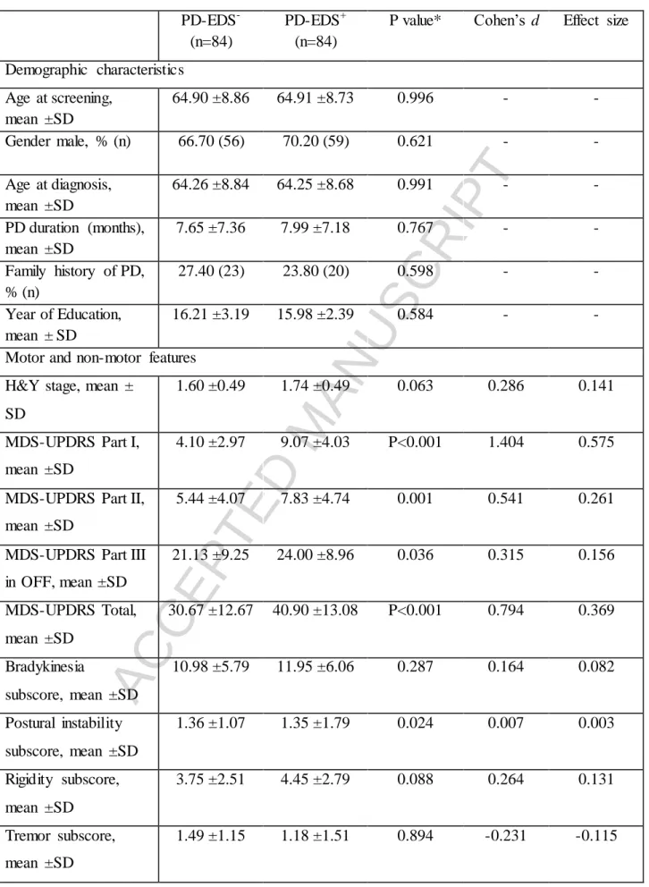
Table 1. Demographic and clinical characteristics for 84 PD patients with and without EDS PD-EDS-
(n=84)
PD-EDS+ (n=84)
P value* Cohen’s d Effect size
Demographic characteristics Age at screening, mean ±SD 64.90 ±8.86 64.91 ±8.73 0.996 - - Gender male, % (n) 66.70 (56) 70.20 (59) 0.621 - - Age at diagnosis, mean ±SD 64.26 ±8.84 64.25 ±8.68 0.991 - - PD duration (months), mean ±SD 7.65 ±7.36 7.99 ±7.18 0.767 - - Family history of PD, % (n) 27.40 (23) 23.80 (20) 0.598 - - Year of Education, mean ± SD 16.21 ±3.19 15.98 ±2.39 0.584 - -
Motor and non-motor features H&Y stage, mean ±
SD
1.60 ±0.49 1.74 ±0.49 0.063 0.286 0.141
MDS-UPDRS Part I, mean ±SD
4.10 ±2.97 9.07 ±4.03 P<0.001 1.404 0.575
MDS-UPDRS Part II, mean ±SD
5.44 ±4.07 7.83 ±4.74 0.001 0.541 0.261
MDS-UPDRS Part III in OFF, mean ±SD 21.13 ±9.25 24.00 ±8.96 0.036 0.315 0.156 MDS-UPDRS Total, mean ±SD 30.67 ±12.67 40.90 ±13.08 P<0.001 0.794 0.369 Bradykinesia subscore, mean ±SD 10.98 ±5.79 11.95 ±6.06 0.287 0.164 0.082 Postural instability subscore, mean ±SD 1.36 ±1.07 1.35 ±1.79 0.024 0.007 0.003 Rigidity subscore, mean ±SD 3.75 ±2.51 4.45 ±2.79 0.088 0.264 0.131 Tremor subscore, mean ±SD 1.49 ±1.15 1.18 ±1.51 0.894 -0.231 -0.115
ACCEPTED MANUSCRIPT
SCOPA-AUT, mean ± SD
8.74 ±5.56 11.96 ±5.99 P<0.001 0.557 0.268
GDS, mean ±SD 1.77 ±1.90 2.54 ±2.55 0.002 0.342 0.169
STAI Total Score, mean ±SD 61.55 ±16.63 65.69 ±19.01 0.135 0.232 0.115 MoCA, mean ±SD 27.42 ±2.14 26.50 ±2.38 0.010 -0.407 -0.199 BJLOS, mean ±SD 12.68 ±2.31 12.81 ±2.13 0.703 0.059 0.029 HVLT Total, mean ±SD 36.51 ±5.67 35.92 ±5.53 0.492 -0.105 -0.053 SFT Score, mean ±SD 48.95 ±11.38 45.73 ±10.11 0.054 -0.299 -0.148 SDMT Score, mean ±SD 41.10 ±9.72 39.06 ±9.69 0.176 -0.210 -0.105 UPSIT, mean ± SD 21.40 ±8.31 20.63 ±7.51 0.527 -0.097 -0.049 ADL, mean ± SD 93.49 ±5.96 91.79 ±5.84 0.063 -0.288 -0.143 [123I]FP-CIT imaging Caudate, mean ±SD 2.06 ±0.53 1.87 ±0.54 0.025 -0.355 -0.175 Putamen, mean ±SD 0.83 ±0.27 0.77 ±0.30 0.164 -0.210 -0.105 Striatum, mean ±SD 1.45 ±0.38 1.32 ±0.39 0.038 -0.338 -0.166
SCOPA-AUT: the scale for outcomes for PD-autonomic function; GDS: 15-item Geriatric Depression Scale, MoCA: Montreal Cognitive Assessment Scale; BJLOS: Benton Judgement of Line Orientation; HVLT: Hopkins Verbal learning Test – revised; STAI: state and train anxiety scale
ACCEPTED MANUSCRIPT
Fig. 1ACCEPTED MANUSCRIPT
Fig. 2ACCEPTED MANUSCRIPT
Fig. 3ACCEPTED MANUSCRIPT
Highlights
Excessive daytime sleepiness is associated with reduced caudate [123I]FP-CIT uptake The severity of sleepiness correlates with decreased [123I]FP-CIT uptake in the caudate Abnormal caudate [123I]FP-CIT uptake and disease duration predict the development of
sleepiness