Procedia Computer Science 89 ( 2016 ) 856 – 863
1877-0509 © 2016 Published by Elsevier B.V. This is an open access article under the CC BY-NC-ND license (http://creativecommons.org/licenses/by-nc-nd/4.0/).
Peer-review under responsibility of organizing committee of the Organizing Committee of IMCIP-2016 doi: 10.1016/j.procs.2016.06.072
ScienceDirect
Twelfth International Multi-Conference on Information Processing-2016 (IMCIP-2016)
Human Skin Region Segmentation Based on Chrominance
Component using Modified Watershed Algorithm
Alak Das
aand Dibyendu Ghoshal
b,∗aGovernment Degree College, Longtarai Valley, Chailengta, Dhalai, Tripura 799 273, India bNational Institute of Technology, Agartala, Jirania, West Triura, Tripura 799 046, India
Abstract
A novel watershed segmentation algorithm has been proposed to segment the human skin region of RGB color images based on Cb component of YCbCr color space in this paper. The conventional watershed segmentation algorithm is not suitable for segmentation of human skin region from color images directly, since it is difficult to decide suitable color information of human skin. At first stage, the RGB image is converted into YCbCr color space which contains three components luminance, blue difference component (Cb) and red-difference component (Cr). The Cb component has been extracted and used for further processing steps. After extraction of Cb component, the modified marker based watershed algorithm has been applied for segmentation of human skin region only from RGB color image. The proposed method has been tested on FRI CVL Face Database. In experimental stage, results show that the proposed technique avoids over segmentation problem and produce good segmentation result which is comparable with the same obtained by other algorithms.
© 2016 The Authors. Published by Elsevier B.V.
Peer-review under responsibility of organizing committee of the Twelfth International Multi-Conference on Information Processing-2016 (IMCIP-2016).
Keywords: Cb Component; Cr Component; Segmentation; Skin Region; Watershed Algorithm.
1. Introduction
Image segmentation and detection are very important techniques in the area of medical image analysis. Now a day, many real time laboratory applications in the area of medical image processing require robust and valid image segmentation for accurate analysis of anatomical structures. A very good number of segmentation on the area of medical image processing will help physicians and patients as those provides important idea for surgical planning, disease detection, segmentation etc. Very large number of segmentation techniques have been proposed for these purposes. Segmentation algorithm has not only been proposed for medical images but also in the area of human face segmentation, facial feature segmentation and facial expression recognition and segmentation of objects from natures etc.
Watershed segmentation approach mainly related to the gradient magnitude which is the first derivative of an image along with a preferred direction of topographic surface. In this technique, the gradient magnitude is computed using a Prewitt or Sobel or any other suitable filter for task. Those pixels whose have the highest gradient magnitude
∗Corresponding author. Tel.: +91 -9436767185.
E-mail address:[email protected]
© 2016 Published by Elsevier B.V. This is an open access article under the CC BY-NC-ND license (http://creativecommons.org/licenses/by-nc-nd/4.0/).
intensities correspond to watershed lines which represent the region boundaries on that image. Watershed segmentation of gray scale image has been seen like a topographic relief. In that case, the gray level of pixel is interpreted like a altitude in the relief. After that, a drop of water flows along a path to reach a local minimum. The low intensity minimum creates catchment basin for representing segmented area on that image.
A few numbers of researchers has proposed watershed technique to segment human face from color images. Human face segmentation is an important task for various applications like face recognition, facial feature recognition, facial expression analysis etc. Wright and Acton1 present a combination of the traditional watershed segmentation;
they applied multi resolution experimentation of the watershed segmentation algorithm. Morphological pyramid or a watershed pyramid has been used to develop a scale space representation. Xinet al.and Wanget al.focused at face detection over recognition. Hence, this algorithm has segmented out larger areas of the human face2, 4. Maliket al.
proposes a conventional watershed transform by improving the image quality in the pre-processing step with the help of random walk algorithm3. Guerfiet al., the normal watershed algorithm is used to the still human face and catchment basins have been taken based of hue component in the HIS color space5. Albertoet al.represented same marker based watershed segmentation technique on face from video frames based on construction of 2-dimensional histogram on Cb and Cr color space images6. Recently, Shylajaet al.introduces improved marker based watershed segmentation as a perquisite for extraction of features in the frontal human face images7. Sobottka and Pitaset al.applied watershed method to a human face where the inter region pixels of the candidate face are sorted according to their gray levels8. The primary regions have been determined using threshold and those regions have been flooded using iterative method to grow the regions with respect to threshold. Numerous research works of related fields have been developed. But, the human face segmentation for both frontal and side face images based on Cb component of YCbCr color space using watershed segmentation algorithm has been found in small number of published. In this paper, watershed segmentation algorithm based on marker has been applied to the Cb component of YCbCr space which has been taken from input RGB human face image. This technique is not only applied for frontal images but also for side images to segment the human skin region.
The structure of this paper is as follows: the Section 2 presents overview of the system. Section 3 presents the discussion of experimental results on proposed database. Finally, Section 4 gives the conclusions.
2. Proposed System
The whole system and step by step of implementation are shown with a block diagram in Fig. 1. At first, the input RGB color input image(IRGB)from taken database are converted into YCbCr color image consisting of three
components such as luminance, blue-difference (Cb) and red-difference (Cr). After converting into YCbCr color image, Cb component are extracted from YCbCr color space. The important extraction step of Cb component(Icb)
from the input RGB color image(IRG B)is called pre-processing step.
After pre-processing step, the Cb component has been used for further processing steps for segmentation of human skin regions. In the next step, the gradient magnitude of IC b has been computed using the application of Prewitt
edge filter. After that, marker based watershed segmentation technique is applied using modified morphological reconstruction such as opening- closing stage. Finally, the superimposed and marker based segmentation result which is labeling by two color (one color for non-skin region and another for skin region) has been found on human face images and medical images. The main goal of our proposed technique is to segment skin region of human or human face from input color images.
2.1 Pre-processing
In this step, the pre-processed image has been obtained for applying step by step. Input image is basically a Red-Green-Blue (RGB) color space which consists of three components named Red component, Green component and Blue component. However, this input color image is not optimal image for image processing because of defused information of intensity level of three components and also is not efficient when dealing with “real world” images. The main problem of RGB color space in applications with images is high correlation between its components about 0:78 for rBR (cross correlation between the B and R channel), 0:98 for rRG and 0:94 for rGB9. For this problem,
Fig. 1. Block Diagram of Proposed System.
Fig. 2. (a) Input Color RGB Image; (b) YCbCr Image; (c) Cb Component Image.
the input image is conveted to the YCbCr color space for extracting the Cb component which is very important component for segmenting color pixels of human skin. Therefore, chroma components (CBandCR) of YCbCr can be
compressed, or otherwise treated separately for improved system efficiency. The RGB imageIRG B is converted into
YCbCr image(IY C bCr). After that, Cb component imageIC bis used for segmentation of pixel regions of skin color
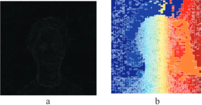
Fig. 3. (a) Gradient Image of Cb Component; (b) Over Segmented Results using Traditional Watershed Algorithm.
2.2 Computation of gradient magnitude
This step involves computation of the gradient image.IC bis the input image generated in step 1. Ig is gradient
magnitude image generated by filteringIC balong with Prewitt masks and calculation of magnitude can be found on
Ig=
|Ih|2+ |Iv|2 (1)
where,IhandIvare images filtered by horizontal and vertical Prewitt mask, respectively.
After generating magnitude image, traditional watershed segmentation algorithm has been performed on Ig. This
algorithm has been started by obtaining all local minima as basins where each and every basin has same label. Then, those basins are grown by testing pixels from each basin’s neighborhood. After that, the lowest gradient neighboring pixel is chosen for test pixel. The test pixel with neigbors which has one level is absorbed by the basin and marked by the corresponding basin’s label. The test pixel has neighbors from different basins is marked as watershed. But, the main disadvantage of this watershed segmentation algorithm is that it will generate over segmented results. The Fig. 3 shows results of gradient image by Prewitt mask and over segmented results using traditional watershed algorithm. 2.3 Generation of skin region marker
To overcome over segmentation problem, the modified marker based technique has been used where the foreground objects (skin regions) and background location (non-skin regions) can be identified for segmentation. There are two markers which have been computed to segment the objects such as foreground markers (skin regions) and background markers (non-skin regions). The foreground markers have been computed in where the resulted images are connected blobs of pixels within skin regions. On the other hand, the background markers have been computed to locate the non-skin regions. The foreground marker has been computed using two very important morphological reconstruction techniques to clean the Cb componentIC b. Morphological reconstruction operations have been used
to create flat maxima inside skin regions. One important MATLAB function in regional max is used to locate the above flat maxima inside skin regions. In this case, the structural element with 25 disk shape has been used for opening and closing operation. After applying morphological reconstruction technique, it has been seen that some of the mostly-occluded and shadowed skin regions pixels are not marked which means that some skin pixel region pixels could not be segmented properly in the end result. However, some foreground markers edge is located on non-skin regions. To overcome this problem, the edges of the marker blobs are cleaned and then shrink them a bit using closing followed by an erosion operation for better results.
2.4 Creation of non-skin region marker
This step is involves compute the non skin regions. In the resulted image obtained from previous step, the dark or black pixels belong to non-skin regions. The thresholding operation has been applied on the image obtained in previous step for compute the non-skin regions. After applying thresholding operation, Euclidean distance has been used for
computation the distance between each pixel and its nearest skin region pixels. This distance transform is used to thin the non-skin region pixels.
2.5 Use of watershed transform
After computing both the skin regions and non-skin regions, the watershed transform has been applied on the resulting image to extract the face image only. The dilation operation is applied to create the face boundary brighter. At last, results have been superimposed through the original input image using label matrix and boundary has been extracted clearly.
3. Experimental Results and Discussions
The whole work of proposed method has been done using MATLAB R2009b platform. The configurations of used machine are 3.00 GHz Intel(R) Core(TM) 2Duo Processor and 3.00 GB of installed memory in a machine. The experiments of our concept is analyzed using some frontal and side view images from FRI CVL Face Database and medical images from internet source.
3.1 Proposed database
FRI CVL FACE Database consists of 7 visual images (frontal and side view). There are 114 no’s of persons present in the database with 798 images total. Images of both male (90%) and female (10%) are present under different lighting conditions. The size of every pixel is 640×480 pixels.
3.2 Results
The experimental results have been discussed in this section. The proposed technique has been applied on images (both frontal and side view) of above database and some medical images from internet source. The average runtime is 3.5 seconds in a proposed machine configuration. At first, the suitable Cb component image has been generated from RGB color input image through YCbCr image in the pre-processing step. Then gradient image of Cb component
Table 1. Skin Region Segmentation Results.
No of Taken Images Correct Segmented Segmented Skin Region Correct Segmentation Rate for Experiment Skin Region with Non Skin Region on Skin Region Only
420 409 11 97.38%
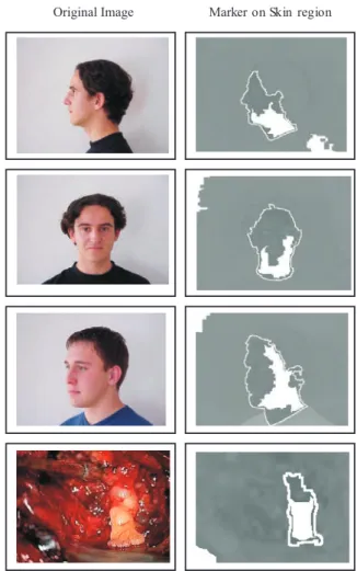
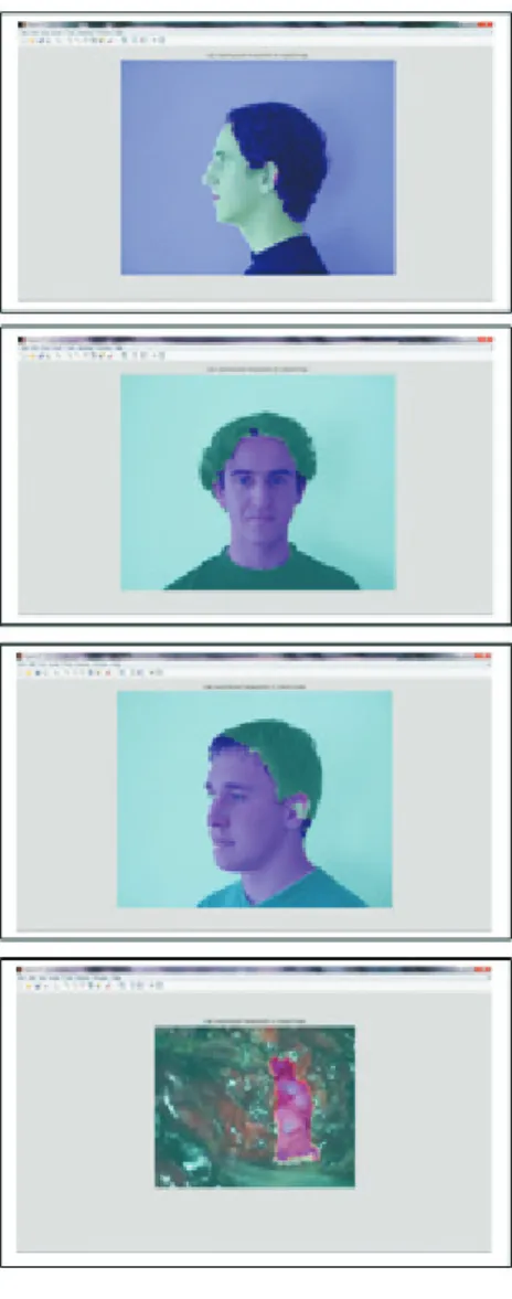
image is computed to segment with the help of watershed algorithm. In order to distinguish between skin and non skin region, marker based watershed segmentation algorithm is applied where those two regions are segmented using two color levels. Lastly, the segmentation result is superimposed on the original RGB image using label matrix. After testing on images, it can be concluded that proposed technique segments the skin and non-skin regions using watershed algorithm on gradient image of Cb component image. Figure 4 shows the original RGB input image in one column and corresponding marker on skin region in another column. The skin regions has been marked only using modified watershed algorithm. The total 420 no of images of 60 different persons have been obtained from the proposed database for experimentation. The results of correct segmentation of skin region only and segmentation of skin region with non skin (hair, clothes) have been reported in Table 1, which implies that the correct segmentation rate on skin region only is 97.38%. The final segmented results using modified marker based watershed algorithm on original image have been shown in Fig. 5.
4. Conclusions
An automatic efficient modified marker based watershed algorithm is presented for segmentation of human skin region. The simulation results show that our technique can segment fontal human face images and also segment side view human face images, critical inner view images of human body. Therefore, the segmented human skin region can be used in several cases like human face recognition, facial feature analysis, facial expression recognition, medical image analysis and detection of critical skin region based area etc in future. However, this technique is very helpful to segment accurate area of skin region. It segments the skin region with very high number of skin region pixels around the border of skin region.
5. Dedication
One of the authors of this paper Alak Das dedicates this research work to the memory to his father late. Anil Das.
References
[1] A. S. Wright and S. T. Acton, Watershed Pyramids for Edge Detection, InProceedings of International Conference on Image Processing, Washington, vol. 2, pp. 578–581, (1997).
[2] H. Xin, H. Ali, H. Chao and D. Tretter, Human Head-Shoulder Segmentation, InProceedings of International Conference on Automatic Face and Gesture Recognition, Santa Barbara, vol. 3, pp. 227–232, (2011).
[3] J. Malik, R. Dahiya and G. Sainarayanan, Harris Operator Corner Detection using Sliding Window Method, International Journal of Computer Applications, vol. 22, pp. 28–37, May (2011).
[4] J. G. Wang, E. T. Lin, R. Venkateswarlu and E. Sung, Stereo Head/Face Tracking and Pose Estimation, InProceedings of International Conference on Control, Automation, Robotics and Vision(ICARCV2002), Singapore, vol. 3, pp. 1609–1914, (2002).
[5] S. Guerfi, J. P. Gambotto and S. Lelandais, Implementation of the Watershed Method in the HIS Color Space for the Face Extraction, InProceedings IEEE Conference on Advanced Video and Signal Based Surveillance, Los Alamitos, pp. 282–286, (2005).
[6] A. Albiol, L. Torres, C. A. Bouman and E. J. Delp, A Simple and Efficient Face Detection Algorithm for Video Databse Application, InProceedings of International Conference on Image Processing, Canada, vol. 2, pp. 239–242, (2000).
[7] S. S. Shylaja, K. N. B. Murthy, S. Natarajan, A. Prasad, A. Modi and S. Harlalka, Feature Extraction using Marker Based Watershed Segmentation on the Human Face, InProceedings of International Conference on Computer Communication and Informatics (ICCCI-2012), Coimbatore, (2012).
[8] K. Sobottka and I. Pitas, Loking for Face and Facial Features in Color Images, InPattern Recognition and Image Analysis: Advances in Mathematical Theory and Applications, Russian Academy of Sciences, vol. 3, (1996).
[9] H. Palus, Chapmann and Hall, Colour Spaces, (1998).
[10] R. C. Gonzalez and R. F. Woods, Digital Image Processing, Second Edition,Addison-Wesley Longman Publishing, Co., (2001). [11] http://web.mit.edu/emeyers/www/face−databases.html
[12] L. P. Son, A. Bouzerdoum and D. Chai, A Novel Skin Color Model in YCbCr Color Space and its Application to Human Face Detection, InProceedings of International Conference on Image Processing, vol. 1, pp. 289–292, (2002).
[13] R. L. Hsu, A. M. Mohamed and K. J. Anil, Face Detection in Color Images,IEEE Transactions on Pattern Analysis and Machine Intelligence, vol. 24, pp. 696–706, May (2002).
[14] M. H. Yang, D. Kriegman and N. Ahuja, Detecting Faces in Images: A Survey,IEEE Transactions on Pattern Analysis and Machine Intelligence, vol. 24, pp. 34–58, January (2001).
[15] E. Hjelm and B. K. Low, Face Detection: A Survey,Computer Vision and Image Understanding, vol. 83, pp. 236–274, September (2001). [16] B. Menser and M. Brunig, Locating Human Faces in Colo Images with Complex Background, Intelligent Signal Processing and
Communications System, pp. 533–536, December (1999).
[17] S. L. Phung, A. Bouzerdoum and D. Chai, Skin Segmentation using Color Pixel Classification: Analysis and Comparison,IEEE Transactions Pattern Analysis Machine Intelligent, vol. 27, pp. 148–154, (2005).