Selection of our books indexed in the Book Citation Index in Web of Science™ Core Collection (BKCI)
Interested in publishing with us?
Contact book.department@intechopen.com
Numbers displayed above are based on latest data collected.For more information visit www.intechopen.com Open access books available
Countries delivered to Contributors from top 500 universities
International authors and editors
Our authors are among the
most cited scientists
Downloads
12.2%
122,000
135M
TOP 1%
154
4,800
Introductory Chapter: Shoulder Joint
Satish B. Sonar and Omkar P. Kulkarni
Additional information is available at the end of the chapter
http://dx.doi.org/10.5772/intechopen.76187
© 2016 The Author(s). Licensee InTech. This chapter is distributed under the terms of the Creative Commons Attribution License (http://creativecommons.org/licenses/by/3.0), which permits unrestricted use, distribution, and reproduction in any medium, provided the original work is properly cited.
Satish B. Sonar and Omkar P. Kulkarni
Additional information is available at the end of the chapter
1. Basic shoulder anatomy
The shoulder is a ball and socket type of synovial joint. It is one of the largest and most complex joints in the body. Its dynamic and hypermobility make it susceptible to many injuries.
The shoulder girdle comprises of glenohumeral joint, acromioclavicular joint, scapulothoracic articulation, and coracoclavicular articulation.
Deltoid, one of the strongest muscles in the body, encircles the shoulder joint all around. It provides the shape and bulk to the shoulder joint. It works in almost all the functions of the joint from forward flexion, abduction, and adduction to rotations. It is supplied by the axil-lary nerve. The pectoralis major and minor, rhomboids, latissimus dorsi, teres major, and trapezius are other major muscles that play an important part in the function and stability of shoulder girdle (Figure 1).
Rotator cuff provides concentric compression, dynamic stability, and smooth arc of motion to the glenohumeral joint. The subscapularis along with the anterior part of the supraspinatus provides excellent anterior stability. The posterior part of the supraspinatus, infraspinatus, and teres minor provides posterosuperior stability and resists superior pull of deltoid. In addition to the glenoid labrum, rotator cuff muscles are the dynamic stabilizers of the shoul-der joint. Injury, dysfunction, or degenerative tears of these muscles hampers the shoulshoul-der function to a great extent.
The subdeltoid bursa cushions and protects the tendons of the rotator cuff. It also provides nutrition and lubrication to the rotator cuff tendons. The subacromial bursa can get inflamed in impingement syndrome, RA, calcific tendinitis, and other subacromial painful pathologies causing severe pain and movement restrictions [1].
© 2018 The Author(s). Licensee IntechOpen. This chapter is distributed under the terms of the Creative Commons Attribution License (http://creativecommons.org/licenses/by/3.0), which permits unrestricted use, distribution, and reproduction in any medium, provided the original work is properly cited.
On the inner most aspect of the joint is an intracapsular structure called glenoid labrum which not only deepens the glenosphere but also provides strong all-round stability to the shoulder joint through the tension and compression it creates through capsular ligaments like superior,
middle, and inferior glenohumeral ligaments in association with rotator cuff. The anterior
part of inferior glenohumeral ligament is the most important anteroinferior stabilizer. Long head of biceps originating from the superior labrum helps in shoulder stability.
The shoulder joint is surrounded by many neurovascular structures like the brachial plexus, axillary nerve, suprascapular nerve, musculocutaneous nerve, brachial artery, and lungs. These structures are always vulnerable to injury in shoulder trauma.
2. Pathoanatomy
Being a major synovial joint of the body and also because of its inherent unstable nature, the
shoulder joint is affected by many pathologies.
Adhesive capsulitis which is commonly called as a frozen shoulder is an inflammatory
response to systemic or local painful pathologies like diabetes mellitus, hypothyroidism, hypertension, etc. As the frozen shoulder progresses, movement in the shoulder can be severely limited. In the later stage as the pain decreases, range of motion improves but never to the original level. Medications, injections, physiotherapy, and home exercises usually help in most of the patients. If it is not, arthroscopic capsular release followed by rehab gives well to excellent results [2].
Primary osteoarthritis of the shoulder is quite rare, but secondary osteoarthritis due to trauma,
rotator cuff insufficiency, RA, gout, etc. is quite common. As we all know, it is painful and is a debilitating condition affecting day-to-day activities. Total shoulder and reverse shoulder are
the modalities of treatment when the patient does not improve by conservative ways. Figure 1. Muscles and nerves.
Rotator cuff tears can be traumatic or degenerative in older age groups from repeated over -use. It causes pain, functional, and motion restrictions. As per the recent research
publica-tions, many patients with full-thickness rotator cuff showed fair to good functional results. These are called compensated rotator cuff tears. On the contrary tear goes on progressing over
the period. Patients who do not improve with all these conservative measures are treated with
either open or arthroscopic repair techniques. Arthroscopic techniques are far better than the open one, giving the patient the benefits of minimally invasive surgery, anatomic repairs, and
rapid recovery (Figure 2).
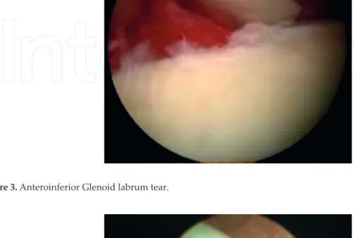
Shoulder dislocation can be anterior, posterior, or multidirectional. It can be traumatic or due to generalized ligament laxity. Traumatic dislocations are usually associated with tear of the labrum, humeral head bony defect, capsular tears, and muscle and nerve injuries. In
emer-gency settings it is reduced under anesthesia, and sling is applied followed by physiother
-apy. Most of the patients do well with this, but if it becomes recurrent due to capsulolabral nonhealing, big humeral bone defect (Hill-Sachs lesion), and/or glenoid bone loss, surgery is indicated. Most of the patients can be managed with arthroscopic repair, but few may require bony procedures like Latarjet, etc. (Figure 3).
Since the biceps plays an important role in shoulder stability and function, many biceps pathologies may cause pain and disability. Biceps tendon problems like tendinopathy or teno-synovitis as well as SLAP lesions compromise optimal shoulder function and may result in impingement. Biceps tenotomy in older population and tenodesis in younger patients are the treatments of choice (Figure 4) [3].
Glenohumeral internal rotation deficit, often referred to as GIRD, is a sport-specific adapta -tion of posterior shoulder structures to chronic excessive overload of these structures during frequent throwing. Burkhart et al. [13] report that GIRD occurs before any other motion adap-tation, suggesting that contracture of the posterior capsule is to blame for this change in range
of motion and is sometimes followed by associated gains in ER. Other researchers believe that GIRD begins in the early years of overhead throwing with a bony adaptation of the humerus. A third hypothesis regarding the cause of GIRD is muscle hypertony in the external rotators due to frequent eccentric loading.
Shoulder bursitis, impingement, and tendonitis are painful conditions due to the involve-ment of narrow subacromial space causing pain with overhead activities or compressive
Figure 3. Anteroinferior Glenoid labrum tear.
forces on upper arm causing impingement. Internal impingement comprises encroachment of the rotator cuff tendons between the humeral head and the glenoid rim. Anterosuperior and posterosuperior glenoid impingements have been described based on its location. The posterosuperior impingement consists of the mechanical encroachment of the rotator cuff tendons, particularly the tendon of the supraspinatus and infraspinatus, between the greater tubercle of the humerus and the posterosuperior rim of the glenoid. This friction occurs spe-cifically during the late cocking position of throwing, which is maximal external rotation, horizontal abduction, and, depending on the specific-sport discipline, a certain amount of abduction. Besides the classification of impingement based on the site of encroachment, a very often impingement is classified based on the cause of the problem, dividing it into pri -mary versus secondary impingement. In pri-mary impingement, a structural narrowing of the subacromial space causes pain and dysfunction, such as acromioclavicular arthropathy, type II acromion, or swelling of the soft tissue in the subacromial space. In secondary impinge-ment, there are no structural obstructions causing the encroachment but rather functional problems, occurring only in specific positions.
Winging of the scapula is a condition where due to insufficiency of scapular muscles, scapu -lar stability is affected and it moves up like a wing. It can mimic as pseudo-instability of the shoulder. Scapular dyskinesia also has been described in relation to impingement symptoms [4]. This is because during arm elevation, impingement may occur if the scapula insufficiently follows the humeral head movements because of a lack of upward rotation, posterior tilt-ing, and external rotation. Neuromuscular stimulation and scapular muscle strengthening improve the condition (Figure 5).
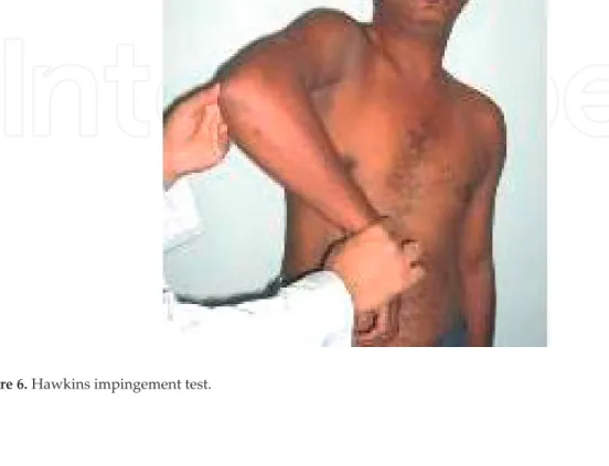
Tractional damage to the suprascapular nerve leads to suprascapular neuropathy caus-ing an achcaus-ing or burncaus-ing pain at the back and/or side of the shoulder joint. Sometimes, a cyst can develop in the region causing symptoms of neural compression and severe shoulder pain. MRI usually diagnoses the condition. It can be treated with arthroscopic decompression.
have pain in the area of superior GH joint or AC joint, and it is an indication for subacromial impingement; the test will be negative in case of internal impingement (Figure 6).
Neer test—This test is carried out in patient with seated and arm at side, palm down (pronated),
Examiner stabilizes scapula and raises the arm (between flexion and abduction). Positive test indicates pain.
Pain at the front of the shoulder is an indication for subacromial impingement, whereas patients with internal impingement will exhibit pain at the posterior aspect of the shoulder.
Instability tests—The apprehension test, load and shift test, crank test, jerk test, sulcus sign test, and the relocation test are some of the most commonly used instability tests of the humeral head.
Apprehension test—The patient is in sitting or standing position and at 90° of abduction; the
examiner applies slight anterior pressure to the humerus and externally rotates the arm. In positive test the patient expresses apprehension.
Relocation test—The test is performed after the positive result on anterior apprehension test.
The patient is in supine or sitting position. The examiner applies posterior force on the proxi -mal humerus while externally rotating the patient’s arm. The test is positive if the patient expresses relief.
Crank test—Shoulder is elevated to 160° in the scapular plane, a gentle axial load is applied through glenohumeral joint with one hand, while other hand does internal and external rota-tion. Positive test is when patient has pain, catching, or clicking in the shoulder. This test is for posterior instability.
3.1. Rotator cuff tests are described in respective chapters in detail
Biceps and SLAP lesion tests—Speed’s test, the O’Brien test, and the biceps load II test are the three most useful tests for biceps pathologies.
Speed’s test—The patient is in sitting or standing position; the examiner asks him to forward flex the shoulder against resistance while maintaining the elbow in extension and the forearm
in supination. In positive test, the patient will have pain into the biceps region and tender in bicipital groove (bicipital tendinitis) (Figure 7).
The O’Brien test—The patient is in sitting position, and the patient’s shoulder is in 90° for-ward flexion, adduction, and internal rotation; the examiner applies downfor-ward force. Positive result is when patient will have pain to the anterosuperior or posterosuperior part of the shoulder indicating superior labral tear.
Biceps load II test—This test is considered positive if the patient complains of pain during the resisted elbow flexion. The patient is in standing position, and the examiner forward flexes the arm to 90°, abducting 15–20° with elbow straight with full internal rotation so the thumb is pointing down, and applies downward force on the arm which the patient resists. Then, the patient externally rotates the arm so that the thumb is pointing up; the examiner applies down-ward force on the arm, and the patient resists it. The test is positive if pain or painful clicking will be elicited with the thumb down and decreased or eliminated with the thumb up (Figure 8).
4. Investigations
Plain X-ray of the shoulder in anteroposterior, axillary lateral, Stryker notch, and 30° caudal view is usually sufficient to diagnose most of the shoulder girdle pathologies like shoulder dis-location, A–C joint injuries, clavicle fracture, Hill-Sachs lesion, acromial spur, etc. (Figure 9) [5]. CT scan—It is very useful to diagnose bony pathologies of the shoulder. It gives excellent three-dimensional imaging of the bony shoulder girdle. Humeral and glenoid bone loss can be accurately calculated. But in case of musculoskeletal injuries, MRI is the investigation of choice (Figures 10 and 11).
MRI and ultrasound are other valuable diagnostic tools because they provide images of the soft tissues without using radiation.
Ultrasonography—It is one of the cheapest and most easily done tests for shoulder
patholo-gies like rotator cuff tear, calcific tendinitis, and biceps tear. But it is very less frequently used
(Figure 12) [6, 9, 10].
MRI—It is the investigation of choice in shoulder joint injuries. It excellently depicts the labral
tear, rotator cuff tear, biceps tear/displacement, and other soft tissue pathologies. The MRI has
a picture that both the clinician and the patient can understand (Figure 13).
Figure 9. X-ray AP view and GT avulsion.
Figure 13. MRI Showing Anterior labral tear.
Figure 11. 3D CT Scan showing Bony Bankart lesion.
Arthroscopy—Though it is mainly a therapeutic and invasive key hole surgery, it can help in accurate diagnosis of many pathologies which are not shown even in MRI. Subscapularis tears, capsular rents, avulsions from the humerus, SLAP tears, etc. can be well diagnosed and treated by shoulder arthroscopy (Figures 14 and 15) [11, 12].
Author details
Satish B. Sonar* and Omkar P. Kulkarni
*Address all correspondence to: stshsonar@gmail.com Dr. PDM Medical College, Amravati, India
Figure 14. Arthroscopy biceps tendon.
British Journal of Surgery. 1938
[6] Cofield RH. Rotator cuff disease of the shoulder. Journal of Bone & Joint Surgery. 1985
[7] Moen MH, de Vos RJ, Ellenbecker TS. Clinical tests in shoulder examination: How to perform them. British Journal of Sports Medicine. 2010
[8] Hegedus EJ, Goode A, Campbell S. Physical examination tests of the shoulder: A sys-tematic review with meta-analysis of individual tests. A Morin-British Journal of Sports Medicine. 2008
[9] Read JW, Perko M. Shoulder ultrasound: Diagnostic accuracy for impingement
syn-drome, rotator cuff tear, and biceps tendon pathology. Journal of Shoulder and Elbow
Surgery. 1998
[10] Roberts CS, Walker 2nd JA. Diagnostic capabilities of shoulder ultrasonography in the
detection of complete and partial rotator cuff tears. American Journal of Orthopedics.
2001
[11] Waldt S, Burkart A, Lange P, Imhoff AB, et al. Diagnostic performance of MR arthrog -raphy in the assessment of superior labral anteroposterior lesions of the shoulder. American Journal of Orthopedics. 2004
[12] Denti M, Monteleone M, Trevisan C. Magnetic resonance imaging versus arthroscopy for the investigation of the osteochondral humeral defect in anterior shoulder instability. Knee Surgery, Sports Medicine and Arthroscopy Journal. 1995
[13] Burkhart SS, Craig MD, Morgan D, Ben Kibler MDW. The disabled throwing shoul-der: Spectrum of pathology part I: Pathoanatomy and biomechanics. The Journal of Arthroscopy and Related Research. April 2003;19(4):404-420