Bell’s Palsy: Diagnosis and Management
JEFFREY D. TIEMSTRA, MD, and NANDINI KHATKHATE, MDUniversity of Illinois at Chicago College of Medicine, Chicago, Illinois
B
ell’s palsy is an idiopathic, acute peripheral-nerve palsy involving the facial nerve, which supplies all the muscles of facial expression. The facial nerve also contains parasympathetic fibers to the lacrimal and salivary glands, as well as limited sensory fibers supplying taste to the anterior two thirds of the tongue(Figure 1). Bell’s palsy is named after Sir Charles Bell (1774-1842), who first described the syn-drome along with the anatomy and function of the facial nerve. The annual incidence of Bell’s palsy is 15 to 30 per 100,000 persons, with equal numbers of men and women affected. There is no predilection for either side of the face. Bell’s palsy has been described in patients of all ages, with peak incidence noted in the 40s. It occurs more commonly in patients with diabetes and in pregnant women. Patients who have had one episode of Bell’s palsy have an 8 percent risk of recurrence.1,2
Clinical Presentation
Patients with Bell’s palsy typically complain of weakness or complete paralysis of all the muscles on one side of the face. The facial creases and nasolabial fold disappear, the fore-head unfurrows, and the corner of the mouth droops. The eyelids will not close and the lower lid sags; on attempted closure, the eye rolls upward (Bell’s phenomenon). Eye irrita-tion often results from lack of lubricairrita-tion and
constant exposure. Tear production decreases; however, the eye may appear to tear excessively because of loss of lid control, which allows tears to spill freely from the eye. Food and saliva can pool in the affected side of the mouth and may spill out from the corner. Patients often complain of a feeling of numbness from the paralysis, but facial sensation is preserved.
Patients with Bell’s palsy usually progress from onset of symptoms to maximal weak-ness within three days and almost always within one week. A more insidious onset or progression over more than two weeks should prompt reconsideration of the diag-nosis. Left untreated, 85 percent of patients will show at least partial recovery within three weeks of onset.3
Etiology and Differential Diagnosis
Bell’s palsy is believed to be caused by inflam-mation of the facial nerve at the geniculate ganglion, which leads to compression and possible ischemia and demyelination. This ganglion lies in the facial canal at the junction of the labyrinthine and tympanic segments, where the nerve curves sharply toward the stylomastoid foramen. Classically, Bell’s palsy has been defined as idiopathic, and the cause of the inflammatory process in the facial nerve remains uncertain. Recently, attention has focused on infection with herpes simplex virus type 1 (HSV-1) as a possible cause because
Bell’s palsy is a peripheral palsy of the facial nerve that results in muscle weakness on one side of the face. Affected patients develop unilateral facial paralysis over one to three days with forehead involvement and no other neurologic abnormalities. Symptoms typically peak in the first week and then gradually resolve over three weeks to three months. Bell’s palsy is more common in patients with diabetes, and although it can affect persons of any age, incidence peaks in the 40s. Bell’s palsy has been traditionally defined as idiopathic; however, one possible etiology is infection with herpes simplex virus type 1. Laboratory evaluation, when indicated by history or risk factors, may include testing for diabetes mellitus and Lyme disease. A common short-term complication of Bell’s palsy is incomplete eyelid closure with resultant dry eye. A less common long-term complication is permanent facial weakness with muscle contractures. Approximately 70 to 80 percent of patients will recover spontaneously; however, treatment with a seven-day course of acyclovir or valacyclovir and a tapering course of prednisone, initiated within three days of the onset of symptoms, is recommended to reduce the time to full recovery and increase the likelihood of complete recuperation. (Am Fam Physi-cian 2007;76:997-1002, 1004. Copyright © 2007 American Academy of Family PhysiPhysi-cians.)
▲
Patient information: A handout on Bell’s palsy, written by the authors of this article, is provided on page 1004.
Bell’s Palsy
research has found elevated HSV-1 titers in affected patients. However, studies have failed to isolate viral DNA in biopsy specimens, leav-ing the causative role of HSV-1 in question.4,5
Many conditions can produce isolated facial nerve palsy identical to Bell’s palsy. Structural lesions in the ear or parotid gland (e.g., cholesteatoma, salivary tumors) can produce facial nerve compression and paral-ysis. Other causes of peripheral nerve palsies include Guillain-Barré syndrome, Lyme dis-ease, otitis media, Ramsay Hunt syndrome (an outbreak of herpes zoster in the facial nerve distribution), sarcoidosis, and some influenza vaccines. Although these
condi-tions can present as isolated facial nerve palsies, they usually have additional features that distinguish them from Bell’s palsy.
Patients with Lyme disease often have a history of tick exposure, rash, or arthralgias. Facial nerve palsies from acute and chronic otitis media have a more gradual onset, with accompanying ear pain and fever. Patients with Ramsay Hunt syndrome have a pronounced prodrome of pain and often develop a vesicular eruption in the ear canal and pharynx, although cases without the vesicular eruption (i.e., zoster sine herpete) have been reported. Polyneuropathies (e.g., Guillain-Barré syndrome, sarcoidosis) will
SORT: KEY RECOMMENDATIONS FOR PRACTICE Clinical recommendation
Evidence
rating References
Patients with Bell’s palsy should be treated within three days of the onset of symptoms with a seven-day course of oral acyclovir (Zovirax) or valacyclovir (Valtrex), plus a tapering course of oral prednisone.
B 15-17 Patients with complete paralysis who do not improve in two weeks on
medication should be referred to an otolaryngologist for evaluation for other causes of facial nerve palsy.
C 19, 20 Patients should be monitored for eye irritation and be prescribed eye
lubrication. Patients with corneal abrasions should be referred to an ophthalmologist.
C 1, 23
A = consistent, good-quality patient-oriented evidence; B = inconsistent or limited-quality patient-oriented evi-dence; C = consensus, disease-oriented evidence, usual practice, expert opinion, or case series. For information about the SORT evidence rating system, see page 922 or http://www.aafp.org/afpsort.xml.
Figure 1. Anatomy of the facial nerve.
Greater petrosal nerve Geniculate ganglion Chorda tympani nerve Internal acoustic meatus Superior salivatory nucleus Motor nucleus of facial nerve Visceral efferent fibers (facial expression muscles, stapedius muscle)
Visceral motor fibers (lacrimal, salivary glands)
Special sensory fibers (supplies taste to anterior two thirds of the tongue)
IL LU ST R A TI O N B Y R EN EE C A N N O N
more often affect both facial nerves. Tumors will present with a more insidious onset of symptoms over weeks or months.
Central nervous system lesions (e.g., mul-tiple sclerosis, stroke, tumor) can also cause facial nerve palsy. However, some motor neu-rons to the forehead cross sides at the level of the brainstem, so the fibers in the facial nerve going to the forehead come from both cere-bral hemispheres (Figure 2). Supranuclear (central) lesions affecting the facial nerve will not paralyze the forehead on the affected side, resulting in a unilateral facial paralysis with forehead sparing. Often, there will be at least some weakness of extremities on the affected side as well. Table 11,6-8 summarizes the
dif-ferential diagnosis of Bell’s palsy.
Influenza vaccines in the past have been associated with peripheral neuropathies. Although influenza vaccines currently avail-able in the United States have not been associ-ated with Bell’s palsy,9-11 a recently developed
Swiss intranasal vaccine was found to have a very high risk of postvaccine facial nerve palsy and has been withdrawn from use.12
Because influenza vaccines change annually,
public health officials should be notified of any cases of Bell’s palsy occurring in the six weeks following vaccine administration.
Evaluation
A patient with an acute onset of unilateral facial weakness most likely has Bell’s palsy. A careful history of the onset and progress of paralysis is important because gradual onset of more than two weeks’ duration is strongly suggestive of a mass lesion. Medical history should include recent rashes, arthralgias, or fevers; history of peripheral nerve palsy; expo-sure to influenza vaccine or new medications; and exposure to ticks or areas where Lyme disease is endemic. The physical examination should include careful inspection of the ear canal, tympanic membrane, and oropharynx, as well as evaluation of peripheral nerve func-tion in the extremities and palpafunc-tion of the parotid gland. In order to assess forehead involvement, physical examination should also include evaluation of cranial nerve func-tion, including all facial muscles.
Laboratory testing is not usually indicated. However, because diabetes mellitus is
pres-A. Facial nerve lesion (Bell’s palsy) B. Supranuclear lesion Nucleus of facial nerve (cranial nerve VII) Lesion in facial nerve Supranuclear lesion Facial nerve
Figure 2. Patients with (A) a facial nerve lesion and (B) a supranuclear lesion with forehead sparing.
IL LU ST R A TI O N B Y R EN EE C A N N O N
ent in more than 10 percent of patients with Bell’s palsy, fasting glucose or A1C testing may be performed in patients with additional risk factors (e.g., family history, obesity, older than 30 years).13 Antibiotic therapy may be
of benefit; therefore, Lyme antibody titers should be performed if the patient’s history suggests possible exposure. Signs and symp-toms atypical for Bell’s palsy should prompt further evaluation. Patients with insidious onset or forehead sparing should undergo imaging of the head. Those with bilateral pal-sies or those who do not improve within the first two or three weeks after onset of symp-toms should be referred to a neurologist
Treatment CORTICOSTEROIDS
Oral corticosteroids have traditionally been pre-scribed to reduce facial nerve inflammation in patients with Bell’s palsy. Prednisone is typically prescribed in a 10-day tapering course starting at 60 mg per day. A 2004 Cochrane review and meta-analysis of three randomized controlled trials comparing corticosteroids with placebo found small and statistically nonsignificant reductions in the percentage of patients with incomplete recovery after six months (relative risk [RR] = 0.86; 95% confidence interval [CI], 0.47 to 1.59) and the percentage of patients with cosmetic complications (RR = 0.86; 95% CI, 0.38 to 1.98).14 However, these trials included only
117 patients; larger prospective trials are needed to establish the benefit of corticosteroids.
ANTIvIRAlS
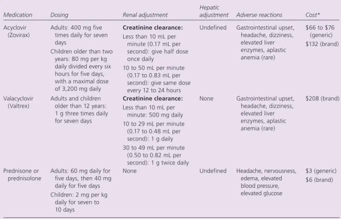
Because of the possible role of HSV-1 in the etiology of Bell’s palsy, the antiviral drugs acy-clovir (Zovirax) and valacyacy-clovir (Valtrex) have been studied to determine if they have any benefit in treatment. Either acyclovir 400 mg can be given five times per day for seven days or valacyclovir 1 g can be given three times per day for seven days. Although a 2004 Cochrane review found insufficient evidence to support the use of these antivirals alone,15 two recent
placebo-controlled trials demonstrated full recovery in a higher percentage of patients treated with an antiviral drug in combination with prednisolone than with prednisolone alone (100 percent versus 91 percent and 95 percent versus 90 percent).16,17 However, no benefit
was seen when treatment was delayed more than four days after the onset of symptoms (86 percent versus 87 percent).17
SPONTANEOuS RECOvERY
It is difficult to establish a statistically signifi-cant benefit of treatment in placebo-controlled trials because Bell’s palsy has a high rate of spontaneous recovery. The Copenhagen Facial Nerve Study evaluated 2,570 persons with untreated facial nerve palsy, including 1,701 with idiopathic (Bell’s) palsy and 869 with
Table 1. Differential Diagnosis for Facial Nerve Palsy
Disease Cause Distinguishing factors
Nuclear (peripheral)
Lyme disease Spirochete Borrelia burgdorferi
History of tick exposure, rash, or arthralgias; exposure to areas where Lyme disease is endemic
Otitis media Bacterial pathogens Gradual onset; ear pain, fever, and conductive hearing loss
Ramsay Hunt syndrome Herpes zoster virus Pronounced prodrome of pain; vesicular eruption in ear canal or pharynx Sarcoidosis or
Guillain-Barré syndrome
Autoimmune response More often bilateral Tumor Cholesteatoma, parotid
gland
Gradual onset
Supranuclear (central) Forehead spared
Multiple sclerosis Demyelination Additional neurologic symptoms
Stroke Ischemia, hemorrhage Extremities on affected side often involved Tumor Metastases, primary
brain
Gradual onset; mental status changes; history of cancer
palsy from other causes; 70 percent had com-plete paralysis. Function returned within three weeks in 85 percent of patients, with 71 per-cent of these patients recovering full function. Of the 29 percent of patients with sequelae, 12 percent rated it slight, 13 percent rated it mild, and 4 percent rated it severe.3 Because of
these findings, some persons have questioned whether treatment for Bell’s palsy should be routinely indicated; however, patients who have incomplete recovery will have obvious cosmetic sequelae and will often be dissatisfied with their outcome.18
Given the safety profile of acyclovir, vala-cyclovir, and short-course oral corticoste-roids, patients who present within three days of the onset of symptoms and who do not have specific contraindications to these medications should be offered combination therapy. Patients who present with complete facial nerve paralysis have a lower rate of spontaneous recovery and may be more likely to benefit from treatment.1-3,19
OThER TREATMENTS
In the past, surgical decompression within three weeks of onset has been recommended for patients who have persistent loss of function (greater than 90 percent loss on electroneurography) at two weeks. How-ever, the most widely cited study supporting this approach only reported results for a total of 34 treated patients at three differ-ent sites, included a nonrandomized control group, and lacked a blinded evaluation of outcome.20
The most common complication of sur-gery is postoperative hearing loss, which affects 3 to 15 percent of patients. Based on the significant potential for harms and the paucity of data supporting benefit, the American Academy of Neurology does not currently recommend surgical decompres-sion for Bell’s palsy.19
Some published studies have reported benefit with acupuncture versus steroids and placebo, but all had serious flaws in study design and reporting.21Table 2
sum-marizes the available treatments.
Table 2. Medications for Treatment of Bell’s Palsy
Medication Dosing Renal adjustment
Hepatic
adjustment Adverse reactions Cost*
Acyclovir (Zovirax)
Adults: 400 mg five times daily for seven days
Children older than two years: 80 mg per kg daily divided every six hours for five days, with a maximal dose of 3,200 mg daily
Creatinine clearance:
Less than 10 mL per minute (0.17 mL per second): give half dose once daily
10 to 50 mL per minute (0.17 to 0.83 mL per second): give same dose every 12 to 24 hours
Undefined Gastrointestinal upset, headache, dizziness, elevated liver enzymes, aplastic anemia (rare) $66 to $76 (generic) $132 (brand) Valacyclovir (Valtrex)
Adults and children older than 12 years: 1 g three times daily for seven days
Creatinine clearance:
Less than 10 mL per minute: 500 mg daily 10 to 29 mL per minute (0.17 to 0.48 mL per second): 1 g daily 30 to 49 mL per minute (0.50 to 0.82 mL per second): 1 g twice daily
None Gastrointestinal upset, headache, dizziness, elevated liver enzymes, aplastic anemia (rare) $208 (brand) Prednisone or prednisolone
Adults: 60 mg daily for five days, then 40 mg daily for five days Children: 2 mg per kg
daily for seven to 10 days
None Undefined Headache, nervousness, edema, elevated blood pressure, elevated glucose
$3 (generic) $6 (brand)
*— Estimated cost to the pharmacist based on average wholesale prices (rounded to the nearest dollar) in Red Book. Montvale, N.J.: Medical Economics Data, 2006. Cost to the patient will be higher, depending on prescription filling fee.
Bell’s Palsy
Complications
Patients with Bell’s palsy may be unable to close the eye on the affected side, which can lead to irritation and corneal ulceration. The eye should be lubricated with artificial tears until the facial paralysis resolves. Permanent eyelid weakness may require tarsorrhaphy or implantation of gold weights in the upper lid. Facial asymmetry and muscular contractures may require cosmetic surgical procedures or botulinum toxin (Botox) injections.In these cases, consultation with an ophthalmologist or cosmetic surgeon is needed.22,23
The Authors
Jeffrey D. TiemsTrA, mD, is an associate professor of clini-cal family medicine at the University of illinois at Chicago College of medicine. He received his medical degree from rush University in Chicago, and completed a family medicine residency at st. Paul University Hospital in Dallas, Tex. NANDiNi KHATKHATe, mD, is the medical director of the family medicine Center and an assistant professor of clini-cal family medicine at the University of illinois at Chicago College of medicine. she received her medical degree from seth G.s. medical College in Bombay, india. Dr. Khatkhate completed general practice and neurosurgery residencies in Ayrshire county, scotland, and a family medicine resi-dency at Cook County Hospital in Chicago.
Address correspondence to Jeffrey D. Tiemstra, MD, Dept. of Family Medicine (M/C 663), University of Illinois at Chicago, 1919 W. Taylor St., Chicago, IL 60612 (e-mail: jtiemstr@uic.edu). Reprints are not available from the authors.
Author disclosure: Nothing to disclose.
REFERENCES
1. Gilden DH. Clinical practice. Bell’s palsy. N Engl J Med 2004;351:1323-31.
2. Morris AM, Deeks SL, Hill MD, Midroni G, Goldstein WC, Mazzulli T, et al. Annualized incidence and spec-trum of illness from an outbreak investigation of Bell’s palsy. Neuroepidemiology 2002;21:255-61.
3. Peitersen E. Bell’s palsy: the spontaneous course of 2,500 peripheral facial nerve palsies of different etiolo-gies. Acta Otolaryngol Suppl 2002:4-30.
4. Linder T, Bossart W, Bodmer D. Bell’s palsy and herpes simplex virus: fact or mystery? Otol Neurotol 2005; 26:109-13.
5. Stjernquist-Desatnik A, Skoog E, Aurelius E. Detection of herpes simplex and varicella-zoster viruses in patients with Bell’s palsy by the polymerase chain reaction tech-nique. Ann Otol Rhinol Laryngol 2006;115:306-11. 6. Makeham TP, Croxson GR, Coulson S. Infective causes
of facial nerve paralysis. Otol Neurotol 2007;28:100-3. 7. Redaelli de Zinis LO, Gamba P, Balzanelli C. Acute otitis
media and facial nerve paralysis in adults. Otol Neurotol 2003;24:113-7.
8. Keane JR. Bilateral seventh nerve palsy: analysis of 43 cases and review of the literature. Neurology 1994; 44:1198-202.
9. Zhou W, Pool V, DeStefano F, Iskander JK, Haber P, Chen RT, for the VAERS Working Group. A potential signal of Bell’s palsy after parenteral inactivated influ-enza vaccines: reports to the Vaccine Adverse Event Reporting System (VAERS)—United States, 1991-2001. Pharmacoepidemiol Drug Saf 2004;13:505-10. 10. Izurieta HS, Haber P, Wise RP, Iskander J, Pratt D,
Mink C, et al. Adverse events reported following live, cold-adapted, intranasal influenza vaccine [Published correction appears in JAMA 2005;294:3092]. JAMA 2005;294:2720-5.
11. Zhou W, Pool V, Iskander JK, English-Bullard R, Ball R, Wise RP, et al. Surveillance for safety after immu-nization: Vaccine Adverse Event Reporting System (VAERS)—United States, 1991-2001 [Published cor-rection appears in MMWR Morb Mortal Wkly Rep 2003;52:113]. MMWR Surveill Summ 2003;52:1-24. 12. Mutsch M, Zhou W, Rhodes P, Bopp M, Chen RT, Linder
T, et al. Use of the inactivated intranasal influenza vac-cine and the risk of Bell’s palsy in Switzerland. N Engl J Med 2004;350:896-903.
13. Adour K, Wingerd J, Doty HE. Prevalence of concurrent diabetes mellitus and idiopathic facial paralysis (Bell’s palsy). Diabetes 1975;24:449-51.
14. Salinas RA, Alvarez G, Ferreira J. Corticosteroids for Bell’s palsy (idiopathic facial paralysis). Cochrane Data-base Syst Rev 2004;(4):CD001942.
15. Allen D, Dunn L. Aciclovir or valaciclovir for Bell’s palsy (idiopathic facial paralysis). Cochrane Database Syst Rev 2004;(3):CD001869.
16. Hato N, Yamada H, Kohno H, Matsumoto S, Honda N, Gyo K, et al. Valacyclovir and prednisolone treatment for Bell’s palsy: a multicenter, randomized, placebo-controlled study. Otol Neurotol 2007;28:408-13. 17. Hato N, Matsumoto S, Kisaki H, Takahasi H, Wakisaka
H, Honda N, et al. Efficacy of early treatment of Bell’s palsy with oral acyclovir and prednisolone. Otol Neu-rotol 2003;24:948-51.
18. Gillman GS, Schaitkin BM, May M, Klein SR. Bell’s palsy in pregnancy: a study of recovery outcomes. Otolaryn-gol Head Neck Surg 2002;126:26-30.
19. Grogan PM, Gronseth GS. Practice parameter: steroids, acyclovir, and surgery for Bell’s palsy (an evidence-based review): report of the Quality Standards Subcommittee of the American Academy of Neurology. Neurology 2001;56:830-6. Accessed April 17, 2007, at: http:// www.aan.com/professionals/practice/pdfs/gl0064.pdf. 20. Gantz BJ, Rubinstein JT, Gidley P, Woodworth GG.
Surgical management of Bell’s palsy. Laryngoscope 1999;109:1177-88.
21. He L, Zhou D, Wu B, Li N, Zhou MK. Acupuncture for Bell’s palsy. Cochrane Database Syst Rev 2004;(1):CD002914. 22. Bulstrode NW, Harrison DH. The phenomenon of the
late recovered Bell’s palsy: treatment options to improve facial symmetry. Plast Reconstr Surg 2005;115:1466-71. 23. Holland NJ, Weiner GM. Recent developments in Bell’s