Department of Medicine, Solna
Karolinska Institutet, Stockholm, Sweden
SURVIVAL AFTER DIFFERENT FORMS OF
BYSTANDER CARDIOPULMONARY RESUSCITATION
IN OUT-OF-HOSPITAL CARDIAC ARREST
“TO BREATHE OR NOT TO BREATHE?”
Gabriel Riva
All previously published papers were reproduced with permission from the publisher. Published by Karolinska Institutet.
Printed by Eprint AB 2019 © Gabriel Riva, 2019 ISBN 978-91-7831-626-7
Survival after different forms of bystander cardiopulmonary
resuscitation in out-of-hospital cardiac arrest
THESIS FOR DOCTORAL DEGREE (Ph.D.)
This thesis is to be defended at Hörsalen, S:t Görans Hospital, Stockholm, Sweden November 22, 2019 at 09:00 a.m. By
Gabriel Riva
Principal Supervisor: Jacob Hollenberg, Associate Professor, M.D. Karolinska InstitutetDepartment of Medicine, Solna Center for Resuscitation Science
Co-supervisor(s):
Leif Svensson,
Professor, M.D.
Karolinska Institutet
Department of Medicine, Solna Center for Resuscitation Science Mattias Ringh,
M.D., Ph.D.
Karolinska Institutet
Department of Medicine, Solna Center for Resuscitation Science Sten Rubertsson,
Professor, M.D.
Uppsala University
Department of Surgical Sciences
Anesthesiology and Intensive Care Medicine
Opponent:
Markus Skrifvars,
Professor, M.D.
Helsinki University
Department of Anaesthesiology Division of Intensive Care
Examination Board:
Angela Bång,
Associate Professor, R.N.
University of Gothenburg
Institute of Health and Care Sciences
Division of Diagnostics, Acute and Critical Care Claes Held,
Professor, M.D.
Uppsala University
Department of Medical Sciences Division of Cardiology
Hans Persson,
Associate Professor, M.D.
Karolinska Institutet
Department of Clinical Sciences Division of Cardiology
“Absence of evidence is not evidence of absence”
Carl SaganABSTRACT
Background: Out-of-hospital cardiac arrest (OHCA) affects more than 6000 people per year in Sweden and only one in ten survive. One of the most important modifiable factors
determining survival is early cardiopulmonary resuscitation (CPR), but different forms of CPR and their association with survival remain inadequately studied. The overall aim of this thesis was to assess the association between different forms of CPR prior to Emergency Medical Services (EMS) arrival and survival in OHCA.
Methods: All patients were EMS treated OHCAs reported to the Swedish register for cardio-pulmonary resuscitation. Study I-III are register based observational cohort studies. Study IV is a feasibility study of a national, investigator-initiated, multicentre, randomized clinical trial (RCT) comparing survival after dispatcher instructions of standard CPR (S-CPR) with
compressions and rescue breaths vs of compressions only (CO-CPR) to trained bystanders in OHCA (TANGO2),
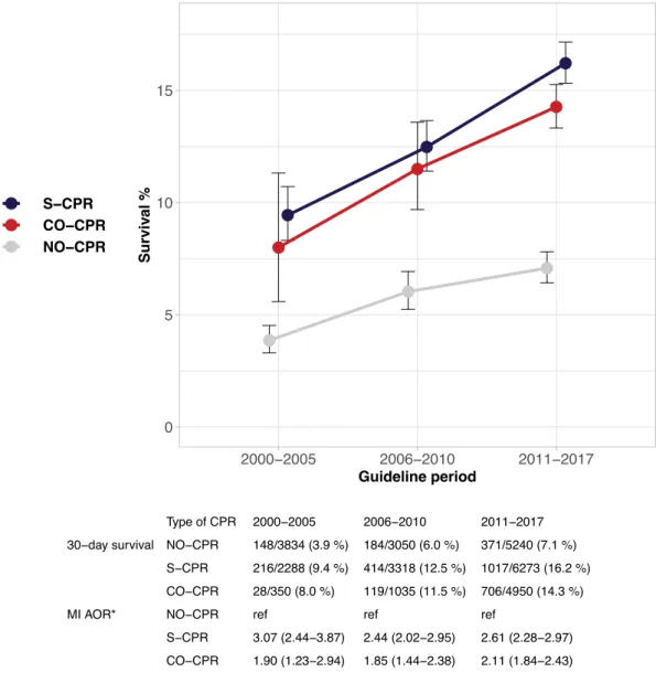
Specific study Aims and Results: In Study I we assessed survival after CPR prior to EMS arrival compared to no CPR prior to EMS arrival. Witnessed OHCA in 1990-2011 were included (N = 30 381). CPR prior to EMS arrival was performed in 51 % of all cases. Survival to 30 days was 10.5 % for patients receiving CPR and 4.0 % when CPR was not performed, odds ratio (OR) 2.80 (95% CI, 2.47 – 3.18), adjusted OR 2.15 (95 % CI, 1.88 – 2.45). The association with survival was greater when the time to the initiation of CPR was short. InStudy II we aimed to describe temporal changes in CPR rates and type of CPR prior to EMS arrival and survival in relation to three time periods of different CPR guidelines in Sweden. Witnessed OHCA in 2000 – 2017 (N = 30 455) were divided into groups reflecting guideline periods (2000 – 2005, 2006 – 2010, 2011 – 2017). Exposure was no CPR, S-CPR or CO-CPR. The proportions of patients receiving CPR prior to EMS arrival changed from 40.8 % to 68.2 % and CO-CPR changed from 5.4 % to 30.1 % between the first and the last guideline period. Adjusted OR for 30-day survival was 2.6 (95 % CI, 2.4–2.9) for S-CPR and 2.0 (95 % CI, 1.8–2.3) for CO-CPR, in comparison with no CPR. S-CPR was superior to CO-CPR (adjusted OR, 1.2; 95 % CI, 1.1–1.4).
InStudy III we aimed to assess survival after CPR with dispatcher instructions compared with no CPR and spontaneously initiated CPR. Lay-bystander witnessed OHCA in 2011 – 2017 were included (N = 15 471). Propensity score matched cohort were used for
comparison. Using dispatcher assisted-CPR as reference, spontaneously initiated CPR was associated with higher survival, OR 1.21 (95 % CI, 1.05–1.39) and no CPR with lower survival, OR 0.61 (95 % CI, 0.52–0.72).
In Study IV we aimed to assess feasibility and intermediate clinical outcomes in the TANGO2 trial. From Jan 1st to Dec 31st, 2017, a total of 729 emergency calls of suspected OHCA were randomized and 381 (51.4 %) of these were EMS treated OHCAs, 185 (48.6%) were assigned to S-CPR and 196 (51.4%) to CO-CPR. CPR instructions were provided in 89.3 % of all calls
and CPR was initiated in 93.4 % of all calls. Median time to CPR instructions was 210 s in the S-CPR group (IQR 140 – 301) and 180 s in the CO-CPR group (IQR 135 – 275), this time difference was not significant (NS). Cross-over from the S-CPR group to CO-CPR instructions was found in 22.3 % (40 calls), and from the CO-CPR group to S-CPR instructions in 16.1 % (30 calls). The number of patients surviving to hospital admission were 17.3% (n = 32) versus 20.4% (n = 40) for S-CPR and CO-CPR respectively (NS).
Conclusions: The current studies confirm the independent association between CPR prior to EMS arrival and survival in OHCA, irrespectively if CPR was performed with compressions and ventilation, compressions only or with dispatcher assistance. There was an almost doubled rate of CPR prior to EMS arrival in Sweden between 1990 – 2017 and a concomitant 6-fold increase in the rate of CO-CPR between 2000 – 2017. The pilot study of the TANGO2 trial was found to be feasible and safe. However, cross-over was found as a limitation.
PRELUDE
Out-of-hospital cardiac arrests is sudden and for the majority of victims difficult to prevent and predict. It affects more than 6 000 people every year in Sweden and only one in ten survive. It is a condition with heterogenous etiology, but underlying cardiac disease is the most common cause.
Once it has occurred the chance of survival is dependent on swift and efficient interventions by witnesses and a coordinated emergency medical response.
Cardiopulmonary resuscitation (CPR) performed before arrival of the ambulance is one of the strongest predictors of survival. This treatment is commonly performed by witnesses to the event, often a relative, partner, friend, colleague or a stranger. Many people might have limited training and little experience of this kind of situation. Nevertheless, their
interventions can be lifesaving.
In 1983 the Swedish Society of Cardiology proposed their first standardized CPR training program. This was done under the supervision of Dr Stig Holmberg.1 The aim was to disseminate CPR knowledge to the general population. Since then, an estimated 5 million attendees have been registered at basic CPR courses in Sweden.2
This work is a tribute to that movement and to the more than 20 000 bystanders, hidden behind the numbers, saving lives.
LIST OF SCIENTIFIC PAPERS
This thesis is based on the following studies which will be referred to by their roman numerals
I. Hasselqvist-Ax I, Riva G, Herlitz J, Rosenqvist M, Hollenberg J, Nordberg P, Ringh M, Jonsson M, Axelsson C, Lindqvist J, Karlsson T, Svensson L.
Early Cardiopulmonary Resuscitation in out-of-hospital cardiac arrest New England Journal of Medicine. 2015; 372: 2307–2315
II. Riva G, Ringh M, Jonsson M, Svensson L, Herlitz J, Claesson A, Djärv T, Nordberg P, Forsberg S, Rubertsson S, Nord A, Rosenqvist M, Hollenberg J.
Survival in out-of-hospital cardiac arrest after standard cardiopulmonary resuscitation or chest compression-only before arrival of emergency medical services
Circulation. 2019;139:2600–2609
III. Riva G, Jonsson M, Ringh M, Claesson A, Djärv T, Forsberg S, Nordberg P, Rubertsson S, Rawshani A, Nord A, Hollenberg J.
Survival after dispatcher-assisted cardiopulmonary resuscitation in out-of-hospital cardiac arrest – a nationwide observational study
Submitted
IV. Riva G, Ringh M, Jonsson M, Claesson A, Nord A, Rubertsson S, Blomberg H, Nordberg P, Forsberg S, Hollenberg J.
Compression-only or Standard Cardiopulmonary Resuscitation for Trained Bystanders in Out-of-Hospital Cardiac Arrest – A Nationwide Randomized PILOT study
CONTENTS
Prelude ... 3 1 Background ... 3 1.1 Historical glance ... 3 1.2 Definition ... 5 1.3 Incidence ... 5 1.4 Etiology ... 6 1.5 Risk factors ... 7 1.6 Predictors of survival ... 8 2 Treatment ... 112.1.1 Early recognition and call for help ... 12
2.1.2 Early CPR ... 13
2.1.3 Early defibrillation ... 13
2.1.4 Advanced Life Support ... 14
2.1.5 Post-resuscitation care ... 14
2.2 Physiological aspects of CPR ... 15
2.2.1 Chest compressions ... 15
2.2.2 Coronary perfusion pressure ... 16
2.2.3 Ventilation ... 16
2.2.4 Compression-only CPR ... 17
2.3 Current recommendations for CPR ... 18
3 Aim of this thesis ... 19
4 Methods ... 20
4.1 design, Studies I - IV ... 20
4.2 Setting ... 21
4.2.1 The Swedish emergency medical services system ... 21
4.2.2 Swedish emergency dispatch organization ... 21
4.3 The Swedish register for cardiopulmonary resuscitation ... 22
4.4 Description of the TANGO2 trial ... 23
4.4.1 Background and aim ... 23
4.4.2 Overview ... 23
4.4.3 Study phases ... 24
4.4.4 Description of the TANGO2 Pilot study (Study IV) ... 24
4.5 Ethical considerations ... 26
5 Results ... 28
5.2 Study II – “Temporal changes in frequency of CPR, type of CPR prior to
EMS arrival and survival during three guideline periods” ... 30
5.3 Study III – “Survival after dispatcher-assisted CPR” ... 32
5.4 Study IV – “Feasibility assessment of the randomized clinical trial TANGO2” ... 33
6 Discussion ... 36
6.1 Main findings ... 36
6.2 Is the association between early CPR and survival causal? ... 36
6.3 Is VT/VF at first rhythm analysis a confounder? ... 37
6.4 What explains the higher rates of CPR during the study period? ... 38
6.5 Is endorsement of CO-CPR associated with higher rates of CPR prior to EMS arrival? ... 38
6.6 Why have we only included witnessed OHCA? ... 39
6.7 Should all OHCA emergency calls be reviewed? ... 39
6.8 Which outcomes should be measured in OHCA? ... 40
6.9 What are the weaknesses and strengths of the Swedish Register for Cardiopulmonary Resuscitation? ... 40
6.10 What is the rationale and potential benefits of CO-CPR? ... 41
6.11 Are there any potential risks with chest compression-only CPR? ... 41
6.12 How reliable is the evidence supporting CO-CPR? ... 42
6.13 Is the survival difference between CO-CPR and S-CPR time-dependent? ... 43
6.14 Why was a pilot study performed in the TANGO2 trial? ... 43
6.15 What are the main findings from the pilot TANGO2 trial? ... 44
6.16 Future perspectives: ... 45
7 Conclusions ... 46
8 Sammanfattning på svenska ... 47
9 Acknowledgements ... 50
LIST OF ABBREVIATIONS
AED AHA ALS
Automated External Defibrillator American Heart Association Advanced Life Support BLS
CI CO-CPR CPC CPP
Basic Life Support Confidence Interval
Compression-only Cardiopulmonary Resuscitation Cerebral Performance Category
Coronary Perfusion Pressure CPR DA-CPR ECG EMS ERC ETI ICD ILCOR ITT IQR mRS NS OHCA OR PEA PP PPV PROM RCT Cardiopulmonary Resuscitation Dispatcher assisted CPR Electrocardiography
Emergency Medical Services European Resuscitation Council Endotracheal Intubation
Implantable Cardioverter Defibrillator
International Liaison Committee on Resuscitation Intention To Treat
Interquartile Range modified Ranking Scale Not Significant
Out-of-Hospital Cardiac Arrest Odds Ratio
Pulseless Electrical Activity Per Protocol
Positive Pressure Ventilation
Patient Reported Outcome Measurements Randomized Controlled Trial
ROSC S-CPR
Return Of Spontaneous Circulation Standard CPR
SCD SRCR TEE TTM VF VT
Sudden Cardiac Death
Swedish Register for Cardiopulmonary Resuscitation Transesophageal Ecography
Targeted Temperature Management Ventricular Fibrillation
1
BACKGROUND
1.1 HISTORICAL GLANCE
One of the first reports of chest compressions as a way of supporting circulation was “Resuscitation technique following cardiac death after inhalation of chloroform” by Dr Friedrich Maas in 1892.3 Dr Maas was a student of the famous surgeon Dr Franz Koenig at the University Hospital of Göttingen, Germany, who in 1883 developed a technique for treating the dreaded “chloroform syncope”. The technique consisted of compressing the xhiphoid and costal margins at the rate of respiration, as a form of assisted ventilation. Dr Maas described a nine-year old boy who became unresponsive with no pulse or breathing after chloroform anesthesia. Dr Koenig´s technique was started, but as the boy did not show any sign of improvement Dr Maas started to compress faster. He then noticed that the compressions produced a palpable carotid pulse. Dr Maas suggested that fast chest compressions could support circulation.3 However, this discovery appears not to have spread outside Germany.4 In 1901 the first open chest “cardiac massage” was performed in Tromsø, Norway, and attention shifted to open cardiac massage for nearly 70 years.
In 1958 Kouwenhoven, Knickerbocker and Jude made an accidental discovery while
performing experimental studies on defibrillation in anesthetized dogs. The found that firm application of the defibrillation pads on the thorax could produce a femoral pulse. After further experiments they concluded that manual compressions over the sternum could induce a circulation and prolong the time window for successful defibrillation. Shortly
afterwards, Jude successfully applied this technique in a patient with cardiac arrest following anesthesia. In 1960 they reported 20 in-hospital cardiac arrest treated by means of closed cardiac chest massage, of whom 14 survived. They stated: “Anyone, anywhere, can initiate cardiac resuscitative procedures. All that is needed is two hands”. 5,6
Successful resuscitation by means of mouth-to-mouth ventilation was described by the Scottish surgeon William Tossach in 1744.1 Although the method was recommended and used successfully during the eighteenth century, other techniques to assist ventilation such as applying external pressure to the thorax or tilting the body gradually attained more attention and the mouth-to-mouth (or mouth-to-nose) technique was almost forgotten.1 In 1946, during a Minnesota polio outbreak Dr James Elam successfully performed mouth-to-nose breathing on a boy with acute bulbar poliomyelitis. He went on to prove that expired air was sufficient for adequate oxygenation by blowing into the tracheal tube of
postoperative surgical patients in 1954.7 James Elan met with Dr Peter Safar and together they performed compelling experiments on anesthetized paralyzed human volunteers. By
1957 they concluded that by tilting a person’s head backwards, most will have an open airway, external chest compression is not enough for adequate ventilation in contrast to mouth-to-mouth breathing and that this technique can be used for resuscitation.8 In 1958 the method was endorsed by the American Medical Association.9
At the Maryland Medical Society meeting on the 16 of September 1960, Kouwenhoven, Jude and Safar presented their findings, connecting chest compressions with mouth-to-mouth breathing, and creating the concept of cardiopulmonary resuscitation (CPR) as we know it today.10
1.2 DEFINITION
A cardiac arrest is usually a sudden event and if not treated death is inevitable. Several definitions of the condition exist and are to some extent overlapping. An international consensus workgroup has defined cardiac arrest as “the cessation of cardiac mechanical activity as confirmed by the absence of signs of circulation”.11
An out-of-hospital cardiac arrest (OHCA) is such an event occurring outside of hospital. If fatal, the term Sudden Death (SD) is defined by the European Society of Cardiology as a “non-traumatic, unexpected fatal event occurring within 1 hour of the onset of symptoms in an apparently healthy subject. If death is not witnessed, the definition applies when the victim was in good health 24 hours before the event”. Sudden cardiac death (SCD) is used when a cardiac condition was known to be present during life, or autopsy revealed a cardiac or vascular anomaly, or no obvious extra-cardiac causes have been identified.12
1.3 INCIDENCE
OHCA is a major cause of death worldwide. It is estimated that a total of 275 000 people in Europe and 180 000 in the US suffers from OHCA treated with resuscitation attempts by the Emergency Medical Services (EMS) each year, with an overall survival rate of 7-10 %.13-15 However, exactly how OHCA contribute to the burden of public health is unknown.
Reported incidences vary widely between and within countries. 13,16,17 This may reflect true regional variances due to differences in population risk factors and socioeconomic factors. But it may also be a result of differences in reporting, organization of EMS systems and epidemiological methods. Also, not all OHCA are treated by the EMS. This could be because of ethical reasons, “do not resuscitate orders” as well as cultural reasons.
In the European Cardiac Arrest Register, a report of cardiac arrests in 27 European countries in one month, revealed an overall incidence of EMS assessed OHCA of 81/100 000 and EMS treated OHCA of 47/100 000 (varying from 19 to 104) with an overall survival to hospital discharge of 10.3 %.17 In Sweden the reported incidence of EMS treated OHCA in 2011 was 52/100 000, with an overall survival to 30-days of 10.7 % in 2011. 18
In 1990 a meeting to establish a system for coherent reporting in resuscitation was held in Utstein abbey, Norway, giving rise to the “Utstein style” templates.19 These offer a base of uniform definitions and suggest core data elements to collect, enabling comparison, quality improvement and research.11,20 These templates have been widely adopted in resuscitation registries globally.17,21-24
1.4 ETIOLOGY
The clinical manifestation of OHCA can arise from numerous conditions ultimately leading to reduced cardiac output and cardiac arrest. These can be divided into medical and non-medical (external) causes.20 Medical causes can be further divided into cardiac and non-cardiac causes. (Table 1)
Cardiac disease is the predominant cause, estimated to account for 2/3 of all OHCA.25-27 However, the determination of the exact cause of a cardiac arrest is difficult, especially in an out-of-hospital setting. Furthermore, information on comorbidities and symptoms prior to the cardiac arrest is often missing. The gold standard for assessing cause of death is autopsy, but far from all patients suffering a fatal cardiac arrest will undergo this investigation.28,29 According to current definitions a OHCA can be presumed to be of cardiac etiology unless it is likely to be caused by trauma, submersion, drug overdose, asphyxia, exsanguination or any other non-cardiac cause as best determined by rescuers.11 Therefore, EMS reports might overestimate the proportion of cardiac etiology.
In a series of 569 cardiac arrest resuscitated and admitted to a hospital in Vienna, cardiac causes accounted for 69 %. A cardiac cause presumed by attending physicians at the emergency department showed a high sensitivity (93 %) but was less specific (77 %). Pulmonary embolism, cerebral disorders and bleeding were among the most frequently overlooked conditions.30 In parallel, in a study of 809 prospectively collected OHCAs in Helsinki, cardiac causes accounted for 65.6 %. Of patients with a non-cardiac causes, the correct cause was suspected in the prehospital setting in only 63.8 %, in the remaining patients the non-cardiac cause of arrest was revealed after in-hospital examination or autopsy.31
Of all patients successfully resuscitated from OHCA more than 50 % will have significant coronary heart disease.32 In a prospective Parisian study of successfully resuscitated OHCA patients 61% had no obvious extra-cardiac cause of arrest. Of those, 70 % had at least one significant lesion (96 % among those with ST-segment elevation and 58 % among those without ST-segment elevation on the initial ECG).32 In a trial of urgent coronary angiography among patients presenting with ventricular fibrillation, without ST-segment elevation on initial ECG, 64.5 % had significant coronary disease, and 17 % had an acute unstable lesion or coronary occlusion.33 Finally, causes of OHCA are different in different age groups. Cardiac causes accounted for 69 % among individuals aged > 65 years, but only 10 % among individuals between 16-40 years of age in the Swedish register for cardiopulmonary
Table 1: Causes of OHCA
Table 1: This list is not intended to be exhaustive and causes are not listed in order of frequency
1.5 RISK FACTORS
Since cardiovascular disease is a dominant cause of OHCA, it naturally shares many risk factors of ischemic heart disease such as age,15 male sex,35 smoking, hypertension36 and lower socioeconomic status.37
Manifest heart disease is a strong marker of an increased risk of SCD, in particular previous myocardial infarction and heart failure. However, and most importantly, the majority of SCD victims have not been previously diagnosed with heart disease.35 There is an inverse
relationship between the number of cases of SCD observed in different risk groups and the individual risks of SCD. The largest number of SCDs in society will occur in the general
population with a low risk. Therefore, risk stratification can be sensitive for a small subgroup of patients, but have a low power to as regards large, low-risk groups.
Figure 1: Proportion of SCD in different subgroups
Figure 1: Proportion of Sudden Cardiac Death (SCD) in different subgroups.
Al-Khatib et al.38 Reproduced with permission from Circulation
A prediction model calculated from two American cohorts of individuals with no previous cardiovascular disease, three-fourths of the participants could be identified as being “low risk” (with a year risk of less than 1 %). However, among the highest risk decile, the 10-years risk was not greater than 5 %.39
Also, among patients with known heart disease the risk assessment of SCD is complex. The risk of ventricular arrythmia after myocardial infarction is time-dependent, with the highest risk in the first 48 hours.40 The risk factors with the best predictability (ischemic heart disease, low ejection fraction, inherited genetic disorders and patients with previous documented sustained ventricular arrythmias or previous cardiac arrest) are susceptible to preventive treatment with implantable cardioverter defibrillators (ICDs). 12 Several other markers of risk have been evaluated such as heart-rate variability, Qintervals dispersion, T-wave alternans and biomarkers such as BNP, without influencing clinical practice.41 Risk stratification of patients with heart disease is extensively described elsewhere,38 and is beyond the scope of this thesis.
1.6 PREDICTORS OF SURVIVAL
There is substantial variability in survival across EMS systems and several factors that influence the probability of survival after OHCA have been studied. These factors can be patient related such as sex,42 age,43 and cause of arrest, 25 or event related. Examples of event related factors are witnessed event, CPR prior to EMS arrival, first recorded rhythm, location of the arrest, and time to defibrillation. Some of these factors will be discussed below.
1.6.1.1 Location
Around 2/3 of all OHCA occur at home and this is associated with lower survival compared to a cardiac arrest in a public location.44 Individuals suffering a cardiac arrest at home are usually older, less frequently receive bystander CPR, have longer EMS response times and a lower incidence of VT/VF as first rhythm.44 However, the association between location and chance of survival is independent, and might reflect unmeasured factors such as
co-morbidities or lower degree of physical activity among patients suffering OHCA at home. 1.6.1.2 Witnessed event
A witnessed event is defined as a collapse seen or heard by another person, or monitored.20 Since time to treatment is crucial, it is only logical that individuals who suffer an
unwitnessed cardiac arrest have a poorer prognosis. 45 Cardiac arrests witnessed by the EMS are a separate group. This group usually have had symptoms prior to the arrest
necessitating call for an ambulance prior to the arrest, and the time to treatment is very short. Cases of OHCA witnessed by the EMS have been found to have better prognosis. 46
1.6.1.3 Sex
Female sex accounts for about 1/3 of all OHCA. Females sex is associated with OHCA at home and a lower percentage of bystander CPR and VT/VF as first rhythm.42 This can explain the lower survival reported for females.42,47 The reason for the lower rate of VT/VF among those with female sex is not entirely clear and might have biological explanations. In contrast, others have found female sex to be associated with higher survival among the subgroup presenting with VT/VF as first rhythm.48
1.6.1.4 Age
The incidence of OHCA increases with age. Advanced age is also associated with lower survival in OHCA. In one study the chance of a positive outcome after OHCA decreased with 22% for each decade of life.49 However, the correlation appears to be weaker than many other event related factors such as initial rhythm.50
1.6.1.5 First recorded rhythm
The arrythmia at the time of collapse is for natural reasons unknown. The first recorded ECG rhythm at EMS arrival or by an AED can be divided into; a) Ventricular Fibrillation (VF) and Ventricular Tachycardia (VT) susceptible to defibrillation, and b) Asystole and Pulseless Electrical Activity (PEA), not susceptible to defibrillation. VT/VF as first recorded rhythm is the strongest predictors of survival in OHCA. 51 For patients found in VT/VF, and treated with a public AED, survival can be as high as 59-71%.52,53
1.6.1.6 Ventricular Fibrillation
VF can be described as an electrical chaos, causing asynchronous activity of cardiac muscle with subsequent cessation of pump function. 38,54 The pathophysiology of VF is complex. Heterogeneity of repolarization and conduction properties in myocardial tissue can cause electrical wave fronts to break up (functional re-entry) into multiple wavelets that cause fibrillation.55 Structural predisposition (cardiac scarring, hypertrophy, myopathies or electrical abnormalities) and transient factors (ischemia, hypoxia, electrolyte disturbances, hemodynamic alternation, neurophysiological factors or drugs) interact to make the myocardium susceptible to VF. 56
If VF is left untreated it will deteriorate from “coarse” to “fine” VF and finally to asystole.57 This explains the association of greater probability of finding a patient in VT/VF with a shorter time interval from cardiac arrest to rhythm analysis.58,59 The proportion of OHCA with VT/VF as first recorded rhythm is around 20 – 30 % for witnessed OHCA.47 However, the incidence of VT/VF as first recorded rhythm has declined during the last decades.60,61 The reasons are not clear but longer EMS response times,18 improved cardiovascular and heart failure care with subsequent reduction in mortality,60 and the implementations of ICD62 are possible reasons.
1.6.1.7 Ventricular Tachycardia
VT is an abnormal cardiac rhythm originating from the ventricle at a rate > 100 beats per minute. VT can originate from myocardial cells with altered conductance and repolarization properties compared with adjacent myocardium that form electrical re-entry circuits, or due to increased automaticity. VT in the setting of myocardial ischemia is often polymorphic whereas sustained monomorphic VT is typically due to a myocardial scar or fibrosis re-entry.63 VT proceeding to VF is thought to be the most common mechanism of SCD among patients with ischemic heart disease.64 In a study of 157 SCD in patients wearing ambulatory ECG-monitoring, VT proceeding to VF was found in 62 %, primary VF in 8 %, Torsade de pointes VT in 13 % and bradyarrhythmia in 17 %.65
1.6.1.8 Asystole
Asystole is defined as cessation of myocardial electrical activity. In a prehospital setting the finding of asystole on initial rhythm analysis usually represents a dying heart with cardiac standstill. Survival in OHCA with asystole as the first recorded rhythm is extremely poor, ranging from 0,2 % to 1.3 %.66-69
1.6.1.9 Pulseless Electrical Activity
PEA is defined as an organised electrical rhythm on ECG but no palpable pulse. PEA can arise from electromechanical dissociation (organized electrical cardiac rhythm but no myocardial pump motion) or mechanical obstruction such as tamponade, pulmonary embolism and hypovolemia causing profound hypotension. The incidence of PEA has increased in recent years, and the prognosis after PEA has been found to be better in comparison with asystole.67-69
2
TREATMENT
Survival after OHCA is highly dependent on a fast response from citizens, emergency
dispatch organisations and EMS-services all working together. A framework for this concept is “the chain of survival” introduced in an AHA statement from 1991.70-72 The links in the chain of survival are (1) early recognition of symptoms and placing an alarm call, (2) early CPR (3) early defibrillation and (4) post-resuscitation care.
Figure 2:
Nolan et al.72 Reproduced with permission from Elsevier
The number of patients susceptible to each intervention decreases for every link in the chain. An alternative demonstration of the chain of survival has been proposed, reflecting the number of patients susceptible to each intervention.73
Figure 3: Revised chain of survival
Deakin et al.73 Reproduced with permission from Elsevier
Figure 3: The revised representation of the chain of survival is adjusted so the area of each link graphically represents the number of patients in each step.
2.1.1 Early recognition and call for help
Early recognition is naturally crucial for the subsequent resuscitation efforts to be effective. A person who is unresponsive and not breathing normally should be considered to be in cardiac arrest. 74 Victims of cardiac arrest often gasp during the first minutes, the so called “agonal breathing”.75 This reflex appears to be triggered by ischemia of the brainstem and decreases with time from cardiac arrest. In experimental studies agonal breathing can be maintained if effective chest compressions are continued,76 and the presence of agonal breathing at EMS arrival is associated with increased survival.75
Emergency dispatchers can play an important role as they can assist in recognising a cardiac arrest. Different protocols to aid dispatchers in cardiac arrest recognition are used.77 The proportion of OHCA recognition during alarm calls ranges from 14 % and 83 %.78-82 The median rate of recognition across dispatch system have been found to be 73 % in a
systematic review.83 Agonal breathing is one of the main barriers for recognition during an alarm call.77,84 Agonal breathing can be present in up to 40 % of all calls,84 and can account for half of the calls with missed diagnosis.82 The importance of the emergency dispatcher together with the caller, forming the “first resuscitation team” is highlighted in European Resuscitation Council (ERC) guidelines.85 When a OHCA is recognised, callers can be offered instruction over telephone on how to perform CPR - dispatcher assisted CPR (DA-CPR).86 Implementation of DA-CPR programs have been found to be associated with higher CPR rates82 and survival.87-90 DA-CPR can be associated with significant time delays, ranging from 138 – 183 s from call to CPR instruction 81,82,91
2.1.2 Early CPR
The aim of cardiopulmonary resuscitation is to create an artificial circulation providing essential blood flow to crucial organs, mainly the brain and heart, until restoration of spontaneous circulation can be achieved. Initiation of bystander CPR prior to EMS arrival is independently associated with survival rates 2-3 fold higher compared with no such initiation.51,92 There are wide differences in incidence of bystander CPR , ranging from 86% in Denmark, 92, 42% in Japan. 23 Factors associated with higher probability of receiving early CPR are among others cardiac arrest in a public location,44 male sex,42 and younger age.43
2.1.3 Early defibrillation
Defibrillation is the only definitive treatment of VT/VF. Time to defibrillation is crucial and survival decreased for every minute without CPR or defibrillation in VF cardiac arrest.93 Traditionally defibrillation is carried out by EMS personnel. However, there are challenges in reducing EMS response time.18 Therefore, alternative strategies to enable faster
defibrillation has emerged. The concept of “dual dispatch” referrers to dispatching first responders such as fire-fighters or Police units equipped with an AED. In a project in
Stockholm, the implementation of a dual dispatch in parallel to the traditional EMS response was associated with higher survival, in spite that time to defibrillation was marginally
reduced.94 Publicly available AED used by bystanders can be associated with survival rates well over 50% in VT/VF arrest.52 However, in spite the fact that the numbers of AED is increasing, they are seldom used.74 Different strategies to optimize AED placements have been proposed.95 The implementation of AED-registers can enable emergency dispatchers to guide callers to the location of a nearby AED.96 Another concept based on AED registers, is to use a smartphone application to dispatch lay volunteers to retrieve an AED to nearby OHCA.97 Finally, in rural areas the concept of drone delivered AED has been proposed.98 In summary, time to defibrillation is the most crucial factor for surviving a VT/VF arrest and new strategies to decrease time to defibrillation are under investigation.
2.1.4 Advanced Life Support
The concept of Advanced Life Support (ALS) in EMS includes CPR and defibrillation, but also advanced airway management, intravenous access and administration of drugs. The benefit of ALS compared to more basic life support (BLS) in the prehospital setting have been debated. In one study in Ontario, the implementation of ALS in the ambulances (opposed to BLS) resulted in higher rates of survival to hospital admission but not to hospital discharge.45 Some components of ALS interventions will be discussed below, but ALS as a whole is beyond the scope of this thesis.
There is controversy about optimal airway management in ALS.99 Several RCT´s have compared Endotracheal Intubation (ETI) to simpler techniques such as bag-mask ventilation,100 laryngeal tube, 101 and supraglottic airway devices.102 Optimal method of airway management might depend on local EMS organization and the training level of EMS staff. Adrenaline is used to increase systemic pressure and coronary and cerebral blood flow during CPR and anti-arrhythmic drugs are recommended for VT/VF not responding
defibrillation.103 The use of drugs in ALS have been found to increase the rate of return of spontaneous circulation (ROSC) and survival to hospital admission,104 but the effect on long term survival and neurological function is less clear. In a recent double-blind RCT including more than 8 000 patients, adrenalin was found to increase survival, but not a favorable neurological outcome.105 In a RCT comparing Amiodarone, Lidocaine and placebo found no difference in survival to discharge, although there was an interaction between drug effect and witnessed status, indicating a potential benefit among cases of witnessed OHCA.106 The routine use of mechanical chest compressions devises has not been found to increase survival.107,108 However, mechanical chest compressions are recommended for prolonged CPR and during transport. In summary, ALS interventions include advanced airway
management and administration of intravenous drugs, but the impact on survival is lower compared to early CPR and defibrillation.
2.1.5 Post-resuscitation care
The goal of post-resuscitation care is to stabilize the circulation and ventilation, find and treat the cause of arrest, and to optimize recovery.109 Depending on the length of the arrest, most patients achieving ROSC are comatose with an indication for intubation and assisted ventilation. Shock is common and can be treated with fluids or vasopressors. Targeted temperature management (TTM) to 32-36 degrees for 24 hours at is recommended for specific subgroups of patients.110 This is based on the results of two trials comparing TTM to 32 degrees with no temperature control.111,112 A larger trial comparing TTM at 33 to 36 degrees did not demonstrate any difference in survival or favorable neurological function,
113 but a recent RCT including non-shockable rhythms found higher survival for TTM 33 degrees compared to normothermia.114 Coronary angiography for patients with ST-segment elevation on initial ECG is recommended, but for patients presenting without ST-segment elevation the benefit of early angiography is unclear.33 Many resuscitated patients will develop a “post cardiac arrest syndrome” with hyperinflammation and sepsis-like features. Targeted goals for intensive-care management and neurologic prognostication exist and are extensively described elsewhere. 110 However, controversies regarding optimal blood-pressure targets, oxygenation and ventilation strategies remain.115
2.2 PHYSIOLOGICAL ASPECTS OF CPR
2.2.1 Chest compressions
Traditional CPR is composed of repetitive chest compressions with interruptions for ventilation. It has been estimated that 25 % of systemic perfusion pressure and 12-27 % of normal cardiac output can be maintained with CPR.116,117
The exact mechanism of how chest compressions generate a forward blood flow is complex. According to the cardiac pump theory the heart is directly compressed between the
sternum and vertebrae, creating a higher pressure in the ventricles than in surrounding compartments, with closure of the mitral valve and opening of the aortic valve, creating a forward blood flow.6,118
This theory has been complemented with the thoracic pump theory, by which pressure fluctuations in all intrathoracic compartments, including the intrathoracic aorta, generate blood flow.119,120 If patent, jugular venous valves or collapsing veins at the thoracic outlet are thought to prevent backward venous flow.121 Opening of both aortic and mitral valves during compression transmits the intrathoracic pressure to extra thoracic arteries.
According to this theory the heart acts as a passive conduit. During decompression the chest wall recoils and intrathoracic pressure drops, enabling venous return from extra-thoracic veins.122
It is likely that both mechanisms apply to chest compressions in clinical practice, and the magnitude of each component might be time-dependent.123 Kim et al performed contrast enhanced transesophageal echocardiography (TEE) in 10 patients during CPR (all in asystole) and found aortic valve opening and mitral valve closing during compression (supporting the cardiac pump theory), and mitral regurgitation in all patients.124 However, they found large variations in retrograde mitral valve flow, and concluded that the cardiac pump theory was predominant although both mechanisms may be contributing.
2.2.2 Coronary perfusion pressure
During CPR coronary perfusion pressure (CPP) is defined as the difference between aortic pressure and right atrial pressure during decompression phase.125 CPP has been shown to correlate well with coronary blood flow in experimental models.126 The probability of ROSC in both animals and humans appears to be low when CPP is below 15 mmHg.127,128 Blood-flow through the coronary vessels is higher during decompression.116 Therefore, the heart is mainly perfused during the decompression phase.129
After onset of VF, the aortic pressure drops and there is a gradual increase in the right-atrial pressure, with distention of the right ventricle. Eventually there is equilibrium between aortic pressure and right-atrial pressure, rendering CPP to zero. Steen et al. found in an experimental study that chest compressions promptly increased extra-thoracic aortic pressure and carotid blood flow but it took up to two minutes of mechanical chest compression to establish an adequate CPP after VF was untreated for 6.5 min.130 The optimal rate of chest compressions has been found to be between 100 – 120 per minute. 131 If compressions are too fast, the left ventricle will not have sufficient time to refill before next compression. The optimal compression depth has been found to be
around 4 – 5.5 cm.132 There appears to be an inverse relationship between compression rate and depth, faster chest compression in real life scenarios will result in inadequate depth.133
2.2.3 Ventilation
Ventilation is the movement of air from outside the body to the alveoli to enable gas exchange. During bystander CPR ventilation is provided by positive-pressure breaths (PPV). Intermittent positive airway pressure can maintain oxygenation, increase carbon dioxide clearance and prevent from lung atelectasis. 134 However, PPV also effects circulation in a complex way. In mechanically ventilated patients increased intrathoracic pressure
“squeezes” blood from the lung capillaries to the left side of the heart, momentarily increasing left ventricular filling pressures and stroke volume.135 On the other hand,
increased intrathoracic pressure reduces venous return to the right side of the heart and has been found to inversely correlate to CPP and cerebral blood flow.136 In summary,
hypoventilation can cause hypercapnia and hypoxia,137 while hyperventilation can have adverse effects on cerebral and myocardial circulation.136 There is arguably a balance between too much and too little ventilation during CPR.
2.2.4 Compression-only CPR
Interruptions in chest compressions can have a deleterious effect on hemodynamics during CPR 138,139 and long interruptions have been found to correlate with a decreased rate of ROSC in ALS.140 The concept of CO-CPR, continuous chest compressions without rescue breaths, has been extensively evaluated in experimental models.76,127,139
There is conflicting evidence from observational human studies. A number of register-based observational cohort studies have been carried out to compare CO-CPR with S-CPR, many of which have shown neutral results.141-143 Differences in favour of S-CPR have been found in connection with paediatric arrests, 144 individuals younger than 18 years,145, and when EMS response-time were longer than 15 minutes.146 Others have found higher survival for CO-CPR among patients found in VT/VF or treated with public AED.147,148 One study showing a higher survival rate with CO-CPR was conducted during a community program promoting CO-CPR. 149 In a Japanese population of OHCA of medical origin, CO-CPR was associated with a higher survival with CPC 1-2 in a propensity score matched cohort (7.2% vs 6.5%, OR 1.12 p < 0.001).150
Three randomized trials have compared dispatcher assisted CO-CPR with S-CPR (in a 15:2 ratio), involving bystanders without previous CPR training, and the results have been neutral. 151-153. In a meta-analysis of these trials there was a modest but statistically significant increase in survival with CO-instructions. (RR 1.22, 95% CI 1.01-1.14)154
Techniques to minimize interruptions in chest compressions during ALS is typically involve placement of an ETI device and positive pressure ventilation (PPV). Alternatives are continuous chest compressions and desynchronised PPV via a bag-mask device, or passive oxygenation with an oropharyngeal airway or oxygen mask.155 This concept was compared to bag-mask ventilation in a 30:2 ratio in a cluster RCT, including 23 711 patients, showing neutral results in the ITT analysis but, in a PP analysis those treated with 30:2 had a favourable neurological outcome.
2.3 CURRENT RECOMMENDATIONS FOR CPR
In 1992 resuscitation scientists formed a forum for collaboration, the International Liaison Committee on Resuscitation (ILCOR), publishing treatment recommendations.156 The ILCOR “consensus statement on science and treatment recommendations” has been periodically updated to serve as a foundation for regional resuscitation guidelines.85,157,158 In 2005, there was a shift in the compression to ventilation ratio from 15:2 to 30:2, and CO-CPR was
introduced as an option if the bystander was unable or unwilling to perform rescue breaths.157 In 2010 CO-CPR was recommended for dispatcher assisted CPR.159 Because of the controversies regarding the optimal compression to ventilation ratio, ILCOR reviewed all available evidence in 2017 and made the following recommendations for bystander CPR.160
“We continue to recommend that bystanders perform chest compressions for all patients in cardiac arrest (good practice statement)”.
“We suggest that bystanders who are trained, able, and willing to give rescue breaths and chest compressions do so for all adult patients in cardiac arrest (weak recommendation, very-low-quality evidence)”.
For dispatcher assisted CPR, CO-CPR is recommended for adults with suspected OHCA.161 This recommendation is cited as a strong recommendation, but based on low quality evidence. The level of evidence was downgraded because of a serious risk of bias.160 In the process of reviewing the current evidence knowledge gaps identified by ILCOR were the effect of delayed ventilation versus 30:2 high-quality CPR, the impact of continuous chest compressions on outcomes of CA arising from noncardiac causes and the ability of bystanders to correctly perform mouth-to-mouth ventilation.
In Summary, guidelines on how to perform CPR have changed. Changes in CPR rates, methods of CPR, and the impact on 30-day survival after these modifications in
recommendations are unclear. Furthermore, there is conflicting data regarding the effect of dispatcher assisted CPR and survival. Finally, to address the question of whether CO-CPR leads to survival no worse than, equally effective or superior to S-CPR for bystanders with previous CPR training the TANGO2 pilot trial was launched in 2017.
3
AIM OF THIS THESIS
The overall aim of this research project was to assess the association between different forms of CPR prior to EMS arrival and survival in cases of OHCA.
Study specific aims:
1. To assess survival after CPR prior to EMS arrival compared with no CPR prior to EMS arrival in OHCA (Study I)
2. To describe temporal changes in CPR rates and type of CPR prior to EMS arrival and survival in relation to three time periods of different CPR guidelines in Sweden (Study II).
3. To assess survival after CPR with dispatcher CPR instructions compared with no CPR and CPR not requiring dispatcher instructions (Study III).
4. To assess feasibility in a pilot study of a national, investigator-initiated, multicentre, RCT designed to compare survival after dispatcher instructions of chest compressions and rescue breaths versus dispatcher instructions of chest compressions only, to trained bystanders in OHCA (study IV).
4
METHODS
4.1 DESIGN, STUDIES I - IV
Studies I-III are observational cohort studies including patients reported to the SRCR. Study IV is a pilot study of a prospective, investigator-initiated, multicenter, randomized clinical trial. The data for Study IV relies on automatically stored event times obtained from the national Swedish emergency dispatch organization SOS Alarm and reviews of audio files of emergency calls. Patients characteristics and follow up are collected through the SRCR.
4.2 SETTING
4.2.1 The Swedish emergency medical services system
The Swedish EMS system is organized in regional EMS agencies. Ambulances are in general staffed with registered nurses with additional training in emergency
medicine/anesthesiology. Ambulances are equipped with AEDs. The ambulance crew performs ALS in accordance with ERC guidelines.85 In some regions fire fighters and/or police are dispatched in parallel to EMS.94,162 These units, in this test referred to as “first responders”, are equipped with AEDs and the personnel trained in BLS. A cardiac arrest typically generates an EMS response of two ambulances, and in some regions a second tier of ALS units, each equipped with an emergency physician.
Rules for termination of resuscitation are defined by local EMS advisory guidelines in adherence to the “universal” termination of resuscitation rule. 103,163 In Stockholm, the criterion for termination of resuscitation is 20 minutes of ALS without electrical activity (asystole). Criteria for early termination of resuscitation are; non-witnessed event, no CPR prior to EMS arrival, > 15 minutes from call to EMS arrival and asystole at first rhythm analysis.
4.2.2 Swedish emergency dispatch organization
Sweden has a population of 10 120 242 inhabitants (31 December 2017), covering an area of 450 000 km2. All emergency calls in Sweden are answered by a SOS Alarm. SOS Alarm is organized in 18 dispatch centers, all operating nationwide. In 2018 SOS Alarm handled 3.2 million emergency calls of which 44 % were related to medical issues.164 Emergency calls are primarily answered by emergency dispatch telecommunicators. For medical emergencies SOS Alarm coordinates the triage and dispatch of EMS according to a criteria-based dispatch protocol.77 (If the caller is situated in one of three regions [Uppsala, Södermanland and Västerås] calls are redirected to local medical dispatch centers run by local EMS
organizations for triage and dispatch). A cardiac arrest should be suspected when a person is described as unconscious with no breathing, or not breathing normally. Dispatchers are instructed to deliver CPR instructions to bystanders untrained or unable to initiate CPR.165 For adult victims of CA instructions are compression-only. For presumed asphyxia-related CA and for children, the instructions are compressions and rescue breaths in a 30:2 ratio. For children CPR is to be initiated by five rescue breaths. In some regions a smart-phone application is used to locate and recruit lay volunteers to perform CPR or retrieve a nearby AED. This system is activated by the emergency dispatchers. 97
4.3 THE SWEDISH REGISTER FOR CARDIOPULMONARY RESUSCITATION
The Swedish Register for Cardiopulmonary Resuscitation (SRCR) is a National Quality Register founded in 1990 in Gothenburg. It has expanded gradually and since 2010 all EMS organizations in all 21 regions in Sweden report to the register. The register is one of the Swedish national quality registers of health-care measurements166 and contain variables in accordance with the “Utstein” guidelines of reporting in OHCA.167 The register is composed of three parts.
1) The first part is completed by EMS crew after attending an OHCA. Reports are entered in the register only if EMS or bystanders attempted resuscitation and the patient was not declared dead at EMS arrival. The variables in the first section describe the circumstances of the OHCA, patient related factors, treatment variables and patient status at the end of the prehospital treatment.
2) The second part contains variables regarding in-hospital treatment and follow up. Survival to 30 days is obtained by linkage with the Swedish National Population Register through the 10-digit personal identification number.168 Neurological function is assessed according to the Cerebral Performance Category (CPC) scale at hospital discharge.169 According to this scale, 1 is good cerebral performance, 2 is moderate disability, 3 is severe disability, 4 is coma or vegetative state and 5 is brain death. This section is completed by a local CPR coordinator.
3) The third part is composed of Patient Reported Outcome Measurement (PROM) and is completed by patients and nurses at follow-up visits. The PROM includes health related quality of life assessment (EQ-5D, EQ VAS) and anxiety assessment (HADS). The PROM evaluation occurs between 3-6 months after the cardiac arrest and have been part of the register since 2013. As of 2018 a total of 52 hospitals (73 %) report PROM to the register. Between 1990 and 2007 reports were written manually on paper, since 2008 data has been reported electronically. The accuracy of inclusion of all cardiac arrests between 1992 to 2010 is estimated to range from 70–100%.18,170 Today, cross-checking of EMS record to identify missing OHCA and retrospective inclusion in the register is performed by a local register coordinator (usually an experienced nurse).
4.4 DESCRIPTION OF THE TANGO2 TRIAL
4.4.1 Background and aim
Current guidelines advocate CPR provided by trained bystanders to be carried out with both rescue breaths and chest compressions. However, if the bystander is untrained, uncertain or unwilling to provide rescue breaths, CO-CPR is recommended.161
There is a lack of conclusive evidence regarding the effect of CO-CPR compared to S-CPR when performed by bystanders with previous CPR training for adult OHCA. 151-153,171,172 This knowledge-gap is highlighted by the International Liaison committee on resuscitation (ILCOR) consensus-report in 2017.160
In the light of the equipoise and the very-low quality of the available evidence the TANGO2 trial aims to investigate whether survival after instructions to perform CO-CPR to bystanders with prior CPR-training is no worse, or better than instructions to perform S-CPR in
witnessed, adult OHCA of presumed cardiac origin.
4.4.2 Overview
TANGO2 is an interventional, prospective, 1:1 open label, multicenter randomized trial (Clinical Trials, NCT02401633). TANGO2 is conducted at the dispatch center. Emergency calls concerning suspected OHCAs (unresponsive patient with no, or agonal breathing) are
eligible for inclusion. Study inclusion criteria are: - Witnessed collapse (seen or heard)
- The caller or anyone at the scene has previous CPR-training - Victim´s age above 18
- No signs of trauma, asphyxia, intoxication or pregnancy
Included calls are randomized 1:1 to instructions to provide either S-CPR (control) or CO-CPR (intervention).
Post-randomization exclusion criteria for final data analysis are: Not EMS treated OHCA. Primary outcome is 30 days survival.
Secondary outcomes are survival to hospital admission, survival to 1-year survival, survival with good neurological outcome at discharge (defined as CPC 1-2) and survival with good neurologically outcome (defined as CPC 1).
4.4.3 Study phases
The overall study project is conducted in three phases: 1) Pre study RUN-IN period
Objective: In order to test the technical inclusion procedures, logistics, feasibility and data collection a pre-study RUN-IN period started in Stockholm during 2015 (completed). 2) PILOT study
The aim of the pilot study was to assess safety and feasibility of the TANGO2 trial, as well as intermediate clinical outcomes.
3. MAIN study
The aim of the main study is to evaluate survival to 30 days following instructions to perform CO-CPR is non-inferior compared to instructions to perform S-CPR bystanders in witnessed adult OHCA of presumed cardiac origin and where the bystander has CPR training. The main trial will include patients from both the pilot and main trial.
4.4.4 Description of the TANGO2 Pilot study (Study IV)
The aim of the pilot study was to assess safety and feasibility of the TANGO2 trial, as well as intermediate clinical outcomes. (Clinical Trials NCT02401633)
Feasibility measures are: evaluation of automated inclusion and randomization, adherence to protocol by dispatchers and callers, validation of data collection and rate of inclusion. Safety measures are: time losses during screening for inclusion and randomization. Time to CPR instructions and time to start of CPR. Correct inclusion and proportion of patients correctly identified as cardiac arrests.
Intermediate clinical outcomes are: proportions of patients with VT/VF as first recorded rhythm, and survival to hospital admission.
4.4.4.1 Screening for inclusion and randomization:
The TANGO2 pilot study is conducted at dispatch centers throughout Sweden, including all SOS Alarm dispatch centrals and the local medical dispatch centrals in Uppsala,
Västmanland and Södermanland.
For the purpose of this trial a screening tool was developed and integrated in the dispatcher’s software to enable fast screening and randomization. The screening tool becomes visible for the dispatch operators only after a call is characterized as a suspected OHCA. The screening tool is composed of fuor checkboxes corresponding to the inclusion criteria described above. If all inclusion criteria are present the call can undergo
randomization. Randomization is performed by activating a web-based random constructor (Windows Int 32), generating a random assignment to either control or intervention. 4.4.4.2 Study intervention:
After randomization a pop-up window appears on the dispatcher’s desktop with instructions to provide to the caller. In the intervention group, dispatchers are instructed to deliver the following phrases to the caller:
- An ambulance is dispatched and is on its way to you. - Perform CPR with chest compressions only.
- Push hard on the chest at a pace of 100/minute without interruptions for rescue breathing.
The intervention continues until arrival of EMS or first responders at the patient’s side. In the control group, dispatchers were instructed to deliver the following phrases to the caller:
- An ambulance is dispatched and is on its way to you.
- Perform CPR with chest compressions and rescue breathing.
- Push hard on the chest 30 times and give two rescue breaths. The pace of the compressions should be 100/minute.
4.4.4.3 Data collection
Automatically generated event times, stored at the dispatch organization ́s computer system, were retrieved for all randomized calls. The randomized assignment for each call was stored in a separate data-file generated by the random sequence constructor. To assess event times not automatically generated at the dispatch organization and to evaluate call processes, all included calls were audited. For this purpose a standardized template was used, 81 with the specific inclusion criteria of the study added as auxiliary variables. All evaluators were blinded to randomized assignments during the call evaluation, and inter-rater reliability was assessed.
Patient characteristics and data on resuscitation efforts were collected from the SRCR. All data was entered in a study-specific database at the Center for Resuscitation Science, Karolinska Institutet, Stockholm. The randomized assignment of each case was blinded during the data collection to avoid any bias in reporting or collecting data.
4.5 ETHICAL CONSIDERATIONS
All studies in this thesis are approved by a regional board of Ethics. Study I was approved by the regional board of ethics in Gothenburg (Dnr 174-96). Study II – IV obtained ethical approval from the regional board of ethics in Stockholm. (Study II & III, Dnr 2016/1532-31, study IV, Dnr 2014/97-31/2 and Dnr 2015/1833-32).
In OHCA, the victim is unconscious and therefore incapable of providing informed consent to participate in a clinical trial or to be part of a national register. OHCA, however is also a medical emergency with a mortality rate of around 90 %. Treatment has to be started immediately, making informed consent by a relative or legally authorized representative impossible for practical reasons. Therefore, introducing new treatments in this population raises ethical questions regarding autonomy.
The TANGO2 trial has ethics approval by the regional board of ethics (Dnr 2014/97-31/2 and Dnr 2015/1833-32). Approval was given without the use of informed consent. The use of deferred consent in emergencies has been used by our and other research groups. It is supported by the Paragraph 30 in the Helsinki declaration of 2013, which states that involving subjects can be done “if the physical or mental condition that prevents giving informed consent is a necessary characteristic of the research group (...) specific reasons for involving subjects with a condition that renders them unable to give informed consent have been stated in the research protocol (…)has been approved by a research ethics
committee.”173All patients who regain consciousness
Potential risk and benefit of the intervention in the TANGO2 trial. (is there equipoise?) Whether or not CO-CPR leads to a survival rate no worse than, equal to, or even superior to S-CPR in situations where the bystander has previous CPR training remains unclear. There is conflicting data from available evidence and meanwhile the CO-CPR method has gained widespread acceptance and is used in about half of all OHCA patients receiving CPR. Also, guidelines on to perform either S-CPR or CO-CPR differ internationally. Therefore, more studies are warranted.
In OHCA, effective chest compressions are an absolute necessity to increase the otherwise low chance of survival. Experimental studies and previous randomized trials have shown that successful CPR can be achieved with CO-CPR.139 In OHCA of cardiac origin, blood oxygenation at the time of cardiac arrest might be good, and therefore, rescue breathing might be less important in the first minutes of CPR.147 Even if trained in CPR, mouth-to-mouth breathing is relatively difficult to perform, and it takes time away from chest
compressions. During this time coronary perfusion pressure and cerebral perfusion pressure are very low. Therefore, there might be a risk that too much time is spent on performing
mouth-to-mouth breathing when the value of uninterrupted chest compressions could be higher. Performing mouth-to-mouth breathing could also be a barrier to initiation of CPR or it could delay CPR start. CO-CPR could lead to an earlier start of CPR, no interruptions in chest-compressions and could therefore be beneficial for a majority of cases.
A small but important group of all OHCAs are due to external causes inducing hypoxia, i.e. drowning, strangulation, hanging or opioid intoxication. These patients are supposed to be excluded from the study as described in the study protocol. We believe there might be a risk that a small group of patients with an OHCA caused by hypoxia, not identified by either the witness or by the dispatcher, could experience harm with this intervention.
A simplified method of CPR could be disseminated in a more cost-effective manner throughout society, in companies and schools and could enhance the care for this patient group nationally and internationally.149,174 The potential benefits of this study are two-fold in the sense that the results can lead to increased survival rates: 1) simplified CPR might be better than traditional CPR for adult OHCA of presumed cardiac origin 2) simplified CPR could lead to more people performing it.
5
RESULTS
5.1 STUDY I – “EARLY CPR IN OUT-OF-HOSPITAL CARDIAC ARREST”
The aim of Study I was to assess survival after CPR prior to EMS arrival compared with no CPR prior to EMS arrival in OHCA.
6.1.1 Main Results
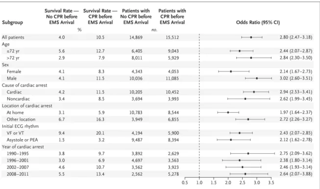
Witnessed OHCA between 1990-2011 were included (N = 30 381). CPR prior to EMS arrival was performed in 51 % of all OHCAs. Survival to 30 days was 10.5 % for patients receiving CPR prior to EMS arrival compared with 4.0 % among those where no CPR was performed (unadjusted OR 2.80; 95 % CI 2.47 – 3.18). Similar associations were found for all subgroups analyzed, as shown in figure 4.
Figure 4: Subgroup analysis of survival rates.
Figure 4 shows the subgroup analysis of survival rates. ECG denotes electrocardiographic, PEA pulseless electrical activity, VF Ventrucular fibrillation, and VT ventricular tachycardia.
Hasselqvist-Ax et al., New England Journal of Medicine, Early Cardiopulmonary Resuscitation in out-of-hospital cardiac arrest, 372, 2307– 2315. Copyright © (2015) Massachusetts Medical Society. Reprinted with permission.
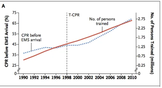
We also found higher proportion of persons who received CPR prior to EMS arrival over time as shown in figure 5.
Figure 5: Changes over Time in CPR Training and the Performance of Early CPR.
Figure 5 shows the cumulative number of attendees at CPR training sessions in Sweden and the proportion of patients receiving CPR prior to EMS arrival. T-CPR denotes the introduction of DA-CPR to untrained witnesses.
Hasselqvist-Ax et al., New England Journal of Medicine, Early Cardiopulmonary Resuscitation in out-of-hospital cardiac arrest, 372, 2307– 2315. Copyright © (2015) Massachusetts Medical Society. Reprinted with permission.
Patients not receiving CPR prior to EMS arrival were older, more frequently of female sex, more likely to have collapsed at home and were less likely to have VT/VF as first recorded rhythm. After adjusting for differences in baseline characteristics, using propensity score as an adjustment factor in a logistic regression model, CPR prior to EMS arrival was associated with a higher rate of survival to 30 days (odds ratio 2.15; 95 % CI 1.88 – 2.45).
5.2 STUDY II – “TEMPORAL CHANGES IN FREQUENCY OF CPR, TYPE OF CPR PRIOR TO EMS ARRIVAL AND SURVIVAL DURING THREE GUIDELINE PERIODS”
In Study II we aimed to describe temporal changes in CPR rates, type of CPR prior to EMS arrival and survival in relation to three time periods of different CPR guidelines in Sweden. Patients were divided into three groups, reflecting different guideline time periods (2000 – 2005, 2006 – 2010, 2011 – 2017) and exposure was divided into three categories; no-CPR before EMS arrival, S-CPR with chest compressions and rescue breaths or CO-CPR.
6.2.1 Main results
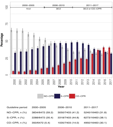
Witnessed OHCA between 2000 – 2017 were included (N = 30 455), 40.0 % received no-CPR, 39.2 % received S-CPR, and 20.8 % received CO-CPR. The overall proportions of patients receiving CPR prior to EMS arrival changed from 40.8 % in the first time period to 68.2 % in the last period. S-CPR changed from 35.4 % to 38.1 % and CO-CPR changed from 5.4 % to 30.1 % respectively.
Figure 6: Percentages of no-CPR, S-CPR, and CO-CPR, by year and guideline period.
Riva et al. Circulation. 2019;139:2600–2609 © 2019 American Heart Association. Reprinted with permission.
Figure 6: CO-CPR indicates chest compression–only cardiopulmonary resuscitation; NO-CPR, no cardiopulmonary resuscitation; and S-CPR, standard cardiopulmonary resuscitation.
2000−2005 2006−2010 2011−2017 15:2 30:2 30:2 or CO−CPR 0 25 50 75 100 2000 2001 2002 2003 2004 2005 2006 2007 2008 2009 2010 2011 2012 2013 2014 2015 2016 2017 Year Per centa ge NO−CPR S−CPR CO−CPR Guideline period NO−CPR, n (%) S−CPR, n (%) CO−CPR, n (%) 2000−2005 3834/6472 (59.2) 2288/6472 (35.4) 350/6472 (5.4) 2006−2010 3050/7403 (41.2) 3318/7403 (44.8) 1035/7403 (14.0) 2011−2017 5240/16463 (31.8) 6273/16463 (38.1) 4950/16463 (30.1)


