Yale Medicine Thesis Digital Library School of Medicine
January 2015
Evaluation Of A 5-Group Classification System For
Severe Sepsis By Ed Vasopressor Use And Initial
Serum Lactate
Kai Swenson
Follow this and additional works at:http://elischolar.library.yale.edu/ymtdl
This Open Access Thesis is brought to you for free and open access by the School of Medicine at EliScholar – A Digital Platform for Scholarly Publishing at Yale. It has been accepted for inclusion in Yale Medicine Thesis Digital Library by an authorized administrator of EliScholar – A Digital Platform for Scholarly Publishing at Yale. For more information, please contactelischolar@yale.edu.
Recommended Citation
Swenson, Kai, "Evaluation Of A 5-Group Classification System For Severe Sepsis By Ed Vasopressor Use And Initial Serum Lactate" (2015).Yale Medicine Thesis Digital Library. 2014.
Evaluation of a 5-‐group classification system for
severe sepsis by ED vasopressor use and initial
serum lactate
A Thesis Submitted to the
Yale University School of Medicine
in Partial Fulfillment of the Requirements for
the Degree of Doctor of Medicine
by
Kai Erik Swenson
2015
Abstract
HYPOTHESIS AND SPECIFIC AIMS: The aim of this study is to characterize the incidence and outcomes of various groups within a novel classification system of severe sepsis and septic shock, for the purpose of informing more accurate risk prediction in the proximal phases of care. Our primary hypothesis is that an early classification system of septic patients categorized by organ dysfunction, initial emergency department (ED) serum lactate, and ED vasopressor utilization will offer accurate mortality prognostication in patients with severe sepsis and septic shock.
METHODS: We performed a retrospective analysis of a prospectively-‐ gathered registry of severe sepsis and septic shock patients presenting to a dual-‐site academic emergency department (ED). In the primary analysis, registry subjects were categorized into five groups by initial ED serum lactate level and vasopressor requirement in the ED: dysoxic shock (vasopressor use + lactate >4 mmol/L), vasoplegic shock (vasopressor use + lactate ≤4 mmol/L), cryptic shock major (no vasopressor use + lactate >4 mmol/L), cryptic shock minor (no vasopressor use + lactate >2 and ≤4 mmol/L), and severe sepsis without lactate elevation (no vasopressor use + lactate ≤2 mmol/L + evidence of ≥1 organ dysfunction). For each group, the 28-‐day mortality rate was evaluated by logistic regression controlling for specific factors associated with sepsis severity.
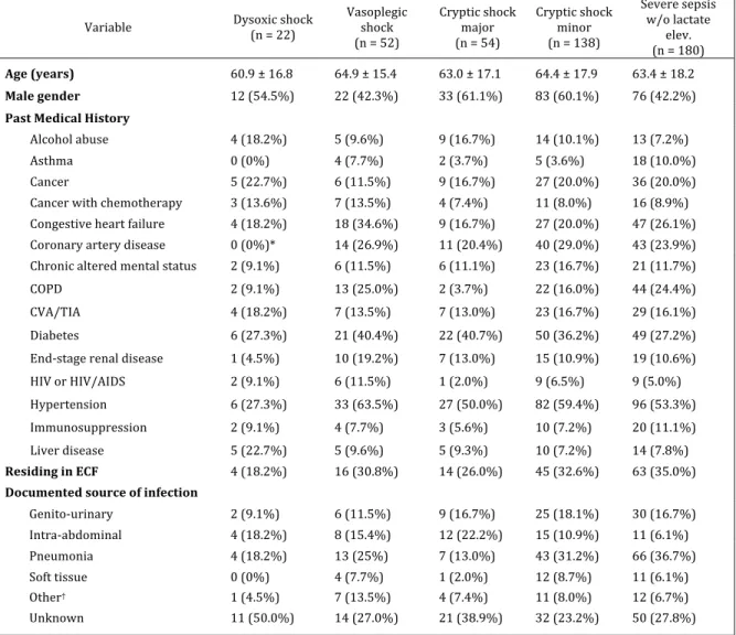
RESULTS: Of 521 registry subjects, 85.6% (n=446) met inclusion criteria. 4.9% (n=22) subjects presented in dysoxic shock, 11.7% (n=52) in vasoplegic shock,
12.1% (n=54) in cryptic shock major, 30.9% (n=138) in cryptic shock minor, and 40.4% (n=180) in severe sepsis without lactate elevation. The 28-‐day mortality rates for these groups were 50.0%, 21.1%, 18.5%, 12.3%, and 7.2%, respectively; this stepwise trend was paralleled by metrics of critical care utilization such as ICU admission, mechanical ventilation, and vasopressor use within 72 hours of admission. After controlling for known risk factors for sepsis severity, the odds ratios for death before 28 days were 15.06 for dysoxic shock, 3.61 for vasoplegic shock, 3.77 for cryptic shock major, and 1.93 for cryptic shock minor, as compared to severe sepsis without lactate elevation.
CONCLUSION: This study suggests that high-‐risk subgroups of severe sepsis and septic shock patients can be identified at presentation and during the emergency department stay. We show that in severe sepsis and septic shock, a proximal-‐phase classification system based on vasopressor requirement in the ED and initial ED lactate level predicts 28-‐day in-‐hospital mortality and may inform prognostication, triage decisions and future sepsis clinical trial design.
Acknowledgements
I would like to express my sincere gratitude to Dr. Charles R. Wira, III, for his guidance and encouragement throughout his mentorship of this thesis. His patience and generosity were as essential in developing and sustaining this project as his research expertise and clinical insights in the field of sepsis. I am also grateful to other investigators, including Melissa Wolan, Martina Sanders-‐Spight and Sundeep Bhat, who were instrumental in creating the Yale Emergency Medicine Sepsis Registry from which this work is derived.
I am indebted to Dr. James Dziura and Jessie Reynolds at the Yale Center for Analytical Studies for their valuable assistance with the statistical analysis. I also wish to acknowledge the generous research funding provided by the Yale School of Medicine and G.D. Hsiung Medical Student Research Fellowships.
Lastly, I would like to thank my parents, Cornelia Schneider and Erik Swenson, and my partner Kara McKinley; their love and support is unconditional, for which I am truly fortunate.
Table of Contents
Introduction ... 1
Epidemiology ... 1
Pathophysiology ... 3
Classification ... 8
Management ... 12
Indicators of sepsis severity ... 24
Alternative classification systems ... 38
Hypothesis and Aims of Research ... 43
Methods ... 44
Setting and design ... 44
Study population and inclusion criteria ... 44
Data extraction ... 46
Protocol and classification system ... 48
Statistical analysis ... 49
Results ... 51
Characteristics of included subjects ... 51
Frequency of presentations, baseline characteristics ... 55
Clinical features ... 58 Treatments ... 61 Hospital outcomes ... 64 Discussion ... 71 Limitations ... 89 Future directions ... 91 Conclusion ... 93 References ... 96 Appendix ... 107
Introduction
Sepsis is a clinical syndrome of immense complexity, an understanding of which is essential for clinicians and researchers who hope to improve sepsis care. The following literature review explores the key epidemiologic and clinical aspects of sepsis that informed the current research project. After a review of the epidemiology and pathophysiology of sepsis, we summarize the current widely-‐ utilized system for classifying sepsis. This is followed by an appraisal of the literature on sepsis severity, evidencing the need for both simpler and more accurate sepsis classification systems. We conclude with a summary of various alternative classification systems for severe sepsis and septic shock, to which our system, described in this thesis, can be compared.
Epidemiology
The clinical entity of sepsis, a systemic inflammatory response to infection, is one of the greatest causes of morbidity and mortality in the United States. Sepsis is the 6th
most common admitting diagnosis in the US [1]. In 2009, 836,000 hospital stays were attributed to a principal diagnosis of sepsis, with another 829,500 having a secondary diagnosis of sepsis during the course of hospitalization; when combined, sepsis is implicated in 4.2% of all hospitalizations [1]. In addition, the incidence of sepsis has been trending upward. Hospital stays with a principal diagnosis of sepsis increased by 153% between 1993 and 2009: a six percent increase in hospitalizations due to sepsis every year [1]. This trend is likely due to an aging
population, increasing burden of other health conditions, a higher proportion of immunocompromised patients from any cause, and perhaps an increased awareness and documentation of sepsis by providers. Sepsis is also extremely costly, in terms of lives as well as care delivered. The mortality rate of all patients with a diagnosis of sepsis in the US was around 16% in 2009: eight times higher than the average mortality rate of all other hospital stays [1]. Sepsis remains the most expensive reason for hospitalization in the US, with estimates of $15.4 billion in aggregate hospital costs per year, representing 4.0% of all hospital costs [1].
Sepsis may also be increasing in severity. The incidence of severe sepsis (defined as sepsis-‐induced organ dysfunction, hypoperfusion or hypotension) is reportedly between 256,000-‐700,000 per year [2-‐4], with an increase of around 13% per year [5]: far faster than the rate of increase of all sepsis diagnoses. One study documented that the percentage of sepsis presentations qualifying as severe sepsis increased from 26% to 44% between 1993 and 2003 [2]. Severe sepsis accounts for between 6 and 15% of all intensive care unit (ICU) admissions, and is estimated to consume up to half of ICU resources [3]. Many of these sicker patients will present through the emergency department (ED); severe sepsis is the suspected diagnosis in 571,000 ED visits annually [6]. Thus, the majority of patients in severe sepsis will be hospitalized through the ED.
Fortunately, in-‐hospital mortality from sepsis may be decreasing. In a recent multicenter observational study involving over 100,000 ICU patients with severe sepsis, the mortality rate decreased between 2000 and 2012, from 35% to 18.4% [7]. This dramatic drop in mortality has been mirrored in other studies, both
observationally after implementation of protocol-‐based care [8, 9] and in the control groups of recent randomized trials [10, 11]. It is unknown whether the decreasing mortality rates seen in these trials represent an improvement in sepsis interventions or an increased recognition of sepsis in infected patients. Nevertheless, given the increasing incidence of sepsis as discussed previously, the absolute number of deaths due to sepsis is likely still on the rise. By some estimates, severe sepsis accounts for around 220,000 deaths per year, making it the 3rd most
common cause of death in the US, after heart disease and malignancy [1].
Although the trend towards improved immediate mortality is heartening, less is known about the long-‐term prognosis of sepsis survivors. In a recent meta-‐ analysis, a discharge diagnosis of sepsis carried an increased risk of recurrent sepsis as well as death after hospital discharge, with a mortality rate between 7 and 43% at one year [12]. Sepsis survivors were more likely to be discharged to acute or long-‐ term care facilities than other discharged patients of similar illness severity; furthermore, they suffered a pronounced decrease in quality of life when matched against their own pre-‐sepsis condition, as well as compared to other patients recovering from critical illness [13, 14].
Pathophysiology
The inflammatory response and microvascular dysfunction
A basic understanding of the pathophysiology of sepsis is germane to both its clinical consequences and management. The presence of infection, usually by bacterial or fungal organisms, triggers an initial immune response via pattern-‐ recognition receptors in the cells of the innate immune system. Occasionally, and
for reasons not well understood, local infection will trigger a systemic response heralding the onset of sepsis. This host response involves both pro-‐ and anti-‐ inflammatory pathways, and imbalances in these pathways, both in time and in scale, can be damaging to the host. In general, the pro-‐inflammatory pathways activate earlier in the course of sepsis and are involved in controlling and clearing the infectious organism; these pathways are mostly implicated in the collateral damage to host organs in severe sepsis and septic shock [15]. The anti-‐ inflammatory pathways are generally associated with repairing tissue damage and may peak later in the course of sepsis. In a weakened host, dampening the immune response can increase the risk of secondary and opportunistic infections [15].
Pro-‐inflammatory processes conspire to orchestrate the dysfunctional and self-‐perpetuating tissue damage that underlies severe sepsis. Activated via innate responses to pathogen motifs, leukocytes infiltrate both infected and non-‐infected tissues, activating complement and releasing damaging substrates such as proteases and reactive oxygen species. When misdirected, these attacks cause damage to normally-‐functioning host cells. The pro-‐inflammatory milieu is also responsible for coagulation abnormalities via the intermediary protease-‐activated receptors (PARs) [15]. Increased tissue factor from damaged endothelial cells, the down-‐regulation of normal anticoagulant mechanisms (protein C, S, and antithrombin), and depression of natural fibrinolysis all contribute to clot formation; the ultimate result is disseminated intravascular coagulation (DIC) [16]. The downstream effect of these pathways, especially in the presence of hypoperfusion, is damage to collateral tissues and organ dysfunction. Furthermore, necrotic cell death releases damage-‐
associated molecular pattern (DAMP) molecules, leading to a vicious cycle of immune activation and tissue destruction [15].
The key intermediary between the damage described above and global tissue underperfusion is the vascular endothelium; microvascular dysfunction lies at the basis of severe sepsis and septic shock. The microvascular circulation is the main controller of end-‐organ perfusion [17]. In severe sepsis, the diffuse endothelial damage, hypercoagulability, and loss of vascular barrier function impair oxygen delivery and cause third-‐spacing of fluids into tissues. The resulting tissue hypoperfusion leads to abnormal systemic vasodilation and a decrease in systemic vascular resistance, likely via uninhibited release of vasoactive mediators such as nitric oxide and prostacyclin. When not corrected, these hypovolemic and distributive mechanisms contribute to hypoperfusion, organ dysfunction, and the shock state.
Organ-‐specific dysfunction
At the level of individual organs, direct tissue damage and hypoperfusion-‐related anoxic injury is initially reversible; however, if not corrected these insults can lead to irreversible loss of function and ultimately to organ failure. The mechanisms and clinical manifestations of failure in each organ are distinct. Acute kidney injury is common, either as a consequence of decreased perfusion or direct vascular injury; tubular necrosis and resultant chronic kidney disease can occur if the insults are not adequately reversed [18]. Similarly, hypoperfusion and damage to the blood-‐brain barrier can cause acute central nervous system (CNS) disturbances, most commonly encephalopathy. Splanchnic hypoperfusion can lead to gut translocation of bacteria
and bacterial toxins, the systemic effects of which are multiplied if hypoperfusion of the liver decreases first-‐pass metabolism [19].
Microvascular damage to the pulmonary circulation can impair barrier function of alveolar capillaries, leading to interstitial and alveolar edema. The clinical effect is ventilation/perfusion mismatching, causing hypoxemia: if severe enough, the acute respiratory distress syndrome (ARDS) develops, as evidenced by a PaO2/FiO2 ratio of less than 300 [20]. Sepsis-‐induced ARDS seems to be much
more common in chronic alcoholics [21]. Sepsis-‐induced cardiomyopathy is another well-‐recognized clinical entity. The mechanism is not entirely elucidated, but certainly involves myocardial depression by inflammatory cytokines; in shock states, cardiac muscle hypoperfusion likely contributes [22]. In one study of 108 severe sepsis and septic shock patients, 64% had evidence of myocardial dysfunction, whether via left ventricular systolic, diastolic, or right ventricular failure [23]. The development of cardiomyopathy also contributes to poor tissue oxygenation and has implications for treatment strategies.
Evidence of systemic hypoperfusion
At the global level, impairment in microvascular circulation and hypotension leads to an imbalance between oxygen delivery and oxygen requirements: a critical juncture on the path to tissue hypoxia, irreversible tissue damage, organ failure, and death. The relationship between global oxygen consumption and its determinants is summarized in the following equation:
(Equation 1) VO2 = CO × Hb × 1.34 × (SaO2 – Sv02)
(VO2: oxygen consumption, CO: cardiac output, Hb: hemoglobin concentration,
SaO2: oxygen saturation of arterial blood, SvO2: oxygen saturation of mixed
venous blood)
When oxygen demand (DO2) outstrips supply, a DO2/VO2 mismatch develops,
and the body attempts to respond to global tissue hypoxia by optimizing certain variables in the equation above [24]. Regulation of some of these variables occurs directly: in brief, cardiac output is regulated by autonomic nervous activity and catecholaminergic stimuli, SaO2 by changes in ventilator drive, and hemoglobin
levels (slowly) by EPO secretion and subsequent RBC production. In contrast, there does not appear to be any direct process by which the body regulates SvO2 [24].
Therefore, changes in SvO2 are most truly representative of a growing gap between
oxygen consumption and oxygen demand by tissues: the DO2/VO2 mismatch. ScvO2,
the saturation of venous blood at the level of the entrance to the right atrium, is considered a surrogate for SvO2; it is usually 3-‐5mmHg higher than SvO2 [24]. As
will be discussed in a later section, ScvO2 is often measured in clinical situations and
utilized as a surrogate for the adequacy of oxygen delivery in severe sepsis.
Another physiologic marker of tissue hypoxia is the serum lactate level. Under hypoxic conditions, lactate is a byproduct of cellular glycolysis to regenerate much-‐needed reducing power (in the form of NAD+). Lactate excreted from the cell into the bloodstream can either be metabolized directly by certain tissues (such as the heart) or converted into glucose via gluconeogenesis in the Cori cycle, mostly in the liver but to a smaller extent in the kidneys [25]. This production of lactate often parallels the increased production and release of protons by metabolically-‐active,
hypoxic cells (a result of a shift in the ratio of ATP to ADP/AMP) [25]. Thus, the serum lactate has traditionally served as a marker for the presence of tissue hypoxia, whether at the local level (in acute mesenteric ischemia) or globally (in various forms of shock). Lactate production could also occur via anaerobic metabolism in the absence of hypoxia, as seen with mitochondrial dysfunction states.
More recently, the accuracy of using lactate levels as an indicator of anaerobic states has been questioned, and alternative hypotheses have been proposed. Lactate may be a byproduct of stress-‐induced muscle metabolism, a hypermetabolic response to the catecholaminergic surge commonly seen in severe sepsis. Alternatively, a decrease in lactate clearance by the liver or kidneys may be responsible for lactate elevation in a subset of septic patients [25]. Some studies even suggest that elevated lactate may play a protective role in sepsis as an alternative metabolic fuel for the heart and brain [26]. Although uncertainty exists regarding its true nature, the serum lactate level continues to be utilized in clinical situations as a surrogate for tissue hypoxia and as a prognostic indicator in sepsis.
Classification
Given the incredible variability of physiologic derangements in sepsis, an inclusive and accurate classification system is essential for aiding clinical diagnosis and treatment decisions, as well as for informing study design. For many years, the medical field lacked definitive criteria for making the diagnosis of sepsis and documenting its severity. In 1992, the American College of Chest Physicians (ACCP) and the Society for Critical Care Medicine (SCCM) jointly developed a classification
system for sepsis in order to standardize diagnosis and to categorize cases along a spectrum of disease severity [27].
The classification system included various definitions for sepsis syndromes. It first defined the systemic inflammatory response syndrome (SIRS), composed of four criteria: fever (>38°C) or hypothermia (<36°C), tachycardia (>90 beats/min), tachypnea (>30 breaths/min) or PaCO2 <32 mmHg, and an abnormal WBC (>12,000, <4,000, or >10% bands). The diagnosis of SIRS requires fulfilling at least 2 of the 4 above criteria [27], shown in Table 1.
Table 1. Criteria for the Systemic Inflammatory Response Syndrome (SIRS)* (1)temperature > 38°C or < 36°C
(2)heart rate > 90 beats per minute
(3)respiratory rate > 20 breaths per minute or PaCO2 < 32mmHg
(4)white blood cell count > 12,000/mm3, < 4,000/mm3,
or > 10% immature (band) forms
* Adapted from ACCP/SCCM Consensus Conference [27].
Using the above criteria for the system inflammatory response, the consensus defined sepsis as the presence of SIRS with a documented or probable infectious source. Severe sepsis was defined as sepsis with organ dysfunction, hypoperfusion, or hypotension; sepsis-‐induced hypotension is a subset of severe sepsis defined by a systolic blood pressure (SBP) less than 90 mmHg, a mean arterial pressure (MAP) less than 65 mmHg, or a decrease in SBP greater than 40 mmHg from baseline. The most severe form of sepsis is septic shock, defined as
sepsis with persistent hypotension or hyperlactatemia (above 4.0 mmol/L) despite adequate fluid resuscitation. These definitions have been shown to correlate stepwise with mortality: in one large series, patients with SIRS, sepsis, severe sepsis and septic shock had hospital mortality rates of 7%, 16%, 20%, and 46%, respectively [28].
The consensus document also categorized the multiple organ dysfunction syndrome (MODS), a severe and deadly consequence of sepsis related to hypoperfusion and endothelial damage. Multiple organ failure in response to acute insult had already been recognized for many years, but the focus of the consensus document on organ dysfunction, before the presence of overt failure, was original. The document defined MODS as “the presence of altered organ function in an acutely ill patient such that homeostasis cannot be maintained without intervention,” and subclassified MODS into primary and secondary etiologies [27]. Primary MODS can be considered a direct reaction to the initial insult; secondary MODS is a consequence of the host response to the original injury. Either type can lead to progressive organ failure and death, sometimes far removed in time and severity from the initial presentation [27]. Although no universally-‐accepted criteria exist for the diagnosis of MODS, it logically requires the continued presence of multiple measures of organ dysfunction.
In 2001, the ACCP/SCCM revisited their consensus document and altered some of their previous definitions, most importantly on the concept of SIRS [29]. Although characteristic of sepsis, the presence of SIRS is also common in other conditions, including trauma, pancreatitis, and burns. However, these criteria are
Table 2. Sepsis definitions, International Sepsis Definitions Conference.*
Infection, documented or suspected, and some of the following:
General variables
Fever (core temperature >38.3°C) Hypothermia (core temperature <36°C)
Heart rate >90 min-‐1 or >2 SD above the normal value for age Tachypnea
Altered mental status
Significant edema or positive fluid balance (>20 mL/kg over 24 hrs)
Hyperglycemia (plasma glucose >120 mg/dL or 7.7 mmol/L) in the absence of diabetes
Inflammatory variables
Leukocytosis (WBC count >12,000 μL-‐1)
Leukopenia (WBC count <4000 μL-‐1)
Normal WBC count with >10% immature forms
Plasma C-‐reactive protein >2 SD above the normal value
Plasma procalcitonin >2 SD above the normal value
Hemodynamic variables
Arterial hypotension (SBP <90 mm Hg, MAP <70, or an SBP decrease >40 mm Hg in
adults or <2 SD below normal for age)
SvO2 >70%
Cardiac index >3.5 L/min/M-‐23 Organ dysfunction variables
Arterial hypoxemia (PaO2/FIO2 <300)
Acute oliguria (urine output <0.5 mL/kg/hr or 45 mmol/L for at least 2 hours)
Creatinine increase >0.5 mg/dL
Coagulation abnormalities (INR >1.5 or aPTT >60 secs) Ileus (absent bowel sounds)
Thrombocytopenia (platelet count <100,000 μL-‐1)
Hyperbilirubinemia (plasma total bilirubin >4 mg/dL or 70 mmol/L)
Tissue perfusion variables
Hyperlactatemia (>1 mmol/L)
Decreased capillary refill or mottling
* Adapted from 2001 SCCM/ESICM/ACCP/ATS/SIS International Sepsis Definitions Conference [29]. WBC, white blood cell; SBP, systolic blood pressure; MAP, mean arterial blood pressure; SvO2, mixed
venous oxygen saturation; INR, international normalized ratio; aPTT, activated partial thromboplastin time.
not exclusive to these disease states but are in fact present in many hospitalized patients; in one recent study of over 100,000 ED encounters, the presence of SIRS correlated with infection in only 26% of cases, whereas 56% of patients with SIRS did not fit into disease states normally associated with these criteria [30]. Given the
increased scrutiny surrounding the tenuous ability of the SIRS criteria to delineate the host response to infection, the ACCP/SCCM redefined sepsis as the presence of documented or probable infection in association with clinical signs/symptoms suggestive of a broader inflammatory response [29]; these consist of clinical and laboratory variables as displayed in Table 2. This definition suspended the explicit need for 2 of 4 SIRS criteria to be met. However, despite their removal from current sepsis guidelines, SIRS criteria are still widely used as convenient objective measures of sepsis in observational trials, in accordance with the original 1992 consensus.
Indicators of sepsis severity
Despite its utility, our current classification system for sepsis belies an incredibly diverse patient population; many factors influence the severity of the illness and alter the prognosis of the individual patient. Recognizing the incredible heterogeneity in sepsis and the many factors affecting outcomes, the 2001 International Sepsis Definitions Conference conceptualized a new system for categorizing sepsis [29]. The PIRO system (which stands for Predisposition, Infection, Response and Organ dysfunction) is loosely designed around known and suspected prognostic factors, and offers a template by which clinicians can conceptualize the severity of an individual patient’s septic presentation. The following sections will discuss known risk factors in each component of the PIRO system. Therapeutic factors also influence outcomes in sepsis, as will be discussed in a later section.
Predisposing characteristics of the host
Well-‐known risk factors for developing sepsis include age, certain demographic factors, and comorbidities. Elderly age is a strong predictor of developing sepsis, with a stepwise increase in risk: in a longitudinal survey of over 30,000 adults, the hazard ratios for developing sepsis in adults between the ages of 55-‐64, 65-‐74, and ≥75 were 1.44, 2.29, and 3.87 respectively when compared to an age range of 45-‐54 [31]. In the same study, the frequency of sepsis episodes also correlated significantly with tobacco use, alcohol use, lower education, and lower income. Women who develop sepsis are generally older and less likely to have severe sepsis than their male counterparts, but are possibly at increased risk of death in the ICU [32, 33]. Chronic comorbid conditions which increase the likelihood of developing sepsis include a history of chronic lung disease, peripheral artery disease, chronic kidney disease, myocardial infarction and diabetes, among others; the presence of multiple comorbidities has an additive effect on the risk of developing sepsis [31]. Lastly, there is likely a large genetic component involving innate and adaptive immunity that exacerbates or mitigates the risk of sepsis [34].
Furthermore, the severity and prognosis of a sepsis episode is influenced by underlying comorbidities. Age above 40 years is a stepwise negative prognostic factor, and the effect of increasing age is most pronounced in the absence of comorbidities [3]. Mortality from sepsis seems to be lowest in previously healthy young adults: in a large retrospective analysis, adults younger than 45 without comorbidities who developed severe sepsis had a mortality rate of less than 5% [7]. Immunocompromised states such as AIDS [35], solid or hematologic malignancy
[36], neutropenia [36], chronic liver disease [3], chronic renal disease [3], and asplenia [37] worsen prognosis to varying degrees in severe sepsis. Alcohol dependence is independently associated with higher mortality in critically-‐ill patients with severe sepsis and septic shock [38].
Characteristics of infection
The prognosis in sepsis is also predicated on the characteristics of the underlying infection. The type of organism responsible may influence outcomes. The largest observational trial of sepsis to date showed that Gram-‐positive organisms account for the majority (52.1%) of sepsis cases with a documented culture, as compared to gram-‐negative organisms (37.6%), polymicrobial infections (4.7%), fungal infections (4.6%), and anaerobes (1.0%) [4]. Other studies have shown that Gram-‐ negative sepsis cases may predominate in the ICU setting [39, 40]. The most common Gram-‐positive organisms are Staphylococcus aureas and Streptococcus pneumonia, whereas Gram-‐negative organisms are usually E. coli, Pseudomonas and Klebsiella subspecies [41]. Although the presence of bacteremia positively predicts the development of SIRS [42], bacteremia itself does not appear to be associated with mortality in sepsis [43]. Bloodstream infections carry a worse prognosis if they are caused by nosocomial pathogens, such as MRSA (OR 2.7), fungal infections (OR 2.32-‐2.66) or pseudomonas (OR 1.6) [44]. However, only one third to one half of septic patients have positive blood cultures [45, 46], and up to 30% of patients have negative cultures from all sites [40]. In some cases, blood cultures may be incorrectly negative due to previous use of antibiotics or the incorrect collection of blood cultures [47].
The source of infection in sepsis likewise influences severity. Respiratory infections are the most common source (35-‐44%), followed by urinary (9-‐37%), occult bacteremia (12-‐17%), abdominal (8-‐19%), wound/soft tissue (5-‐7%), device-‐ related (2-‐6%), and other sources (including endocarditis, CNS, bone and joint infections) [3, 45, 48]. Sepsis develops commonly in the setting of pneumonia; in an observational study of 1,339 patients admitted with community-‐acquired pneumonia (CAP), severe sepsis developed in 48% [49].
Sepsis from a genitourinary source (urosepsis) is generally considered to have a better prognosis than other causes of sepsis, especially pneumonia [3, 50]. In a study cohort of almost 200,000 patients in the United States, Angus et al. found that patients who developed severe sepsis from a lung infection had a mortality of 32.9%, whereas the mortality of urosepsis was 16.1% [3]. In a more recent, large-‐ scale cohort from Australia, the mortality from urosepsis in 2012 was 6.7% compared to 17.0% in sepsis from a non-‐urinary source [7]. The range of prognosis in sepsis from abdominal sources seems to be wider; although one study showed a mortality rate of 55% in septic patients with abdominal foci, larger studies quote mortality rates closer to urosepsis [3]. This may be due to differences in severity between sources within the abdomen. In one study of around 8,000 patients with septic shock, mortality rates depended on the underlying diagnosis including ischemic bowel (77.9%), spontaneous bacterial peritonitis (76.4%), perforated viscus (55.6%), pancreatitis (50.0%), cholecystitis/cholangitis (38.3%), and enterocolitis/diverticulitis (28.0%) [51]; these rates roughly reflect the clinical severity of the underlying disease process. In the same study, mortality from skin,
soft-‐tissue and bone infections was high (42.0-‐52.5%). Furthermore, the presence of multiple sources of infection seems to portend a poor prognosis [52]. However, distinctions in mortality between different infectious sources are not universally demonstrable [43].
Features of the host response
Many clinicians are intuitively aware that a formidable host response to infection, as exhibited by abnormalities in multiple SIRS criteria, is clinically relevant and has prognostic information. In one study of the SIRS criteria, in-‐hospital mortality increased steadily, from 14% with 2 criteria, 26% with 3 criteria, and 36% with 4 SIRS criteria met [42]. In keeping with this data, most of our current therapies seek to counteract the excessive host response to infection that occurs in sepsis. However, comparatively little is known about the specific pathways of host response that correlate with severity in sepsis. Biomarkers of immune activation may hold promise as risk stratifiers, and over 150 have been proposed. C-‐reactive protein is the most widely available marker of the inflammatory response; it is released by hepatocytes after stimulation from cytokines including IL-‐6 as part of the acute phase response. However, given its frequent elevation in non-‐septic conditions, its specificity is too poor to be of use as a prognostic indicator.
Procalcitonin is one of the best markers, not only for its high negative predictive value in ruling out infection but also as a prognosticator of severity [53]. Although not specifically performed in sepsis, one study of 472 critically-‐ill patients demonstrated an increased risk of death with an initial elevated procalcitonin level or an increase over 24 hours [54]. The ability of other biomarkers, including IL-‐6
and sTREM-‐1, to risk-‐stratify sepsis patients remains undefined. It is likely that, given the complexity of the host immune response, combinations of biomarkers will allow for better risk stratification than any single marker alone. However, there is as yet no evidence that incorporation of sepsis biomarkers, including procalcitonin, into sepsis alerts has improved outcomes.
Impairment in certain host endocrine pathways may exacerbate hypoperfusion in sepsis by augmenting systemic vasodilatation. A relative deficiency of endogenous cortisol, called critical illness-‐related corticosteroid insufficiency (CIRCI), is commonly seen in sepsis; this syndrome is likely an effect of inflammatory mediators on both suppression of the hypothalamic-‐pituitary-‐adrenal axis and peripheral tissue corticosteroid resistance [55]. The prevalence of CIRCI may be as high as 60% in patients with septic shock [56]. A relative deficiency in vasopressin in septic shock patients may also contribute to hypotension [57].
Organ dysfunction
As was discussed in the previous section, severe sepsis (which by definition involves sepsis-‐induced organ dysfunction) carries a worse prognosis than the sepsis syndrome. However, the type of organ or organ system involved also seems to affect outcomes. In a retrospective analysis of over 3,000 patients presenting to an emergency department with sepsis, Shapiro et al. found higher odds ratios for mortality in patients with hematologic (OR 4.5), cardiovascular (3.6), or respiratory (3.6) dysfunction [58]. In particular, coagulation dysfunction is considered one of the worst organ dysfunctions in severe sepsis; the presence of DIC is an independent predictor of organ failure and mortality, with a rate of up to 77% in
one study [16]. Higher DIC scores also correlate directly with mortality [59]. Even before overt DIC has developed, abnormal coagulation tests including thrombocytopenia and elevated INR predict increased mortality in infected patients presenting to the ED [60].
The number of dysfunctional organs is also a strong predictor of increased risk. In the same study by Shapiro et al., increasing number of organ dysfunctions was correlated stepwise with increased mortality: 1.0% with no organ dysfunction, 5.9% with 1 organ dysfunction, 12.5% with 2, 25.9% with 3, and 53.3% with 4 or more [58]. Similar, though lower, mortality rates were seen in a previous analysis of the registry used in this study; specifically, the mortality rate of 5-‐6 organ dysfunctions was only 17.7%, whereas 7 or more was 55.9% [61]. These discrepancies may be a result of differences in how one defines organ dysfunction. Although the 2003 Surviving Sepsis Campaign guidelines describe a strict set of definitions for organ dysfunction based on objective criteria, these thresholds clearly exist on a spectrum of organ health from normal functioning to complete failure. Furthermore, they are not inconclusive of all categories of organ dysfunction induced by sepsis, including altered mental status and cardiac dysfunction. For example, neurologic dysfunction can variably be defined with a specific threshold Glasgow Coma Scale <12 or, alternatively, any change from baseline mental status. Many studies both before and after 2003 have incorporated their own unique measures of organ dysfunction, and these discrepancies in definition by different investigators limit the comparability of various study populations.
Risk scoring systems
Given the massive heterogeneity of the patient population in sepsis, and the presence of many known and presumed prognostic influences, much research has been focused on finding a more universal system of prognostication. One method is to create objective scores to signify increased risk of a poor outcome. Some scoring systems, such as the Pneumonia Severity Index (PSI) or CURB-‐65 for pneumonia, are limited to a single type of presentation and are not generalizable. The APACHE II score was originally formulated to be used 72 hours into admission for critically-‐ ill patients, and incorporates 12 physiologic variables [62]. In a study of fewer than 100 critically-‐ill patients presenting to the ED, Nguyen et al. found that the initial APACHE II predicted mortality at 12 hours, and that the largest changes in the score over hospitalization occurred in the first few hours within the emergency department [63]. However, these data are not exclusive to septic patients, and the score is too cumbersome to be utilized clinically in the emergency department. Scores dedicated to assessing organ dysfunction in sepsis are widespread in the research literature. Two of the most prominent are the Multiple Organ Dysfunction Score (MODS) and the Sequential Organ Failure Assessment (SOFA). MODS was developed in 1995, after an extensive search of the previous sepsis literature to identify the most optimal descriptors of organ failure [64]. The MODS included six components of organ dysfunction: respiratory (PaO2/FiO2), renal (creatinine level), hepatic (bilirubin levels), hematologic (platelet count), CNS (Glasgow Coma Scale), and cardiovascular (pressure-‐adjusted heart rate, a unique variable devised for the score). Each dysfunction was scored from 0 to 4, for a total
MODS score out of 24 points. In the validation cohort, the MODS score was predictive of ICU mortality in a graded fashion: mortality was 25% with scores of 9-‐ 12, 50% with scores of 13-‐16, 75% with scores of 17-‐20, and 100% with scores greater than 20 [64].
The SOFA score is another method of characterizing organ dysfunction. Similarly to the MODS, the SOFA score incorporates assessments of respiratory (PaO2/FiO2), hepatic (bilirubin level), renal (creatinine level or urine output), neurologic (Glasgow Coma Score), cardiovascular (hypotension) and hematologic (platelet count) function. In the ICU setting, the initial, peak and mean SOFA scores all correlate well with mortality [65]. In a population of 248 emergency department patients with severe sepsis and evidence of hypoperfusion, the initial SOFA score was found to be moderately effective at predicting in-‐hospital mortality [66].
However, risk in sepsis depends on more than merely the presence of organ dysfunction, as the PIRO model shows. Howell et al. developed a scoring system based on the full extent of risk variables involved in sepsis severity; their PIRO score incorporated predisposing traits (age, COPD, liver disease, malignancy, and nursing home residency), infection characteristics (pneumonia, cellulitis, or other), host response criteria (tachypnea, tachycardia, and bandemia) and various objective measures of organ dysfunction [67]. In the derivation cohort as well as in two validation cohorts, the PIRO score had a high degree of accuracy in predicting sepsis mortality [67]. Because it incorporates known risk factors from each component of the PIRO system, the PIRO score is intuitively attractive as a prognostic tool; however, its complexity may make it unwieldy in the emergency department.
Responding to the need for a PIRO-‐based, prognostic tool for sepsis patients in the emergency setting, Shapiro et al. developed the Mortality in Emergency Department Sepsis (MEDS) score. As shown in Table 3, the MEDS score incorporates nine clinical variables that are immediately available from the patient history, physical exam, and early laboratory results. In the validation set of the initial study, the MEDS score correlated impressively with in-‐hospital mortality, in a graded fashion: 1.1% with MEDS 0-‐4, 4.4% with MEDS 5-‐7, 9.3% with MEDS 8-‐12, 16% with MEDS 12-‐15, and 39% with MEDS >15 [68]. The score and its stepwise correlation with 28-‐day mortality has been externally validated by other investigators [69], it performs similarly to the more complex PIRO score, and it may outperform organ dysfunction scores (such as SOFA) originally formulated for
Table 3. Mortality in Emergency Department Sepsis (MEDS) Score. Criterion Points
Rapidly terminal co-‐morbid illness* Age > 65 years
Bands > 5%
Tachypnea or hypoxemia Septic shock
Platelet count < 150,000 mm3 Altered mental status
Nursing home resident Lower respiratory infection
6 3 3 3 3 3 2 2 2
Adapted from Shapiro et al. [68].
* Terminal illness is defined as metastatic cancer or a disease condition with a >50% likelihood of predicted fatality within 30 days.
hospitalized patients [70]. One potential problem with the MEDS score exists in the intermediate range (scores 5-‐15) where MEDS may underestimate mortality [71]. Another criticism of MEDS is that it places a great deal of weight (6 points) on the assessment of a terminal chronic condition, which is somewhat subjective and often outweighs other components of the score.
Serum lactate as prognostic measure
The best prognostication tool would ideally be an objective measure, readily available and easily interpretable upon patient presentation. One such measure is the lactate level. As described in a previous section, the serum lactate level may represent tissue hypoperfusion, a hypermetabolic state with activation of skeletal muscle, mitochondrial dysfunction, decreased clearance by the liver or kidneys, or a combination of these mechanisms. An elevated lactate is also not specific for severe sepsis and occurs in many other disease states, such as in other forms of shock, liver failure, drugs and toxins (especially when interfering with mitochondrial activity), diabetic ketoacidosis, and regional forms of ischemia [25]. However, regardless of etiology, the serum lactate has been used for decades as a marker of poor prognosis and mortality in sepsis [72, 73]. In 2005, Shapiro et al. reported a study of 1,278 patients presenting to an emergency department with an infection-‐related diagnosis; 28-‐day mortality rates for normal (<2.5 mmol/L), intermediate (2.5-‐4 mmol/L) and high (>4 mmol/L) lactate levels were 4.9%, 9.0% and 28.4% respectively. Furthermore, the specificity of a high lactate for death within 3 days was 91%, although the sensitivity was poor [74].
Many other studies have supported these findings [75-‐77]. In one series of over 800 patients presenting to the emergency department, both intermediate (2-‐4 mmol/L) and high (>4 mmol/L) lactate levels correlated with 28-‐day mortality; these associations persisted in both shock and non-‐shock groups after controlling for the severity of other organ dysfunction [75]. Howell et al. found that in severe sepsis, an initial lactate level greater than 2.5 mmol/L was associated with an odds ratio for hospital mortality of 7.1 compared to a lactate less than 2.5 mmol/L [78]. After admission, sustained elevations in lactate is a poor prognostic sign: time-‐ weighted lactate levels >2 mmol/L, also known as the “lac-‐time” [79], is associated with an in-‐hospital mortality OR of 4.8 [76]. Even lactate levels within the normal range (defined in one study as <2.3 mmol/L) are predictive of organ dysfunction and mortality in septic shock [77].
Given its very clear prognostic value, some clinicians have considered entirely replacing the more difficult and invasive measurements of hypoperfusion (such as ScvO2) with serial lactate measurements. One possible goal is lactate
clearance, defined as a decrease in the serum lactate level during sepsis resuscitation. Nguyen et al. calculated 6-‐hour lactate clearance in 111 septic patients presenting to an emergency department and found that a low lactate clearance (<10% decrease or any increase from initial lactate level) had a 67.6% accuracy of predicting in-‐hospital mortality [80]. The specificity of this cutoff was 84%, although the sensitivity was very low. Furthermore, the authors estimated that the risk of death decreased by around 11% for each 10% increase in lactate clearance at 6 hours. Arnold et al. published a similar trial showing that lactate non-‐