The extracellular matrix in bronchopulmonary dysplasia:
the role of the lysyl oxidase family
Inaugural Dissertation
submitted to the Faculty of Medicine
in partial fulfillment of the requirements for the PhD Degree
of the Faculties of Veterinary Medicine and Medicine of the Justus Liebig University Giessen
by
Mižíková Ivana
of
Košice, Slovakia
1
Director / Chairman: Prof. Dr. Klaus-Dieter Schlüter
Department of Physiology
Faculty of Medicine of the Justus Liebig University Giessen
First Supervisor and Committee Member:
Prof. Dr. Werner Seeger
Second Supervisor and Committee Member:
Prof. Dr. Dr. Thomas Braun
Committee Members:
Prof. Dr. Werner Seeger
Prof. Dr. Dr. Thomas Braun
Prof. Dr. Anja Sterner-Kock
2
If you eliminate the impossible, whatever remains, however
Improbable, must be the truth.
3
I Table of contents
I Table of contents ... 3
II. List of figures ... 6
III. List of tables ... 8
IV. List of abbreviations ... 9
V. Summary ... 13
VI. Zusammenfassung ... 14
1 INTRODUCTION ... 16
1.1 Development and structure of the lung ... 16
1.1.1 Early (prenatal) lung development ... 16
1.1.2 Late (postnatal) lung development ... 19
1.2 Bronchopulmonary dysplasia ... 20
1.3 The extracellular matrix in context of normal and aberrant lung development ... 22
1.3.1 Key components of extracellular matrix in the context of lung development ... 24
1.4 The lysyl oxidase family ... 27
2 AIMS OF THE STUDY ... 31
3 MATERIALS AND METHODS ... 32
3.1 Materials ... 32
3.1.1 Equipment ... 32
3.1.2 Reagents ... 35
3.1.3 Purchased primary cells and cell-lines ... 39
3.2 Methods ... 40
3.2.1 Animal experiments ... 40
4
3.2.3 Cell culture and cell treatments ... 47
3.2.4 Gene expression analysis ... 51
3.2.5 Analysis of protein expression, activity and deposition ... 56
3.2.6 Cloning and lentiviral production ... 59
3.2.7 Statistical analysis ... 63
4 RESULTS ... 64
4.1 Exposure of immature mouse lungs to 85% O2 dysregulates lysyl oxidase expression ... 64
4.2 Inhibition of lysyl oxidase activity in the lungs of developing mice exposed to 85% O2 ... 66
4.3 Cross-linking of collagen and elastin in the lungs of developing mouse pups upon hyperoxia exposure and treatment with β-aminopropionitrile ... 68
4.4 The impact of hyperoxia exposure and β-aminopropionitrile administration on elastin fibers formation in the parenchyma of the developing mouse lung... 72
4.5 Treatment of hyperoxia-exposed mouse pups did not preserve normal lung architecture ... 75
4.6 Expression profile of lysyl oxidase family members in various cell-types present in the lung ... 81
4.7 Knockdown of Lox, Loxl1 and Loxl2 expression impacts the transcriptome of primary mouse lung fibroblasts ... 82
4.8 Lentiviral overexpression of lysyl oxidase family members impacts the gene expression in murine fibroblasts ... 88
4.9 Role of lysyl oxidase enzymatic activity in lysyl oxidase-mediated gene regulation ... 91
4.10 The expression of LOX, LOXL1 and LOXL2target-genes in a murine model of aberrant lung development ... 94
5
5.1 The expression of lysyl oxidases is dysregulated in animal models of
bronchopulmonary dysplasia ... 98
5.2 Lysyl oxidase activity is essential for early postnatal development ... 99
5.3 Normalization of lysyl oxidase activity improves the formation of elastin and collagen cross-links and elastin foci structure in mouse model of bronchopulmonary dysplasia ... 100
5.4 Normalization of lysyl oxidase activity does not improve alveolarization and septal formation in mouse model of bronchopulmonary dysplasia ... 101
5.5 Lysyl oxidase family members have gene regulatory roles in lung fibroblasts ... 102
5.6 Gene regulatory functions for lysyl oxidases in the context of in vivo animal model of bronchopulmonary dysplasia ... 104
6 CONCLUSIONS ... 106 7 REFERENCES ... 107 8 DECLARATION ... 113 9 ACKNOWLEDGEMENTS ... 114 10 CURRICULUM VITAE ... 115 11 APPENDIX ... 117
6
II. List of figures
Figure 1. Progression of prenatal and postnatal lung development. ... 17 Figure 2. Histological structure of the lung of a patient with bronchopulmonary dysplasia
and a healthy age-matched control lung. ... 21 Figure 3. Elastin deposition during lung formation in bronchopulmonary dysplasia and in
the hyperoxia-based mouse model of bronchopulmonary dysplasia... 23 Figure 4. Chemical reaction catalyzed by lysyl oxidase. ... 28 Figure 5. Visiopharm NewCast counting tools used in the stereological analysis of lung
structure. ... 46 Figure 6. The expression of lysyl oxidase family members is dysregulated during aberrant
late lung development. ... 65 Figure 7. Effect of β-aminopropionitrile administration on mouse viability. ... 67 Figure 8. Lysyl oxidase enzymatic activity was normalized by the administration of
β-aminopropionitrile. ... 68 Figure 9. Effect of hyperoxia exposure and β-aminopropionitrile administration on the
levels of collagen and formation of collagen cross-links in the lungs of
developing mouse pups at postnatal day 19.5. ... 70 Figure 10. Effect of hyperoxia-exposure and β-aminopropionitrile administration on the
levels of elastin and elastin cross-links in the lungs of developing mouse pups at postnatal day 19.5. ... 72 Figure 11. Hyperoxia-exposure and β-aminopropionitrile administration alter the formation
of elastin foci in developing alveolar septa. ... 74 Figure 12. Impact of the hyperoxia-exposure and β-aminopropionitrile administration on
the lung architecture at postnatal day 9.5. ... 77 Figure 13. Impact of the hyperoxia-exposure and β-aminopropionitrile administration on
the lung architecture at postnatal day 19.5. ... 80 Figure 14. Relative expression of lysyl oxidase family members in various mouse lung
cell-types cultured in vitro. ... 81 Figure 15. Short interfering RNA-mediated knockdown of lysyl oxidases in primary mouse
7
Figure 16. Microarray analysis validation following the short interfering RNA-mediated knockdown of lysyl oxidases in primary mouse lung fibroblasts. ... 85 Figure 17. Lentivirus-mediated overexpression of lysyl oxidases in the mouse NIH/3T3
fibroblast cell-line... 90 Figure 18. Inhibition of lysyl oxidase enzymatic activity in primary mouse lung fibroblasts. 92 Figure 19. Microarray analysis validation following the β-aminopropionitrile
administration in primary mouse lung fibroblasts. ... 94 Figure 20. Gene expression of Lox and selected LOX target-genes in a mouse model of an
arrested lung development... 96 Figure 21. Gene expression of Loxl1 and Loxl2, as well as selected LOXL1and LOXL2
8
III. List of tables
Table 1. Buffers required for treatment of lungs and embedding in Technovit 7100. ... 43
Table 2. Antibodies used for alveolar epithelial type II cells isolation. ... 49
Table 3. Media used for alveolar epithelial type II cells isolation and culture. ... 49
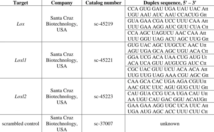
Table 4. List of siRNA oligonucleotides used in knockdown experiments. ... 51
Table 5. Primers employed for sex determination. ... 53
Table 6. Primers employed for a real-time RT-PCR analysis. ... 55
Table 7. Primary and secondary antibodies used for western blot analysis. ... 58
Table 8. Primers used for amplification of Lox, Loxl1 and Loxl2 sequences. ... 60
Table 9. Effect of hyperoxia exposure and β-aminopropionitrile administration on deposition of collagen in the developing mouse lungs at postnatal day 19.5. ... 69
Table 10. Effect of hyperoxia exposure and β-aminopropionitrile administration on deposition of elastin in developing mouse lungs at postnatal day 19.5. ... 71
Table 11. Stereo-morphometric parameters of developing mouse lungs after hyperoxia exposure and β-aminopropionitrile administration assessed at postnatal day 9.5. ... 76
Table 12. Stereo-morphometric parameters of developing mouse lungs after hyperoxia exposure and β-aminopropionitrile administration assessed at postnatal day 19.5. ... 78
Table 13. List of differentially up- and down-regulated genes after a Lox knockdown as assessed by microarray analysis. ... 84
Table 14. List of differentially up- and down-regulated genes after a Loxl1 knockdown as assessed by microarray analysis. ... 86
Table 15. List of differentially up- and down-regulated genes after a Loxl2 knockdown as assessed by microarray analysis. ... 87
Table 16. List of differentially up- and down-regulated genes after β-aminopropionitrile administration in primary mouse lung fibroblasts as assessed by microarray analysis. ... 93
9
IV. List of abbreviations
AECII Alveolar epithelial type II cell
BAPN β-aminopropionitrile
bp Base pair
BPD Bronchopulmonary dysplasia
cDNA Complementary DNA
CE Coefficient of error
CLD Chronic lung disease
Ct Threshold cycle
CV Coefficient of variation
ddH2O Double distilled water
Des Desmosine
DHLNL Dihydroxylysinonorleucine
DMEM Dulbeco’s modified Eagle medium
DNA Deoxyribonucleic acid
E Embryonic day
EC Epithelial cell
ECM Extracellular matrix
EDTA Ethylenediaminetetraacetic acid
EGTA Ethylene glycol-bis (β-aminoethyl ether)-N,N,N',N' -tetraacetic acid EMT Epithelial-to-mesenchymal transition
EtOH Ethanol
FC Fold change
10
FiO2 Fraction of oxygen in the inspired air
g Gravitational acceleration
HBSS Hank’s balanced salt solution
HEK Human embryonic kidney
HEPES 4-(2-hydroxyethyl)-1-piperazineethanesulfonic acid HHL Histidinohydroxylysinonorleucine HLNL Hydroxylysinonorleucine HP Hydroxylysylpyridinoline IgG immunoglobulin G Isodes Isodesmosine i.p. Intraperitoneally kg Kilogram
LTBP Latent TGF-β binding protein
mg Milligram
ml Milliliter
MLI Mean linear intercept
mM Millimolar
MMP Matrix metalloproteinase
MuLV Murine leukemia virus
MΦ Macrophage
µg Microgram
µl Microliter
µM Micromolar
n Number
N Normality, equivalent concentration
11
n.s. Not significant
P Postnatal day
PASMC Pulmonary arterial smooth muscle cell
PBS Phosphate buffered saline
PCR Polymerase chain reaction
PDGF Platelet-derived growth factor
PF Pulmonary fibroblast
PFA Paraformaldehyde
PLOD Procollagen-lysine, 2-oxoglutarate 5-dioxygenase (Lysyl hydroxylase)
PVDF Polyvinylidene difluoride
P (corr) Corrected P value
P/S Penicillin/streptomycin
RDS Respiratory distress syndrome
RFU Relative fluorescence units
RNA Ribonucleic acid
rpm Rotations per minute
RT Room temperature
RT-PCR Reverse transcription-PCR
scr. siRNA Scrambled siRNA
SD Standard deviation
SE Standard error
siRNA Short interfering RNA
αSMA α-smooth muscle actin
SMC Smooth muscle cell
TAE Tris-acetate-EDTA
12
TEMED Tetramethylethylenediamine
TGF-β Transforming growth factor-β
TGM Transglutaminase
TIMP Tissue inhibitors of matrix metalloproteinase
Tm Melting temperature
Tris Trisaminomethane
tRNA Total RNA
UrAc Uranyl acetate
UV Ultraviolet
WFI Water for injection
13
V. Summary
Extracellular matrix (ECM) formation and remodeling play a central role in the processes of lung alveolarization and secondary septation. A key component of ECM structure is a complex collagen and elastin network of fibers. Formation and maintenance of this crucial structure is perturbed in aberrant lung development in patients with bronchopulmonary dysplasia (BPD) as well as in experimental animal models of BPD. Lysyl oxidases comprise a family of five members, which facilitate the covalent cross-linking of collagen and elastin molecules, thus controlling ECM structural homeostasis. Moreover, alternative non-matrix gene regulatory roles for these enzymes have been proposed. Recently, lysyl oxidases have been implicated in the pathogenesis of various lung diseases, including cancer, fibrosis, pulmonary hypertension and BPD. Although several studies report perturbations to lysyl oxidase expression, and elastin and collagen deposition associated with the arrest of alveolarization in BPD, a causal role for lysyl oxidases in this processes has not yet been fully addressed.
In the study presented here, lysyl oxidase activity was neutralized in vivo in a murine hyperoxia-based model of BPD. In addition, non-matrix roles for three of the five family members, Lox, Loxl1 and Loxl2 were explored on the primary mouse fibroblasts background.
In the first part of the study, an arrest in alveolarization in developing mouse lungs was induced by hyperoxia exposure. Damage to alveolar formation was accompanied by an increase in lysyl oxidase activity, and an increase in the abundance of collagen and collagen cross-links. In contrast, the abundance of the insoluble elastin and elastin cross-links desmosine and isodesmosine was decreased, resulting in a substantial shift in collagen-to-elastin ratio. Normalization of lysyl oxidase catalytic activity partially restored normal levels of collagen. The abundance of elastin cross-links and the formation of elastin foci was also improved. However, no significant improvement in lung alveolarization was observed. In the second part of the study, a microarray analysis after small interfering (si)RNA knockdown in primary mouse lung fibroblasts revealed a dysregulation of expression of a large group of genes, including several ECM-relevant players, such as Mmp3, Mmp9, Rarres1 or Eln. Moreover, the expression patterns of these genes in an in vivo animal model of BPD correlated with observations made in vitro. Importantly, the gene regulatory roles of lysyl oxidases were independent of lysyl oxidase catalytic activity.
14
VI. Zusammenfassung
Die Entstehung und Umwandlung von extrazellulärer Matrix (EZM) spielt eine zentrale Rolle bei der Lungenalveolarisierung und der sekundären Septierung. Die Schlüsselkomponente der EZM Struktur ist ein komplexes Netzwerk aus Kollagen- und Elastinfibrillen. Die Entstehung und Aufrechterhaltung dieser wichtigen Strukturen ist bei Patienten, die an bronchopulmonaler Dysplasie (BPD) leiden, genauso wie in tierexperimentellen Modellen von BPD, im Sinne einer gestörten Lungenentwicklung, verändert. Lysyloxidasen sind eine Enzymfamilie, die aus fünf Mitgliedern besteht und für die Kontrolle der strukturellen Homöostase der EZM zuständig sind, indem sie die kovalente Quervernetzung von Kollagen- und Elastinmolekülen übernehmen. Zudem wurden weitere nicht-EZM bezogene genregulatorische Funktionen dieser Enzyme vermutet. Kürzlich konnte gezeigt werden, dass Lysyloxidasen eine Rolle in der Pathogenese verschiedener Lungenerkrankungen, einschließlich Tumorerkrankungen, Fibrose, pulmonale Hypertonie und BPD, spielen. Obwohl verschiedene Studien von der Veränderung der Lysyloxidasenexpression und der veränderten Einlagerung von Kollagen und Elastin im Zusammenhang mit dem Alveolarisierungsstopp bei BPD berichten, wurde die kausale Rolle der Lysyloxidasen in diesen Prozessen bisher nicht untersucht.
In der hier vorgestellten Studie, wurde die Aktivität der Lysyloxidase in vivo in einem Hyperoxie basierten Mausmodell der BPD ausgeschaltet. Außerdem wurde die nicht Matrix bezogene Rolle von drei der fünf Familienmitglieder Lox, Loxl1 und Loxl2 in primären Mausfibroblasten untersucht.
Im ersten Teil der Studie wurde der Alveolarisierungsstopp in sich entwickelnden Mauslungen durch Hyperoxie Exposition ausgelöst. Der Schaden an der alveolären Formation wurde von einer gesteigerten Lysyloxidasenaktivität begleitet, genauso wie von einer erhöhten Kollagenmenge und vermehrten Kollagenquervernetzungen. Im Gegensatz dazu, waren der Überschuss von unlöslichem Elastin und die Anzahl der Elastinquervernetzungen mit Desmosin und Isodesmosin verringert, was in einer deutlichen Veränderung des Kollagen/Elastin-Verhältnisses resultierte. Die Ausschaltung der enzymatischen Aktivität der Lysyloxidase konnte die Kollagenmenge immerhin teilweise normalisieren. Ebenso verbesserten sich die Zahl der Elastinquervernetzungen und die Entstehung der Elastinfoci. Dennoch konnte kein förderlicher Effekt auf die
15
Lungenalveolarisierung beobachtet werden. Im zweiten Teil der Studie zeigte eine Microarray Analyse nach Ausschaltung der Enzyme durch small interfering (si)RNAs in primären murinen Lungenfibroblasten die Dysregulation einer großen Gruppe von Genen, inklusive einiger EZM-relevanter Gene, wie Mmp3, Mmp9, Rarres1 oder Eln. Zudem korrelierten die Expressionsmuster dieser Gene in vitro mit Daten aus einem in vivo Modell der BPD. Interessanterweise waren diese genregulatorischen Eigenschaften der Lysyloxidasen unabhängig von ihrer katalytischen Aktivität.
16
1
INTRODUCTION
1.1 Development and structure of the lung
The primary function of the lung of all air-breathing mammals is the transport of inspired oxygen from the atmosphere into the bloodstream and simultaneous clearance of accumulated carbon dioxide from the blood. This process, which occurs in smallest respiratory units of the lung, the alveoli, takes place across a double alveolo-capillary barrier, composed of the alveolar epithelium, capillary endothelium and adjacent structures of the extracellular matrix (ECM). In order to achieve maximal efficiency of gas exchange, this barrier must be as thin as possible and the alveolar surface area must be as large as possible. Therefore, the main objectives of lung development, namely late lung development, are the formation of a complex structure comprising a large number of alveoli and the formation of thin alveolo-capillary barrier facilitating an effective gas exchange [1-6].
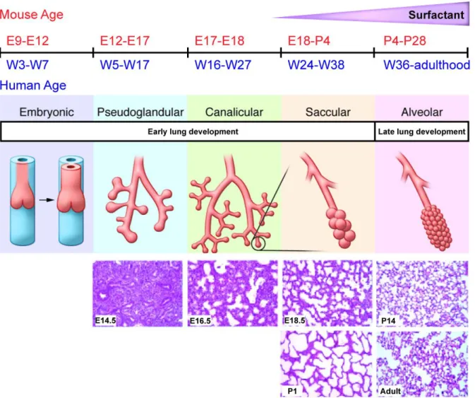
The process of lung development consists of five developmental stages, which are traditionally divided into two phases - early (mainly prenatal) and late (mainly postnatal) lung development (Figure 1) [7, 8].
1.1.1 Early (prenatal) lung development
In order to produce a fully-functional organ, enabling breathing and gas exchange in postnatal environment, the fetal lung must undergo rapid anatomical changes within a relatively short period of time. Based on their morphological and histological characteristics, four developmental stages are usually distinguished within early lung development in humans (three stages in mice) [6].
Embryonic stage
The core lung structure is derived from the endoderm of the primitive foregut at approximately three to seven weeks post-conception in humans (embryonic day [E]9–E12 in the mouse) and undergo main conducting airway branching during the embryonic stage of the early lung development. First, epithelial cells migrating into the surrounding mesenchyme form a primitive trachea, which further branches into two main, epithelium-lined, bronchial
17
buds. This early left and right main bronchus subsequently divides into further lobar and segmental bronchi [1, 6, 9, 10].
Figure 1. Progression of prenatal and postnatal lung development.
Schematic illustration of major stages of early and late lung development and their timing in human and in mouse. E, embryonic day; P, postnatal day; W, week. In addition, representative lung sections from mouse lungs at E14.5, E16.5, E18.5, P1, P14 and adult mice are illustrated. Synthesized from several sources [5-7, 9, 11-13].
18 Pseudoglandular stage
Airway branching continues during the pseudoglandular stage, which occurs between the 5th and 17th week post-conception in humans (E12–E17 in mice) and the formation of the conducting airways and terminal bronchioles is fully completed. Pulmonary arteries and veins are developed during this period, and the cartilage begins to form around the large airways. The first ciliated epithelial cells and immature, cuboidal type II cells appear [3, 6, 9, 14].
Canalicular stage
The canalicular stage, which occurs between the 16th and 27th week post-conception in humans, is mainly characterized by the thinning of the interstitial tissue and formation of acinar units, marking the beginning of the alveolarization process. The thinning of the interstitial mesenchyme leads to closer contact between the forming capillaries and the differentiating epithelium and the formation of the thin air-blood barrier. The primitive cuboidal epithelium is now visibly differentiated into flattened type I alveolar epithelial cells, and the lamellar bodies-containing type II alveolar epithelial cells. The presence of surfactant protein can be detected by week 24 [6, 9, 13]. Due to advances in perinatal medicine, the canalicular stage became the earliest possible period for a successful preterm birth in humans [9, 15]. Canalicular stage is also the last period of prenatal lung development in mice, spanning E17 and E18 [5].
Saccular stage
The last fully prenatal stage of lung development in humans and the first stage of postnatal lung development in mice (24–38 weeks post-conception in humans, and E18-postnatal day [P]4 in mice) is characterized by the enlargement of the peripheral airways, formation of saccular units, further thinning of air-blood barrier and the start of secondary septation.
19 1.1.2 Late (postnatal) lung development Alveolar stage
This stage of alveolar formation is the last stage of lung development in both humans and mice. Although the alveolar stage in humans starts prenatally (36 weeks post-conception), it is largely a postnatal process, taking place during the first 36 months of postnatal life (and possibly beyond). Unlike in humans, alveolarization in mice is entirely postnatal process, taking place between P4-P28 [6, 7, 9, 16]. Alveolar stage is characterized by two, largely interconnected events: secondary septation and microvascular maturation.
The main contribution of secondary septation is increasing the number and reducing the size of the alveoli, eventually leading to an increase of the gas exchange surface area of the lung [5, 16]. While a human lung at birth contains up to 50 million alveoli [17-19], this number increases greatly postnatally, with an average of 480 million alveoli (range of 300-800 million) reported in an adult lung [7, 20, 21]. The dramatic increase in the number of alveoli and overall surface area happens through the formation of secondary septa. At this stage of lung development, distal airspaces are lined by thick walls consisting of two external capillary layers and a single central connective tissue layer. Ridges of secondary septa (or crests) arise from primary septa just before term in humans, and at P7 in mice. Initiation sites of these crests are typically rich in elastic fibers and myofibroblasts expressing α-smooth muscle actin (αSMA), which secrete numerous proteins of the extracellular matrix (ECM) facilitating crest protrusion [1, 5, 7, 13, 22].
The process of microvascular maturation occurs in parallel with alveolarization and is mainly concluded within the first two to three years of age. During this process double capillary layer matures into a single capillary network facilitating the faster gas diffusion [7, 13]. It is largely accepted, that once the single capillary network is formed, no more alveolarization can occur in the lung [9].
The process of alveolarization remains poorly understood but is thought to be carefully coordinated by the combined action of various gene expression programs (comprised of transcription factors and epigenetic effects) and both contact-mediated and growth factor-mediated cell-to-cell communication. The development and maturation of the lung structure is also driven in part by physical forces caused by breathing motions and the production and remodeling of the ECM scaffold [1, 2, 23-26].
20
1.2 Bronchopulmonary dysplasia
Multiple diseases are either caused by, or by themselves cause disturbances to lung development resulting in malformation of the lung architecture, and defects in gas exchange. Depending on the severity of the damage, serious disturbances to respiratory function, as well as long-term consequences may occur [3].
The most common neonatal form of chronic lung disease (CLD) is bronchopulmonary dysplasia (BPD) [27], which was first described in study by William Northway in 1967, in the lungs of prematurely born infants, treated for the severe respiratory distress syndrome (RDS) [28]. Oxygen supplementation and positive pressure mechanical ventilation received by these patients, however lifesaving, was associated with oxygen toxicity and disturbed postnatal lung development, leading to a consecutive injury, characterized by an interstitial and alveolar edema, persistent inflammation and parenchymal fibrosis [5, 28, 29].
Characteristics of BPD have gradually changed over the past several decades. Improvements in the medical management of BPD led to a considerable increase in the survival of infants with extremely low birth weight, resulting in an increase in BPD incidence. On account of these changes, the definition as well as diagnostic criteria of BPD have been eventually altered [3, 27, 29, 30]. Nowadays, the disease, also known as “new BPD”, is clinically defined by the need for supplemental oxygen and/or mechanical ventilation for more than 28 days, or at the 36th week postmenstrual age [27]. Lungs of BPD patients are now histologically characterized less by fibrosis and fibroproliferative airway damage. Instead, the lung structure is more often characterized by defects in alveolar and septal formation (leading to alveolar hypoplasia), thickening of alveolar walls, a dysmorphic pulmonary circulation and ECM remodeling. In comparison to “old BPD”, BPD today is therefore described largely as a disease of arrested lung development (Figure 2) [3, 27, 31, 32].
Despite improvements in clinical care, BPD still remains one of the major causes of morbidity and mortality of infants in a neonatal intensive care unit [3, 27, 29, 30]. Moreover, studies suggest possible long-term consequences of BPD persisting later into the adulthood [33-35]. Although the pathogenic pathways involved in BPD are not well-understood, several hypotheses have been formed about the processes involved in the development of BPD. Processes known to play role in the pathology of BPD include: abnormal shifts in physical forces, failures in the development of pulmonary vasculature, altered cellular composition of
21
the developing alveoli, increase of inflammatory cell infiltration, perturbed gene expression and growth factor signaling, aberrant cell-to-cell communication, and disfunctional ECM architecture and metabolism, particularly changes in the abundance and organization of collagen and elastin fibers and deregulation of ECM-remodeling enzymes [5, 36-40].
Figure 2. Histological structure of the lung of a patient with bronchopulmonary dysplasia and a healthy age-matched control lung.
Comparison of hematoxylin and eosin stained peripheral lung structure in a 12-year old male patient with bronchopulmonary dysplasia (A) and a comparable area of lung tissue from an age-matched control lung with normal alveolarization (B) [41]. Asterix marks abnormally enlarged air space. BR, bronchiole.
22
1.3 The extracellular matrix in context of normal and aberrant lung
development
Branching morphogenesis and alveolarization are characterized by intensive growth and remodeling of newly forming parenchymal structures. This process requires precisely timed mesenchymal-to-epithelial interactions, which cannot occur in the absence of a functional ECM compartment [42, 43].
The first observations regarding the remodeling of ECM structures during lung development, namely collagen and elastin, were made in the 1970’s and 1980’s [44, 45]. While the ECM plays a mainly supporting role in adult lung, it provides a crucial mechanical support for the developing epithelium, mesenchyme and the capillary network [4, 5]. During the alveolar stage of lung development, collagen is mainly present within the alveolar walls, while elastin is deposited more specifically in areas of so-called “foci” at the tips of protruding secondary septa, suggesting the importance of elastin in the process of septal formation [13, 46] (Figure 3). The expression of collagen and elastin is strongly regulated throughout the course of lung development in various experimental animals, including mice [46-49]. While expression of both basement membrane and fibrillary collagens peaks at P7, the peak of elastin expression coincides with the burst of secondary septation around P14 [5, 48]. The importance of collagen and elastin in postnatal lung maturation was further proven in studies using lathyrogens, pharmaceutical lathyrism-inducing agents, namely β-aminopropinitrile (BAPN), which disrupted normal ECM deposition and led to an arrest in alveolarization [50, 51].
A failure of alveolar septation in both a clinical and an experimental setting is accompanied by changes in ECM production and deposition. Perturbed ECM structures have been repeatedly reported in disorders of lung development, including BPD. Collagen fibers in the lung parenchyma in these studies are often described as “disorganized, tortuous, and thickened” [37, 38]. Elastin fibers also exhibit an abnormal structure formation both in the parenchyma [52-54] and the vasculature [55] of patients with BPD, and animal models of BPD alike (Figure 3).
Despite the considerable effort made in order to understand the changes in ECM production and metabolism in the developing lung parenchyma, the underlying reasons for these perturbations are still not fully known [5]. However, ECM homeostasis throughout lung development and a balance between ECM production by cells and degradation and
23
remodeling of ECM components by enzymatic machinery seems to play a key role in processes of alveolar and septal formation.
Figure 3. Elastin deposition during lung formation in bronchopulmonary dysplasia and in the hyperoxia-based mouse model of bronchopulmonary dysplasia.
(A-C) Schematic representation of elastin deposition and capillary network formation during normal and aberrant alveolarization. (A) Initial elastin deposition within the alveolar wall, prior to the formation of secondary septa in the normally developing lung. A double capillary network is present. (B) Elastin localizes to the top of newly forming secondary septa in the normally developing lung. A single capillary network is present. (C) Deposition of elastin at the top of secondary septa is disturbed during aberrant alveolar formation associated with bronchopulmonary dysplasia (BPD). Elastin is partially deposited within thickened septa. The alveolar microvasculature is malformed [13]. (D-E) Elastin foci in the hyperoxia-based mouse model of BPD. (D) Elastin fibers form foci at the top of secondary septa in mice exposed to 21% O2. (E) Elastin fibers fail to form foci in the lungs of mice exposed to 85% O2 from the day of birth up to, and including postnatal day 28. Elastin fibers appear disorganized and malformed. Elastin visualized by Hart’s stain [46].
24
1.3.1 Key components of extracellular matrix in the context of lung development
The ECM forms a complex, large network of multiple heterogenous components with diverse structural, mechanical and biochemical properties [56]. Within these structural elements of ECM the most abundant are collagen and elastin which represent 50% [57] and 18% [58], respectively, of the lung ECM. Other components include microfibrils, glycoproteins fibrillin [59] and fibulin [60], integrins and integrin ligands [61].
The ECM structure, which serves as a scaffold, is constantly remodeled during lung development [42, 43]. Therefore, the production of structural components which form ECM, as well as the enzymatic systems involved in regulation of ECM deposition and stability, must be considered. This machinery include members of families of enzymes involved in the post-translational modulations of various ECM components on one hand, and degradation of the ECM on the other hand. Together this actions of ECM-remodeling enzymes enable natural processes of the ECM renewal [5].
Collagen
The most abundant protein within the interstitial ECM, collagen, is produced by fibroblasts [62-64]. Collagen fibers in the lung, which are represented predominantly by collagen types I and III, are found mainly within blood vessels, bronchi and alveolar septa [65-67]. Quantity of collagen in the lung fluctuates throughout the period of lung development, with a peak in expression at P7 in mice. Parenchymal collagen forms a fine network of fibers. Collagen has been attributed with a major role in processes of alveolar septation [38, 48].
Numerous studies using various animal models of BPD and emphysema have demonstrated the presence of abnormal collagen structures and failures in integrity of collagen network. These changes are thought to be associated not only with defects in formation of new alveoli, but also in the destruction of pre-existing alveoli [38, 46, 68]. Furthermore, studies performed in animal models of BPD report an increase in collagen production accompanied with a formation of thicker collagen fibers, which was associated with decrease in lung elasticity [46, 68].
Observations along this line were also made in clinical studies. Lungs of the patients diagnosed with BPD exhibited an increase in the abundance of fibrillar collagens (collagen I and III) and collagen I/collagen III ratio. In addition the number of collagen-positive cells
25
was also increased [65, 69]. Thickened, disorganized and misshapen collagen fibers were also observed in lungs of patients diagnosed with BPD who received positive pressure ventilation. It is possible that the increase in the size of the alveoli due to the ventilation causes a compression and damage to ECM network surrounding the alveoli, eventually corrupting the normal course of septation [38].
In vitro, collagen production can be regulated by growth factors, including transforming growth factor (TGF)-β [70, 71]. Supporting the findings in vitro are the reports of increased levels of TGF-β in preterm infants with BPD [72]. Moreover, TGF-β was casually implicated in arrest of alveolar formation in animal model of BPD where an increase in TGF-β activation led to an increase in a collagen production by fibroblasts and in the consecutive collagen deposition [46, 73, 74]. Similarly, TGF-β over-expressed in the lungs in utero led to pulmonary hypoplasia, collagen deposition was increased in the developing alveolar septa and animals developed thickened collagen fibers [75]. Furthermore, it has been shown that TGF-β modulates expression levels of multiple matrix-remodeling enzymes, further regulating collagen formation and metabolism [70, 76-78].
Elastin
Elastic fibers are formed by thoroughly cross-linked microfibrils of elastin and fibrillin associated with several accessory molecules, including latent TGF-β-binding protein (LTBP), fibulins and emilins [59]. Elastin in the developing lungs is produced by fibroblasts and smooth muscle cells (SMC). Elastic fibers in the lung are present predominantly in the conducting airways, alveolar ducts, alveoli and in the developing lung vasculature [62, 64, 79]. The expression of elastin and formation of elastin network in lungs begins in the pseudoglandular stage. Elastin (Eln) expression peaks between the saccular and alveolar stage and remain high throughout the period of secondary septation (P5–P15 in mice). Throughout the alveolar stage, elastin is found specifically at the tips of the developing septa, where it forms elastin foci. This pattern of deposition indicates that elastin could play a role in the formation of secondary septa and in the alveolarization [13, 46, 80]. The expression of elastin eventually descreases after the process of alveolarization is completed [48, 81].
Although the expression of elastin in adult lungs is low, reactivation of elastin synthesis can occur in various pathological situations. For example, disorganized and unusually shaped elastic fibers were described in emphysema and pulmonary fibrosis [39,
26
81]. Similarly, abnormal elastin fiber structure and deposition was observed within the parenchyma of prematurely-born ventilated neonates [82].
The importance of elastin in the development of the lung is further emphasized by studies on elastin deficient (Eln-/-) or haploinsufficient (Eln+/-) mice. Complete deletion of elastin causes perinatal lethality and lungs of Eln-/- mice show an impaired distal airway development [83]. While retaining a normal lung development and lung structure, elastin haploinsufficient mice express 50% lower elastin levels, and two-fold increase in levels of collagen and lysyl oxidase (LOX). This observations were accompanied by an increase in lung compliance and decrease in stiffness when compared to wild-type (WT) mice [84, 85]. Along the same line, transgenic mice with elastin expression decreased to 37% of normal levels, had good survival, but exhibited a significant increase in lung size and a pronounced arrest in alveolar formation [85].
Over the past decade, elastin has been studied at length in multiple animal models of lung development and BPD. Both perinatal hyperoxia and mechanical ventilation were shown to increase elastin production. Elastin fibers in these lungs are often described as disturbed and fragmented and are largly localized within alveolar walls instead of septal tips [46, 64, 86-88]. Abnormalities in elastin fibers formation probably leads to a further fiber disintegration and malfunction.
Although the mechanisms behind the elastin regulation are not fully understood, multiple studies highlight the possible roles of TGF-β and platelet-derived growth factor (PDGF) in these processes. While TGF-β seems to be involved in induction of elastin expression [76, 89, 90], certain forms of PDGF suppressed elastin production [91]. This data from experimental models supports observations made in clinical studies, where increase in the abundance of TGF-β [72] and decrease in abundance of PDGF were observed in BPD patients [92].
While both, studies employing mechanical ventilation, and perinatal hyperoxia models report an increase in elastin mRNA levels, controversy exists about the status of mature elastin, where different observations have been made in respect to the methods employed (analysis of tropoelastin by immunoblot, vs. histological staining, vs. analysis of elastin cross-links) [88, 93-95].
27 Extracellular matrix remodeling enzymes
Although many studies focus on ECM components synthesis and production, only few studies to date have explored the subsequent ECM processing and remodeling. However, due to the long half-life of ECM fibers in the lung (up to several years in mice), more importance has recently been given to formation of elastin and collagen fibers, their remodeling and maturation, rather than the gene and protein expression per se [4, 46, 96-98].
It is likely that the formation of elastin and collagen fibers with abnormal structure and properties is a result of dysregulated activity of large amount of matrix-remodeling enzymes. Possible candidate enzymes include: matrix metalloproteinases (MMPs), tissue inhibitors of matrix metalloproteinases (TIMPs), and members of transglutaminase (TGM), lysyl hydroxylase (Procollagen-lysine, 2-oxoglutarate 5-dioxygenase; PLOD) and the lysyl oxidase family. Interestingly in addition to an ability to modulate expression levels of elastin and collagen itself, TGF-β was also shown to impact the expression levels of above-listed matrix remodeling enzymes [70, 76-78, 97, 98]. Therefore, it has been proposed that an arrest in lung alveolarization and proper matrix formation may occur as a consequence of a protease/anti-protease imbalance and changes in collagen/elastin ratio introduced by increased levels of TGF-β [99].
1.4 The lysyl oxidase family
Several recent studies have revealed the importance of matrix remodeling enzymes in ECM deposition and remodeling during both normal and aberrant lung development associated with BPD [46, 58, 97, 98]. Among these are elastin and collagen cross-linking enzymes of the lysyl oxidase family.
The lysyl oxidase family comprises of five copper-dependent amine oxidases: lysyl oxidase (LOX) and four lysyl oxidase-like enzymes (LOXL1-LOXL4) [100, 101]. All members of the family catalyze the process of oxidative deamination of lysine and hydroxylysine residues (Figure 4). Deamination generates semialdehydes, which are highly reactive and spontaneously form intramolecular and intermolecular covalent cross-links in elastin and collagen molecules [100, 101].
28
Figure 4. Chemical reaction catalyzed by lysyl oxidase.
Schematic representation of lysyl oxidase (LOX)-mediated oxidative deamination. Primary amines in elastin and collagen molecules are oxidized by LOX and other members of lysyl oxidase family to reactive semialdehydes. Reactive semialdehydes further form covalent intramolecular and intermolecular cross-links [102].
Lysyl oxidases play a crucial role in the processes of normal lung development as demonstrated by studies in knock-out animals. Lysyl oxidase-deficient mice exhibit perinatal lethality due to aortic aneurism and severe cardio-respiratory dysfunction. Lungs of Lox -/-mice are characterized by an arrest in alveolarization and elastic and collagen fibers in these lungs were described as “fragmented and disperse” [58, 103, 104]. In addition, desmosine (a measure of elastin cross-links) and hydroxylysinonorleucine (HLNL) and hydroxyproline (HP) crosslinks (both measures of collagen cross-linking status), were severely decreased in lungs of Lox-/- mice when compared to wild-type mice [104]. Moreover, lysyl oxidase enzymatic activity was rapidly decreased in the absence of Lox expression. This suggests that LOX alone might serve as the main contributor to overall catalytic activity of this enzymatic family. Unlike Lox-/- mice, mice deficient in Loxl1 survived to adulthood. However Loxl1-/- mice are characterized by decreased connective tissue strength leading to a formation of redundant skin and pelvic organ prolapse. Lungs of Loxl1-/- mice also had enlarged airspaces and exhibited a decrease in desmosine, but not HP levels, suggesting that LOXL1 plays a role exclusively in elastin (but not collagen) metabolism [105].
29
The role of lysyl oxidases have been implicated in the development of several lung diseases, including pulmonary arterial hypertension, lung adenocarcinoma, lung fibrosis, and BPD [46, 106-108]. The role of lysyl oxidases in BPD has also been investigated in several animal models. Gene expression of Lox and Loxl1 was increased in the lungs of pre-term ventilated lambs [109]. Although the expression of Lox in the lung was similarly increased in mechanically ventilated mouse puops, Loxl1 levels were decreased [93]. An increase in lung gene expression and protein abundance of LOX, LOXL1, LOXL2 and LOXL3 was noted in the hyperoxia-based mouse model of BPD, accompanied by increase in overall lysyl oxidase enzymatic activity [46]. Exposure of developing mouse pups to increased concentrations of oxygen was additionally associated with arrest in alveolar formation and a severe defect in elastin foci formation. Similarly, Lox and Loxl1 levels were increased in lung of patients diagnosed with BPD [46].
In addition, localization of LOX in the nuclei of fibrogenic [110] and LOXL2 in the nuclei of epithelial cells [111] has recently been discovered. This findings suggests the existence of “non-matrix” roles for at least some members of lysyl oxidase family [112]. For example, LOX could drive the transcription of collagen IIIα1 (Col3a1) gene in fibroblasts [113]. LOX was also found to increase the activity of Eln promoter in human embryonic kidney (HEK) 293T cells [114]. Furthermore, LOXL2 was associated with Snail1, suggesting a possible role in Cdh expression regulation [111]. LOXL2 was also found to catalyze the deamination of trimethylated Lys4 in histone H3, therefore modulating epigenetic effects in the nucleus [115], which was associated with epithelial-to-mesenchymal transition (EMT).
It is therefore possible that lysyl oxidases play a dual role in the processes of lung development and pathogenesis of BPD. It is likely that disregulation in the gene and protein expression of lysyl oxidases (and potentially expression of other matrix remodeling enzymes) leads to formation of over cross-linked elastin and collagen fibers in the parenchyma of the developing lung. This way excessively stabilized ECM network would be unable to undergo native processes of ECM remodeling leading to an increase in lung stiffness. This would impact the elastic properties of the lung, possibly causing an arrest in alveolarization and formation of secondary septa. Alternatively, unbalance in expression of lysyl oxidases could drive further changes in the expression of potential lysyl oxidase target genes, including essential components of ECM network Eln and Col, contributing even more to the disturbances in ECM formation and remodeling. However, the causal role of lysyl oxidases in
30
formation of ECM structures and perturbed gene regulation in the context of BPD has not yet been fully adressed.
31
2
AIMS OF THE STUDY
Bronchopulmonary dysplasia presents a common complication of preterm birth with significant morbidity and mortality. Pathogenic pathways behind the development of BPD are not well understood, however, the importance of proper formation and metabolism of the ECM in the context of normal and aberrant lung development is well known. Abnormal formation and deposition of two main structural components of ECM, collagen and elastin fibers, represent a pathological feature of both clinical and experimental BPD.
The correct temporal and spatial coordination of the activity of matrix remodeling enzymes plays a crucial role in elastin and collagen metabolism and remodeling. Among these are lysyl oxidases, amine oxidases catalyzing the formation of covalent crosslinks in elastin and collagen molecules. Dysregulated expression and activity of lysyl oxidases have been reported to play a role in pathogenesis of BPD, as well as in various animal models of BPD.
In this context, the main aims of this study were:
i.) To characterize the expression profile of lysyl oxidase family members in mouse lungs during normal and aberrant lung development.
ii.) To investigate a causal role for the lysyl oxidase family in experimental hyperoxia-based mouse model of BPD.
iii.) To assess the impact of lysyl oxidase activity on the formation of elastin and collagen fibers in mice lungs during normal and aberrant lung development.
iv.) To assess the impact of lysyl oxidase activity on lung alveolarization in mice lungs during normal and aberrant lung development.
32
3
MATERIALS AND METHODS
3.1 Materials
3.1.1 Equipment
Name Company
Autoclave Systec, Germany
Agar embedding moulds custom made
Agar cutting mould custom made
Bacteria culture incubator Heareus, Germany
Blotting membrane, Trans-Blot® TurboTM Transfer Pack Bio-Rad, Germany
Camera, D5300 NIKON, USA
Cell counter, Countess® Invitrogen, UK
Cell culture dish, 100 mm Greiner Bio-One, Germany
Cell culture dish for bacteria Greiner Bio-One, Germany
Cell culture flask, 250 ml Greiner Bio-One, Germany
Cell culture incubator Thermo Fisher Scientific, USA
Cell culture plates, 6-well Greiner Bio-One, Germany
Cell culture plates, 6-well, Snapwell Permeable Support Costar, USA
Cell culture sterile working bench Thermo Fisher Scientific, USA
Cell scrapers, 25 cm Sarstedt, Germany
Cell strainers, 100, 40 μm BD Biosciences USA
Cover slides Roth, Germany
Digital slide scanner, NanoZoomer-XR C12000 Hamamatsu, Germany
EcoRI enzyme Promega, USA
Electrophoresis chambers, Wide Mini-Sub® Cell GT Bio-Rad, Germany Electrophoresis chambers, Mini Protean® Tetra system Bio-Rad, Germany
Espresso personal microcentrifuge VWR, USA
Feather® trimming blade Pfm medical, Germany
FIA 96-well plate, black Greiner Bio-One, Germany
Filter tips, 10 µl, 100 µl, 200 µl, 300 µl, 1000 µl Sarstedt, Germany
Fully Automated Rotary Microtome Leica Biosystems, Germany
33
Name Company
Heating plate Medax, Germany
High speed micro centrifuge Hitachi, Japan
Histobloc Heareus, Germany
Histoform Q mould Heareus, Germany
Homogenizing kit for soft tissue, Precellys® PEQLAB, Germany Homogenyzer, Precellys® 24-Dual homogenizer PEQLAB, Germany
Imager ImageQuant® LAS 4000 GE health care, USA
Infinite® 200 PRO multimode reader TECAN, Switzerland
Inoculation loops, 10 µl Sarstedt, Germany
InoLab® pH meter WTW, Germany
Knife holder NZ RM2200 silver Leica Biosystems, Germany
Light microscope Leica, Germany
Magentic Dynabeads® , streptavidin-coupled Thermo Fisher Scientific, USA
Magnetic separator Thermo Fisher Scientific, USA
MicroAmp® Optical 96-Well Reaction Plate Thermo Fisher Scientific, USA Microcentrifuge tubes, 0.5, 1.5, 2 ml Eppendorf, Germany
Microplate reader,VersaMax ELISA Molecular devices, USA
Microscope slides, SUPERFROST ULTRA PLUS® Thermo Fisher Scientific, USA
Microtome blade S-35 pfm, Pfm medical, Germany
Microtome knife, 16cm long, profile d, steel assy Leica Biosystems , Germany
Multifuge 3 S-R centrifuge Heraeus, Germany
NanoDrop® ND-1000 spectrophotometer Thermo Fisher Scientific, USA
Nitrocelulose membrane, 0.2 µm Bio-Rad, Germany
Objective, AF-S DX Micro NIKKOR, 85mm NIKON, USA
Parrafin embedding station Leica Biosystems, Germany
Pasteur pipette, 3.5 ml Sarstedt, Germany
PCR-thermocycler, peqSTAR VWR, USA
Pipetboy® Integra, Switzerland
Pipettes, automatic, 10 µl, 100 µl, 300 µl Eppendorf, Germany Pipettes, manual, 10 µl, 20 µl, 100 µl, 200 µl, 1000 µl Eppendorf, Germany
34
Name Company
Pipettes, serological, 2 ml, 5 ml, 10 ml, 25 ml, 50 ml Falcon, USA Polyvinylidene difluoride (PVDF) membranes, 0.2 µm Bio-Rad, Germany
Real-Time PCR system, StepOnePlus™ Applied Biosystems, USA
Refrigerated microcentrifuge CT15RE VWR, USA
Rotilabo®embedding cassettes Roth, Germany
Routine Stereomicroscope Leica Biosystems, Germany
Single use needles, FINE-JECT® HENKE SASS WOLF, Germany
Snap-cap vials, Rotilabo® Roth, Germany
Snap-on lids Roth, Germany
Standard analog shaker VWR, USA
Standard Clamp w/Adapter 40×40 mm Silver Leica Biosystems , Germany Surgical instruments – scissors, tweezers F.S.T., Germany
Thermo shaker, MS-100 Universal labortechnik, Germany
Test tubes, 15 ml, 50ml Greiner Bio-One, Germany
Tissue processor Leica Biosystems, Germany
Ultraviolet (UV) Transilluminator, Gel Imager Intas, Germany
Unneedled polyamide suture, SUPRAMID SERAG, Germany
VisiopharmNewCast software Visiopharm, Denmark
Vortex mixer VWR, USA
Waterbath, for cell culture Lauda, USA
Water bath, for histological slides Vogel, Germany
Western blot transfer system, Trans-Blot® TurboTM Bio-Rad, Germany
35 3.1.2 Reagents
Name Company
Accutase® solution Sigma-Aldrich, Germany
Acetone, > 99.7% (vol/vol) Roth, Germany
Acrylamide solution, Rotiphorese Gel 30 Roth, Germany
Agar for microbiology (Agar-agar) Sigma-Aldrich, Germany
Agarose Promega, Germany
Agarose, low melting point Sigma-Aldrich, Germany
Ammonium persulfate (APS) Promega, Germany
Ampicillin sodium salt Sigma-Aldrich, Germany
Azure II Sigma-Aldrich, Germany
Borax, di-Natriumtetraborate Decahydrate Roth, Germany
Bovine serum albumin (BSA) Thermo Fisher Scientific, USA
Collagenase Sigma-Aldrich, Germany
Complete macrophage medium CellBiologics, USA
Complete mouse endothelial cell medium CellBiologics, USA
Complete® protease inhibitor Roche, Germany
Dispase BD Biosciences, USA
1,4-Dithiothreitol (DTT) Promega, USA
DNase I Serva, Germany
Dublecco’s modified Eagle medium (DMEM), Thermo Fisher Scientific, USA 1 g/l glucose
Dulbecco’s modified Eagle's medium (DMEM), Thermo Fisher Scientific, USA 4.5 g/l glucose
Dulbeco’s phosphate buffered saline (DPBS) Sigma-Aldrich, Germany
Elastase Sigma-Aldrich, Germany
Ethanol, 70% (vol/vol) Roth, Germany
Ethanol, absolute Roth, Germany
Ethidium bromide Promega, USA
Ethylenediaminetetraacetic acid (EDTA) Sigma-Aldrich, Germany
Ethylene glycol-bis Sigma-Aldrich, Germany
36
Name Company
Fetal bovine serum (FBS) PAA Laboratories, Austria
Gelatin-Based Coating Solution CellBiologics, USA
GeneRullerTM, DNA ladders Thermo Fisher Scientific, USA
Glutaraldehyde Serva, Germany
Glycine Roth, Germany
Hank’s balanced salt solution (HBSS) PAA Laboratories, Austria
Hematoxylin Waldeck, Germany
HEPES Sigma-Aldrich, Germany
Isofluran-CP®, 1ml/ml Cp-pharma®, Germany
LB–Agar, Lurria/Muller Roth, Germany
4× Laemmli Sample Buffer Bio-Rad, Germany
Lipofectamine® 2000 Thermo Fisher Scientific, USA
Liquid nitrogen Air Liquide, Germany
Magnesium chloride (25 nM) Applied Biosystems, USA
Magnesium chloride (50 nM) Thermo Fisher Scientific, USA
2-Mercaptoethanol Bio-Rad, Germany
Methanol Sigma-Aldrich, Germany
Methylene blue Roth, Germany
MuLV reverse transcriptase Applied Biosystems, USA
m-Xylol, ˃ 98.5% (vol/vol) Roth, Germany
NARCOREN®, Pentobarbital Sodium Merial GmbH
Non-fat dry milk powder Roth, Germany
Nuclease-free water Ambion, USA
OptiMEM® medium Thermo Fisher Scientific, USA
Osmium tetroxide Roth, Germany
Paraformaldehyde (PFA) Sigma-Aldrich, Germany
Paraplast®, paraffin Leica, Biosystems, Germany
PCR-Buffer II 10× Applied Biosystems, USA
PCR Nucleotide Mix (10 mM) Promega, USA
pCRTM2.1-TOPO® Gateway Entry Thermo Fisher Scientific, USA
37
Name Company
Pertex, mounting media Medite, Germany
Phosphate buffered saline (PBS) 1×, 10× Sigma-Aldrich, Germany Platinum® SYBR® Green qPCR SuperMix UDG kit Invitrogen, USA
pLenti7.3/V5DESTTM Thermo Fisher Scientific, USA
Polybrene, hexadimethrine bromide Sigma-Aldrich, Germany
Ponceau S Sigma-Aldrich, Germany
Precision Plus Protein® Dual color standard Bio-Rad, Germany
Propane/Butane Campin Gaz, Germany
2-Propanol Merck, Germany
Proteinase K Promega, USA
QIAquick gel extraction kit Qiagen, Netherlands
QIAquick PCR purification kit Qiagen, Netherlands
QIAprep®SpinMiniprep/Maxiprep kit Qiagen, Netherlands
QuickStart® Bradford 1×Dye Reagent Bio-Rad, Germany
Random hexamers, 50 µM Thermo Fisher Scientific, USA
Resorcin-Fuchsin Waldeck, Germany
RIPA® buffer Sigma-Aldrich, Germany
RNase free water Qiagen, Germany
Roti® -Histol Roth, Germany
RNase inhibitor Applied Biosystems, USA
Select agar Sigma-Aldrich, Germany
SmGm-2 Supplements Lonza, Switzerland
Smooth muscle growth medium (SmGM)-2 Lonza, Switzerland
Sodium cacodylate Serva, Germany
Sodium dodecyl sulfate (SDS), 10× solution Promega, USA
Sodium orthovanadate Sigma-Aldrich, Germany
Spectinomycin dihydrochloride pentahydrate Sigma-Aldrich, Germany
Super Optimal Broth (S.O.C) Thermo Fisher Scientific, USA
SuperSignal® West femto maximum sensitivity substrate ThermoFisher Scientific, USA
Tris-acetate-EDTA (TAE) buffer, 50× Roth, Germany
38
Name Company
Technovit 3040 HeareusKulzer, Germany
Technovit 7100 HeareusKulzer, Germany
Technovit, universal liquid Heareus Kulzer, Germany
Tetramethylethylenediamine (TEMED) Bio-Rad, Germany
Tris Roth, Germany
Trypsin-EDTA ThermoFisher Scientific, USA
Trypan blue solution 0.4% (vol/vol) ThermoFisher Scientific, USA
TurboFectTM ThermoFisher Scientific, USA
Tween® 20 Sigma-Aldrich, Germany
Uranyl acetate Serva, Germany
Van Gieson stain Roth, Germany
39 3.1.3 Purchased primary cells and cell-lines
NIH/3T3, fibroblasts, mouse embryonic cells American Type Culture Collection, USA
HEK 293T, human embryonic kidney cells, American Type Culture Collection, USA epithelial cells
TheTransform One Shot®DH5α™-T1R, ThermoFisher Scientific, USA TOP10 Competent Cells, E.coli cells
Bone marrow macrophages, Cell Biologics, USA
bone marrow derived mouse macrophages
Primary lung vascular endothelial cells, Cell Biologics, USA primary mouse cells
40
3.2 Methods
3.2.1 Animal experiments
All animal procedures were approved by the local authorities, Regierungspräsidium Darmstadt (approval numbers B2/329; B2/1029 and B2/277).
3.2.1.1 Pilot study
Prior to intervention studies, a pilot study was performed in order to determine the tolerance of mice to BAPN treatment. Two different doses of BAPN, as well as control injections with vehicle were employed, and animals were observed for duration of five days. Three animals per group were injected intraperitoneally (i.p.) daily with either vehicle [1× phosphate buffered saline (PBS)], or BAPN (diluted in vehicle) at a dose of 15 or 150 mg·kg-1·day-1. All animals were euthanized at P5.5 with an overdose of pentobarbital (500 mg·kg-1, i.p.).
3.2.1.2 Normobaric hyperoxia-based mouse model of BPD
An arrest in the alveolarization in the lungs of newborn mouse pups was induced by continuous exposure of animals to normobaric hyperoxia, more precisely 85% O2 (FiO2 0.85) from the day of birth (P1). This model of BPD recapitulates a several major hallmarks seen in human BPD, including morphological changes, such as a decrease in the number of alveoli, an associated decrease of overall gas-exchange surface area and an increase in the septal thickness, as well as inflammation and perturbations to ECM deposition and remodeling.
Newborn C57BL/6J mice were randomized to equal-sized litters (average eight pups per litter) and placed into either a normoxic (21% O2) or a hyperoxic (85% O2) environment within 2 h after birth. Newborn pups were exposed from the day of birth (P1.5), continuously up to and including P9.5 or P19.5, when experiments were terminated.
In order to minimize oxygen toxicity and potential confounders of milk production caused by hyperoxia, nursing dams were rotated between normoxia and hyperoxia on 24 h : 24 h cycle. Mice were maintained on 12 h : 12 h light-dark cycle and received food ad libitum. All animals were euthanized at P9.5 or P19.5 with an overdose of pentobarbital (500 mg·kg-1, i.p.).
41
3.2.1.3 Treatment of hyperoxia-exposed newborn mice with β-aminopropionitrile
Based on the observations made during the pilot study BAPN was administered at a dose of 15 mg·kg-1·day-1 in a hyperoxia-based BPD model. Newborn C57BL/6J mice were randomized to equal-sized litters and placed into either a normoxic (21% O2) or a hyperoxic (85% O2) environment within 2 h after birth. Both groups were further subdivided into two groups, treated with daily i.p. injections of either vehicle (1×PBS), or BAPN (diluted in vehicle) at a dose of 15 mg·kg-1·day-1 for either 9 or 19 days. Experiments were terminated on P9.5 or P19.5.
3.2.1.4 Organ isolation
All animals were euthanized with an overdose of pentobarbital (500 mg·kg-1, i.p.). Subsequently, the abdomen was opened and the diaphragm was punctured to allow the lungs to retract. The chest wall was opened by cutting through sternum, but ribs remained intact in their original position in order to preserve the natural size of the chest cavity. Lungs were isolated for further stereological analysis, RNA and protein analysis, collagen and elastin measurements or tissue staining.
Lungs isolated for embedding in plastic resin and stereological analysis
In order to preserve the blood in the tissue, lungs of animals used for stereological analysis were not perfused and animals were not exsanguinated. Instead, lungs were instillation-fixed via a tracheal cannula under a 20 cm H2O hydrostatic pressure with fixation solution containing 1.5% (wt/vol) paraformaldehyde (PFA) and 1.5% (wt/vol) glutaraldehyde in 150 mM HEPES-PBS. Lungs were excised and stored at 4 °C in fixation solution for 24 h.
Lungs isolated for future RNA and protein analysis
Animals were exsanguinated and lungs were perfused with 1×PBS via the right heart ventricle. Lungs were removed and immediately frozen in liquid nitrogen. Samples were kept at -80 °C for further processing.
42
Lungs isolated for collagen and elastin crosslinks analysis
Animals were exsanguinated and lungs were perfused with 1×PBS via the right heart ventricle, excised, transferred to 1.5 ml tube containing 1×PBS and frozen in liquid nitrogen. Samples were stored at -80 °C for further processing.
Lungs isolated for embedding in paraffin and staining of elastin fibers
Animals were exsanguinated and lungs were perfused with 1×PBS via the right heart ventricle. The trachea was cannulated and lungs were instillation-fixed under a 20 cm H2O hydrostatic pressure with 4% (wt/vol) PFA in 1×PBS. Lungs were excised and stored at 4 °C in fixation solution for 24 h prior to paraffin embedding.
3.2.1.5 Embedding in agar and lung volume estimation
Following 24 h of fixation, lungs were cleaned of any remains of the trachea, thymus, heart or blood clots and embedded in toto in 2% (wt/vol) agar-agar for at least 6 h. Agar blocks were then cut into 2-mm thick sections and always the same side of each section was photographed for calculations of lung volume. Individual pieces of each lung were retrieved from agar slices for further embedding in plastic resin.
The reference lung volume was estimated from photographs of lung sections embedded in agar by the Cavalieri principle using Visiopharm NewCast computer-assisted stereology system. Briefly, point counting was performed on a point grid. Counting was performed within 100% of each lung section. Volume of the lung was estimated based on the number of points counted, size of the surface area/point and thickness of the section.
3.2.1.6 Embedding in plastic resin
All individual pieces of one lung were transferred into Snap-cap vials and further processed inside a fume hood. Buffers required for treatment of the lungs prior to embedding are listed in Table 1. Briefly, lungs were washed 4 × 5 min with 0.1 M sodium cacodylate buffer and treated with 1% osmium tetroxide (OsO4) in 0.1 M sodium cacodylate buffer for 2 h. Afterwards lungs were washed 4 × 5 min with 0.1 M sodium cacodylate buffer, washed 4 × 5 min with ddH2O. Samples were then protected from light and treated overnight with half-saturated uranyl acetate (UrAc) buffer. Afterwards, samples were washed with double
43
distilled (ddH2O) and dehydrated in increasing concentration of acetone. Lungs were first incubated overnight in 1:1 solution of 100% acetone and Technovit 7100-Hardener I and eventually Technovit 7100-Hardener I solution. Finally, lungs were incubated in Technovit 7100-Hardener I-Hardener II solution for 5 min. Over this time vials with samples were constantly stirred. Individual pieces of lung, together with the buffer were transferred into a Histoform Q mould and allowed to solidify for at least 48 h. Plastic blocks were removed from moulds with Technovit universal liquid and Technovit 3040 using the Histobloc adaptors.
Table 1. Buffers required for treatment of lungs and embedding in Technovit 7100.
Buffer Dilution
Sodium cacodylate 0.1 M, in ddH2O
Osmium tetroxide 2% (wt/vol) stock solution, in water for injections 1% (wt/vol) working solution, in 0.1 M sodium cacodylate Uranyl acetate saturated stock solution, in ddH2O
half-saturated working solution, in ddH2O
Acetone 70% (vol/vol), 90% (vol/vol), 100% (vol/vol), in ddH2O
Technovit 7100 undiluted solution
Technovit 7100-Hardener I 1% (wt/vol) Hardener I, in Technovit 7100 Technovit 7100-Hardener I-Hardener II 200 µl Hardener II
in 3 ml Technovit 7100-Hardener I Technovit 3040-Technovit
universal liquid 2:1 ratio
3.2.1.7 Embedding in paraffin
Lungs were instillation-fixed with 4% (wt/vol) PFA in 1×PBS for 24 h. Afterwards lungs were cleaned of any remains of trachea, thymus, heart or blood clots, transferred in embedding moulds and stored in 1×PBS for up to 1 week prior to processing. Tissue was then dehydrated by ethanol, cleared by xylene to remove alcohol and infiltrated with molten paraffin wax. Finally, tissue was embedded in moulds in paraffin and allowed to solidify on a cooling plate. Material was stored at 4 °C for further use.
44 3.2.1.8 Paraffin and plastic sectioning
Paraffin and plastic embedded lungs were sectioned with fully automated rotary microtome. For paraffin embedded lungs, 3-µm sections were cut using a Feather microtome blade and collected on Superfrost Ultra Plus adhesion slides. Slides were allowed to dry on a heating plate at 40 °C overnight.
For plastic embedded lungs, blocks were cut into 2-µm sections using a, profile d steel assy microtome knife. Forty sections were cut and every tenth section was collected for staining. In addition, for the estimation of the alveolar number, three consecutive sections were cut and first and third sections were collected on the same adhesion slide. Slides were allowed to dry on a heating plate at 65 °C.
3.2.1.9 Hart’s elastin staining
The deposition of elastin fiber structures was visualized in 3-µm thick lung tissue sections from paraffin embedded mouse lungs by Hart’s elastin stain. Shortly, paraffin sections were brought to water via xylene and ethanol and incubated in Hart´s solution overnight. Subsequently, the slides were washed and counterstained with iron hematoxylin and van Gieson solution. Finally sections were dehydrated with ethanol, clear with xylene and covered with cover slips using a mounting medium.
3.2.1.10 Richardson’s staining
Lung structure was visualized in 2 µm thick lung tissue sections from plastic-embedded mouse lungs by Richardson’s staining. Briefly, plastic sections were stained in warm Richardson’s stain (65 °C) for 30 s, washed briefly in cold tap water and rinsed in hot tap water. Subsequently, sections were washed briefly in ddH2O and rinsed in Xylol. All slides were scanned at 60× magnification using a NanoZoomer-XR C12000 digital slide scanner.
45 Richardson’s stain – stock solution:
ddH2O
0.25% (wt/vol) methylene blue
0.22% (wt/vol) Borax (di-Sodium tetraborate) 0.5% (wt/vol) Azur II
The stock solution must be filtered through filter paper and diluted 1:1 with tap water prior to use.
3.2.2 Design-based stereology
Methods of lung structure analysis were based on the American Thoracic Society/European Respiratory Society recommendations for quantitative assessment of lung structure [116] and according to systematic uniform random sampling for stereological analysis.
Analyses were performed using Visiopharm NewCast computer-assisted stereology system. Analysis of lung structure included four main parameters: gas-exchange surface area, septal thickness, mean linear intercept (MLI) and number of alveoli. In addition, lung volume was estimated by the Cavalieri principle using the same software. The parenchymal fraction, as well as airspace and septal fraction within the parenchyma were counted by point counting on a corresponding point grid (Figure 5A-5B). Surface density was assessed by point and intersection counting on a “line grid” of known length (Figure 5E-5F). Approximately 40-60 lung sections were evaluated on total of four scanned slides and 2-3% of each section was analyzed. The alveolar density was assessed by counting of newly appearing alveolar bridges using a physical dissector, where the height of the dissector was 4 µm (Figure 5C-5D). Approximately 10 sections were evaluated for each lung and 2% of each section was analyzed. In order to determine the absolute number of alveoli and absolute size of gas-exchange surface area in the lung, alveolar density and surface density, respectively were normalized to reference lung volume.
For each stereological parameter, the coefficient of error (CE), the coefficient of variation (CV), and the squared ratio between both (CE2/CV2) were calculated in order to confirm the measurement precision. The quotient threshold was set at 0.5.