Supplementary Materials and Methods
Study subjects and data collection
The subjects of the present study include 420 IIM cases and 402 non-IIM cases with definitive diagnosis of IIM or other diseases made by experts including the authors at 19 institutes participating in this national project between 2007 and 2013. The project was approved by research ethics committees of individual institutes, and carried out by expert rheumatologists, neurologists, dermatologists, and epidemiologists in these institutes. As inclusion criteria, the clinical data of allocated number of IIM cases assigned to each facility were retrospectively collected in the order of new patient visits from medical records using questionnaires, which were distributed to the experts. There were no exclusion criteria.
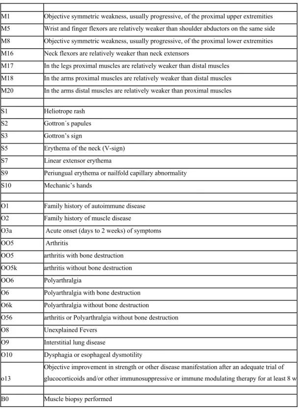
The data collection sheet employed in the study contained diagnostic information, patient backgrounds, seven items on muscle findings, seven items on skin findings, seven items on laboratory findings, 10 items on autoantibody tests, 11 pathological findings from muscle or skin biopsy, three electromyogram (EMG) findings, and three magnetic resonance imaging (MRI) findings (online supplementary Table S2).
Non-IIM cases were also collected from each institute, in the order of patient visits. Non-IIM included 39 rheumatologic, neurologic, and dermatological disorders (online supplementary Table S3). Several disorders, such as hand eczema and Sweet syndrome, were added to the original non-IIM disease list of IMCCP [1, 2]. Diagnosis of all cases was validated by an expert rheumatologist, neurologist, and dermatologist.
Methodology of new EULAR/ACR classification
The new EULAR/ACR classification criteria show the probability of a patient having IIM for use in clinical and research settings [1, 2]. In brief, the new criteria consist of the muscle findings, skin findings, and laboratory measures [1, 2]. Each item is assigned a weighted score, and the aggregated scores are calculated by summing the score points. The aggregated scores can be converted into a probability of IIM using the following formulas:
Probability of IIM = 1 / [1 + exponential (6.49– score)] when muscle biopsy data are present
or,
Probability of IIM = 1 / [1 + exponential (5.33– score)] when muscle biopsy data are absent
Unknown or missing data were regarded as “score 0”. Among the information included in the questionnaires, only the criteria items were used for classification of patients and comparators. The web-based calculator and the Excel file that can be downloaded from criteria web page were not utilized in this study. The correlation between total aggregated scores and varying probability of having IIM are shown in the previous paper [1, 2]. A patient is classified as an IIM case if the patient’s probability is above a specified cut-off value. The recommendation in the new criteria was as follows: 1) Muscle biopsy is required for patients without pathognomonic skin manifestations (heliotrope rash, Gottron’s papules and/or Gottron’s sign) as determined by a consensus of expert opinion within the IMCCP steering and working committees. This is because characteristic skin rashes of DM contributed with high weight to the probability score. A skin biopsy is recommended for patients with suspicion of DM without muscle involvement so to ascertain correct diagnosis [3, 4]. 2) A probability cut-off of 55%, corresponding to a score of 5.5 (6.7 if muscle biopsy data are available), has the best sensitivity/specificity and is recommended as a minimum in order to classify a patient as having IIM. 3) Patients with a probability of between 55% and 90% (a total aggregate score of 7.5 without muscle biopsy data and 8.7 with muscle biopsy data) are designated “probable IIM”. The probability of ≥90% corresponds to “definite IIM” while the probability ≥50% to <55% is termed “possible IIM”.
Furthermore, a patient classified as IIM by the EULAR/ACR classification criteria can be further sub-classified by a classification tree into six groups: PM, DM, ADM, juvenile DM (JDM), other juvenile myositis, and IBM [1, 2].
Using the data from the Japanese cohort, we calculated the sensitivity and specificity of the new EULAR/ACR classification criteria for IIM. To compare the performance of the new criteria with other published criteria, the criteria described by Tanimoto and by
Limitation of the study
The present study has some limitations, observation before inclusion may not be performed in some of IIM and non-IIM cases. Retrospectively collecting skin variables from charts is not reliable and a true prospective study collecting variables from live patients would be helpful. Our questionnaire data sheet used for the validation did not include questions about ‘Finger flexor weakness’ and ‘Response to treatment: not improved’, which was also not included in the early phase of the IMCCP study, but is required for the distinction of PM (IMNM) and IBM in the classification tree. Accordingly, the classification between PM (IMNM) and IBM may be inadequate in this study. However, because number of IMNM or IBM is small in Japan, the influence of this limitation is considered to be marginal. Similarly, the study of sensitivity and specificity of Tanimoto’s revised criteria was derived from data of patients including those with ADM. Since the revision was meant primarily for inclusion of ADM, effects of the correction or subtle changes in definition of the skin rashes, electromyographic findings and autoantibodies were not evaluated.
As one of the features of the new criteria, a web calculator of the new criteria is available, and there is an Excel file that can be downloaded from criteria web page (http://www.imm.ki.se/biostatistics/calculators/iim). They give probability ranges, minimum to maximum probability. The largest possible value is obtained by replacing all the unobserved values with ‘present’. On the other hand, in this paper, unknown or missing data were regarded as “score 0” according to the original papers, which corresponds to the smallest possible value calculated by replacing all the unobserved items with ‘absent’ in web calculator [1, 2]. Because it was the doctor’s decision on what variables to measure and what to leave blank, the web calculator and the Excel file may be more useful in a clinical setting.
A further limitation is that we set the cut-point of probability at 55% according to the original papers [1, 2], which considered sensitivity and specificity as equally important. We also regard both higher sensitivity and specificity as clinically and epidemiologically important in the classification criteria of IIM. On the other hand, as another feature of the new EULAR/ACR IIM classification criteria, it permits setting of
the cut-off to specific values to optimize sensitivity or specificity to meet the specific needs of any given clinical or epidemiological setting [5].
References
1. Lundberg I, Tjärnlund A, Bottai M, et al. 2017 European League Against
Rheumatism/American College of
Rheumatology Classification Criteria for Adult and Juvenile Idiopathic Inflammatory
Myopathies and Their Major Subgroups. Ann Rheumatol Dis. 2017; 69:2271-82.
2. Bottai M, Tjärnlund A, Santoni G, et al. EULAR/ACR classification criteria for
adult and juvenile idiopathic inflammatory myopathies and their major subgroups: a
methodology report. RMD Open. 2017; 3:e000507.
3. Hsiung SH, Chan EF, Elenitsas R, et al. Multicentric reticulohistiocytosis
presenting with clinical features of dermatomyositis. J Am Acad Dermatol. 2003; 48(2
Suppl):S11-14.
4. Fett N, Liu RH. Multicentric reticulohistiocytosis with dermatomyositis-like
features: a more common disease presentation than previously thought. Dermatology.
5. Zhou XH, Obuchowski NA, McClish DK. Measures of diagnostic accuracy. In:
Zhou XH, Obuchowski NA, McClish DK, eds. Statistical methods in diagnostic medicine.
Table S1. Tanimoto’s revised criteria proposed in 2016
(1) Rashes – Heliotrope rash, Gottron's papules or Gottron's sign (2) Proximal weakness, upper or lower extremity and trunk (3) Muscle pain on grasping or spontaneous muscle pain (4) Elevation of CK or Aldolase
(5) Electromyographic findings of myositis (6) Non-destructive arthritis or arthralgias
(7) Systemic inflammatory signs (fever > 37˚C at axilla, elevated CRP or ESR) (8) Anti-aminoacyl-tRNA synthetasesautoantibody
(9) Muscle biopsy evidence of myositis
Definite PM= any 4 of 9 without rash Definite DM = rashes + 4 others
An amyopathic patient whose histopathological findings of the eruptions are compatible with DM can be diagnosed as having ADM.
Table S2. Questionnaire data sheet used for the validation
M1 Objective symmetric weakness, usually progressive, of the proximal upper extremities M5 Wrist and finger flexors are relatively weaker than shoulder abductors on the same side M8 Objective symmetric weakness, usually progressive, of the proximal lower extremities M16 Neck flexors are relatively weaker than neck extensors
M17 In the legs proximal muscles are relatively weaker than distal muscles M18 In the arms proximal muscles are relatively weaker than distal muscles M20 In the arms distal muscles are relatively weaker than proximal muscles
S1 Heliotrope rash S2 Gottron´s papules S3 Gottron’s sign
S5 Erythema of the neck (V-sign) S7 Linear extensor erythema
S9 Periungual erythema or nailfold capillary abnormality S10 Mechanic’s hands
O1 Family history of autoimmune disease O2 Family history of muscle disease
O3a Acute onset (days to 2 weeks) of symptoms OO5 Arthritis
OO5 arthritis with bone destruction OO5k arthritis without bone destruction OO6 Polyarthralgia
O6 Polyarthralgia with bone destruction O6k Polyarthralgia without bone destruction
O56 arthritis or Polyarthralgia without bone destruction O8 Unexplained Fevers
O9 Interstitial lung disease
O10 Dysphagia or esophageal dysmotility
o13
Objective improvement in strength or other disease manifestation after an adequate trial of glucocorticoids and/or other immunosuppressive or immune modulating therapy for at least 8 w
B1 Necrosis of type I and type II muscle fibers, phagocytosis, degeneration of myofibers B3 Endomysial infiltration of mononuclear cells surrounding, but not invading, myofibers B4 Non-necrotic fibers surrounded and invaded by mononuclear cells
B5 Perimysial and/or perivascular infiltration of mononuclear cells B6 Perifascicular atrophy
B8 Rimmed vacuoles
B11 Immunohistochemistry data available
B12 MHC Class I antigen present on scattered or more muscle fibers
L1r Elevated serum levels of CK L2r Elevated serum levels of LDH L3r Elevated serum levels of AST L4r Elevated serum levels of ALT L5 Serum Aldolase activity
L6 Erythrocyte sedimentation rate (ESR) L7 C-reactive protein (CRP)
L8 Autoantibodies
L9_1 ANA
L9_2 Anti-Jo-1 (anti-Histidyl-tRNA synthetase) autoantibody positivity L9_9 Anti-SSA L9_12 Anti-La/SSB L9_13 Anti-ribonucleoprotein (RNP)-70K (U1snRNP) L9_16 Anti-Centromere B (ACA) L9_17 Anti-Topoisomerase-1/Scl70, L9_19 Anti-Sm L9_24 Anti-CCP
Other, please specify below
EMG EMG performed
EMG1
Electromyogram (EMG) - Increased insertional and spontaneous activity in the form of fibrillation potentials, positive sharp waves, or complex repetitive discharges
EMG2
EMG - Morphometric analysis reveals the presence of short duration, small amplitude, polyphasic motor unit action potentials (MUAPs)
MRI1 Muscle edema on STIR or T2-weighted magnetic resonance imaging (MRI)
MRI2
Muscle atrophy and/or increased muscle fat content on T1-weighted MRI scanning consistent with myositis
L13yn Skin biopsy performed
Table S3. The list of non-IIM 1. Becker's dystrophy 2. Duchenne's dystrophy 3. Facioscapulohumeral dystrophy 4. Limb-girdle dystrophy 5. Myotonic dystrophy 6. Other dystrophy 7. Dysferlinopathy 8. Bacterial myopathy 9. Cushing syndrome
10.Drug or toxin associated myopathy (5)
11.Exogenous steroid myopathy (5)
12.Familial periodic paralysis
13.Hypereosinophilic syndrome (5)
14.Hyperthyroidism (5) 15.Hypokalemia (5)
16.Rimmed vacuolar distal myopathy (RVDM)
17.Juvenile idiopathic arthritis 18.Metabolic myopathy
19. Mitochondrial myopathy (5) 20. Mixed connective tissue disease 21.Systemic lupus erythematosus (SLE)
(5) 22.Systemic sclerosis (5) 23.Systemic vasculitis 24. Viral myopathy (5) 25. Seborrheic dermatitis (5) 26. Contact dermatitis (5) 27. Hand eczema (5) 28. Sweet syndrome 29. Thyroid dysfunction 30. Photosensitive dermatitis 31. Erysipelas 32. Drug eruption (5)
33. cutaneous or intermediate lupus erythematosus (5)*
34. Angioedema 35. Adult Still's disease 36. Sarcoidosis
37. Verruca vulgaris (5) 38. Multicentric reticulohistiocytosis 39. lymphoma
The figures on the right of each disease (in parenthesis) are upper limit of case number in each institute. *patients with erythematosus who did not fulfill diagnostic criteria of SLE.
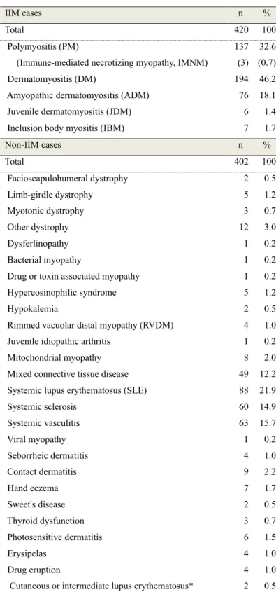
Table S4. The number of IIM cases and non-IIM cases in the study population
IIM cases n %
Total 420 100
Polymyositis (PM) 137 32.6
(Immune-mediated necrotizing myopathy, IMNM) (3) (0.7)
Dermatomyositis (DM) 194 46.2
Amyopathic dermatomyositis (ADM) 76 18.1 Juvenile dermatomyositis (JDM) 6 1.4 Inclusion body myositis (IBM) 7 1.7
Non-IIM cases n % Total 402 100 Facioscapulohumeral dystrophy 2 0.5 Limb-girdle dystrophy 5 1.2 Myotonic dystrophy 3 0.7 Other dystrophy 12 3.0 Dysferlinopathy 1 0.2 Bacterial myopathy 1 0.2
Drug or toxin associated myopathy 1 0.2 Hypereosinophilic syndrome 5 1.2
Hypokalemia 2 0.5
Rimmed vacuolar distal myopathy (RVDM) 4 1.0 Juvenile idiopathic arthritis 1 0.2
Mitochondrial myopathy 8 2.0
Mixed connective tissue disease 49 12.2 Systemic lupus erythematosus (SLE) 88 21.9
Systemic sclerosis 60 14.9 Systemic vasculitis 63 15.7 Viral myopathy 1 0.2 Seborrheic dermatitis 4 1.0 Contact dermatitis 9 2.2 Hand eczema 7 1.7 Sweet's disease 2 0.5 Thyroid dysfunction 3 0.7 Photosensitive dermatitis 6 1.5 Erysipelas 4 1.0 Drug eruption 4 1.0
Angioedema 2 0.5
Adult Still's disease 32 8.0
Sarcoidosis 14 3.5
Verruca vulgaris 2 0.5
Lymphoma 5 1.2
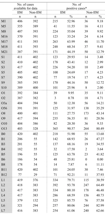
Table S5. The frequency of each variable in IIM cases and non-IIM cases
No. of cases available for data
No. of positive cases*
IIM Non-IIM IIM Non-IIM
n n n % n % M1 406 392 215 52.96 36 9.18 M5 355 389 41 11.55 16 4.11 M8 407 393 224 55.04 39 9.92 M16 370 391 123 33.24 24 6.14 M17 411 393 263 63.99 46 11.70 M18 411 393 248 60.34 37 9.41 M21 387 391 171 44.19 50 12.79 S1 411 402 123 29.93 14 3.48 S2 410 402 178 43.41 12 2.99 S3 417 402 226 54.20 10 2.49 S5 405 402 100 24.69 17 4.23 S7 390 402 77 19.74 17 4.23 S9 385 393 170 44.16 50 12.72 S10 389 400 101 25.96 8 2.00 O1 392 384 39 9.95 35 9.11 O2 387 382 3 0.78 18 4.71 O3a 404 394 50 12.38 56 14.21 O56 391 391 125 31.97 138 35.29 O8 400 401 111 27.75 173 43.14 O9 417 394 235 56.35 81 20.56 O10 404 386 82 20.30 36 9.33 O13 403 328 365 90.57 264 80.49 B0 420 402 218 51.90 55 13.68 B1 206 55 142 68.93 26 47.27 B3 201 55 137 68.16 19 34.55 B4 182 55 32 17.58 2 3.64 B5 196 55 125 63.78 23 41.82 B6 186 54 48 25.81 0 0.00 B8 178 54 14 7.87 6 11.11 B11 420 402 101 24.05 30 7.46 B12 77 29 71 92.21 11 37.93 L1 419 376 334 79.71 115 30.59 L2 418 383 392 93.78 247 64.49 L3 417 383 334 80.10 178 46.48 L4 417 383 286 68.59 156 40.73 L5 379 132 325 85.75 76 57.58 L6 321 294 257 80.06 244 82.99 L7 416 383 254 61.06 240 62.66
L8 420 402 416 99.05 358 89.05 L9_1 399 346 224 56.14 230 66.47 L9_2 402 171 49 12.19 0 0.00 L9_9 366 293 77 21.04 96 32.76 L9_12 312 256 12 3.85 14 5.47 L9_13 356 246 23 6.46 89 36.18 L9_16 222 161 9 4.05 17 10.56 L9_17 258 176 9 3.49 24 13.64 L9_19 270 231 6 2.22 40 17.32 L9_24 204 161 18 8.82 13 8.07 EMG 420 402 285 67.86 61 15.17 EMG1 238 58 143 60.08 20 34.48 EMG2 262 60 171 65.27 31 51.67 MRI 420 402 295 70.24 57 14.18 MRI1 294 57 236 80.27 26 45.61 MRI2 269 57 69 25.65 16 28.07 L13yn 420 402 174 41.43 87 21.64 L13 169 87 144 85.21 9 10.34
The definition of each variable is described in Table S2.
*In B0, B11, L8, EMG, MRI, and L13yn, the number of cases in whom each test was performed was shown.
Supplementary figure legend
Supplemental Figure S1
Distribution of the probabilities of both IIM and non-IIM in all Japanese cases (a), cases without muscle biopsy data (b), and cases with muscle biopsy data (c).
Supplement Figure S2
(A) Receiver operating characteristic (ROC) curves for all Japanese cases (a), cases without muscle biopsy data (b), and cases with muscle biopsy data (c). AUC: areas under curves.