Molecular Sciences
Review
Growth Hormone (GH) and Cardiovascular System
Diego Caicedo1ID, Oscar Díaz2, Pablo Devesa3and Jesús Devesa4,*ID1 Department of Angiology and Vascular Surgery, Complejo Hospitalario Universitario de Pontevedra, 36701 Pontevedra, Spain; diego.caicedo.valdes@sergas.es
2 Department of Cardiology, Complejo Hospitalario Universitario de Pontevedra, 36701 Pontevedra, Spain; oscar.diaz.castro@sergas.es
3 Research and Development, The Medical Center Foltra, 15886 Teo, Spain; pdevesap@foltra.org 4 Scientific Direction, The Medical Center Foltra, 15886 Teo, Spain
* Correspondence: jesus.devesa@usc.es or devesa.jesus@gmail.com; Tel.: +34-981-802-928 Received: 25 December 2017; Accepted: 12 January 2018; Published: 18 January 2018
Abstract:This review describes the positive effects of growth hormone (GH) on the cardiovascular system. We analyze why the vascular endothelium is a real internal secretion gland, whose inflammation is the first step for developing atherosclerosis, as well as the mechanisms by which GH acts on vessels improving oxidative stress imbalance and endothelial dysfunction. We also report how GH acts on coronary arterial disease and heart failure, and on peripheral arterial disease, inducing a neovascularization process that finally increases flow in ischemic tissues. We include some preliminary data from a trial in which GH or placebo is given to elderly people suffering from critical limb ischemia, showing some of the benefits of the hormone on plasma markers of inflammation, and the safety of GH administration during short periods of time, even in diabetic patients. We also analyze how Klotho is strongly related to GH, inducing, after being released from the damaged vascular endothelium, the pituitary secretion of GH, most likely to repair the injury in the ischemic tissues. We also show how GH can help during wound healing by increasing the blood flow and some neurotrophic and growth factors. In summary, we postulate that short-term GH administration could be useful to treat cardiovascular diseases.
Keywords:cardiovascular diseases; atherosclerosis; oxidative stress; angiogenesis and arteriogenesis; endothelial dysfunction; growth hormone; IGF-I; wound healing
1. Introduction
ThehGH gene family is composed by two growth hormone (GH) genes (GH-Nand GH-V), and three placental genes that are located in the chromosome 17 [1]. It has been considered that the GH-V gene is expressed only in the placenta, although some studies indicated that this gene, or some otherGHgene, still unknown, could also be expressed in the human pituitary gland [2,3]. In the case of GH-N, it is already well known that in addition to its pituitary expression, which is responsible for the actions of the hormone at the endocrine level, the hormone is also expressed in numerous cells and tissues, where it acts in an auto/paracrine manner [4]. Perhaps the heart is an exception to this peripheral expression of GH, as we will see later.
The regulation of GH pituitary expression is very complex, since in the last few years the classical knowledge of a positive regulation by GHRH and negative by somatostatin [5], has been changed after the knowledge of a series of factors that are decisively involved in that regulation [6]. This is the case, for instance, of the orexigenic Ghrelin, released by the empty stomach, or the postulated anti-senescence factor Klotho, mainly expressed in the kidney, but also in the brain and in the own somatotroph cells where it would act in an auto/paracrine manner for directly regulating GH secretion [7]; in addition, the growth differentiation factor 15 (GDF15), synthesized and released
by cardiomyocyte, inhibits GH-induced hepatic expression of Insulin-like Growth Factor I (IGF-I), therefore inhibiting the IGF-I effect on hypothalamic somatostatin release and the direct negative effect of IGF-I on pituitary somatotrophs, thus acting as a coordinator between cardiac function and body growth or other IGF-I dependent GH effects on the human body [8].
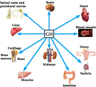
Although the regulation of GH expression is not the aim of this review, perhaps the complexity of its regulation would explain the fact that far beyond of the concept that GH is mainly a metabolic hormone that is responsible for the longitudinal growth of the organism before puberty ends, the hormone exerts many other actions on practically all of the organs and tissues in the human body [4], as schematized in Figure1.
Int. J. Mol. Sci. 2018, 19, 290 2 of 40
cardiomyocyte, inhibits GH-induced hepatic expression of Insulin-like Growth Factor I (IGF-I), therefore inhibiting the IGF-I effect on hypothalamic somatostatin release and the direct negative effect of IGF-I on pituitary somatotrophs, thus acting as a coordinator between cardiac function and body growth or other IGF-I dependent GH effects on the human body [8].
Although the regulation of GH expression is not the aim of this review, perhaps the complexity of its regulation would explain the fact that far beyond of the concept that GH is mainly a metabolic hormone that is responsible for the longitudinal growth of the organism before puberty ends, the hormone exerts many other actions on practically all of the organs and tissues in the human body [4], as schematized in Figure 1.
Figure 1. Growth hormone (GH) is a pleiotropic hormone acting on many tissues and organs in the human organism. Blue arrows show some of the most important territories in which the hormone produces positive effects. For a better understanding of this schema, see reference [4]. AGs: Adrenal glands.
In this review, we will focus on the effects of GH on the cardiovascular system; but before, it will be analyzed the role of the vascular endothelium as an internal secretion gland, as well as the main pathologies that affect the cardiovascular system, to subsequently assess the effect that GH can play in its treatment.
1.1. The Role of the Vascular Endothelium as an Internal Secretion Gland and the Effects of GH on It
Histologically, the vascular endothelium is a single unicellular layer that covers the internal surface of blood vessels and forms the wall of capillaries. However, despite its simplicity, this layer is very complex in physiological terms, since its location allows it to be able to detect alterations in the hemodynamic forces acting on the vascular wall (shear stress forces), as well as changes in circulating chemical signals, responding to all this by releasing vasoactive compounds, able to act oppositely depending on the signals received. For instance, at the level of hemostasis, the vascular endothelium can produce both anti-hemostatic factors (protein C, prostacyclin PGl2, tissue plasminogen activator, nitric oxide), or factors that favor hemostasis (von Willebrand factor, tissue Factor III, plasminogen activator inhibitor, thromboxane A2). The same occurs with the vascular tone, since vasodilators such as nitric oxide (NO) or prostacyclin (PGl2), and vasoconstrictors such as angiotensin II, endothelin (ET), thromboxane II and superoxide anion, can be released from the vascular endothelium to contribute to vascular homeostasis. Moreover, the vascular endothelium produces many growth factors, such as vascular endothelial growth factor (VEGF), platelet derived growth factor (PDGF), basic fibroblast growth factor (bFGF), and ET; but also inhibitory growth factors, as transforming growth factor-β (TGF-β). Even, the vascular endothelium can participate in Figure 1.Growth hormone (GH) is a pleiotropic hormone acting on many tissues and organs in the human organism. Blue arrows show some of the most important territories in which the hormone produces positive effects. For a better understanding of this schema, see reference [4]. AGs: Adrenal glands.
In this review, we will focus on the effects of GH on the cardiovascular system; but before, it will be analyzed the role of the vascular endothelium as an internal secretion gland, as well as the main pathologies that affect the cardiovascular system, to subsequently assess the effect that GH can play in its treatment.
1.1. The Role of the Vascular Endothelium as an Internal Secretion Gland and the Effects of GH on It
Histologically, the vascular endothelium is a single unicellular layer that covers the internal surface of blood vessels and forms the wall of capillaries. However, despite its simplicity, this layer is very complex in physiological terms, since its location allows it to be able to detect alterations in the hemodynamic forces acting on the vascular wall (shear stress forces), as well as changes in circulating chemical signals, responding to all this by releasing vasoactive compounds, able to act oppositely depending on the signals received. For instance, at the level of hemostasis, the vascular endothelium can produce both anti-hemostatic factors (protein C, prostacyclin PGl2, tissue plasminogen activator, nitric oxide), or factors that favor hemostasis (von Willebrand factor, tissue Factor III, plasminogen activator inhibitor, thromboxane A2). The same occurs with the vascular tone, since vasodilators such as nitric oxide (NO) or prostacyclin (PGl2), and vasoconstrictors such as angiotensin II, endothelin (ET), thromboxane II and superoxide anion, can be released from the vascular endothelium to contribute to vascular homeostasis. Moreover, the vascular endothelium produces many growth factors, such as vascular endothelial growth factor (VEGF), platelet derived growth factor (PDGF), basic fibroblast growth factor (bFGF), and ET; but also inhibitory growth factors, as transforming growth factor-β (TGF-β). Even, the vascular endothelium can participate in immunological responses by producing interleukins (IL-1, IL-6 and IL-18), tumor necrosis factor-α (TNF-α), monocyte chemoattractant
protein-1 (MCP-1), vascular cell adhesion molecule-1 (VCAM-1), intercellular adhesion molecule-1 (ICAM-1) and selectins E and P.
Most of these factors act locally by auto/paracrine mechanisms, so that they allow, as stated above, to regulate the vascular homeostasis.
In general, the vascular endothelium decreases the vascular tone, inhibits platelet adhesion and aggregation, decreases the activation of the coagulation system, stimulates fibrinolysis, decreases capillary permeability and inhibits the adhesion and migration of neutrophils and inflammation-generating macrophages. Therefore, endothelial dysfunction, a primary event in the development of atherosclerosis, is associated with increased smooth muscle vascular tone with arterial rigidity and elevated intima-media thickness.
Interestingly, while there are clear evidences that receptors for GH (GHR) and IGF-I (IGF-IR) are expressed in the vascular endothelium and myocardium [9–12], the possibility exists that GH itself is expressed in this territory, as in vitro studies reflect [13]. In any case, there are not doubts about the fact that GH plays a very important role on vascular endothelium. This statement is supported by early studies carried out in children with GH-deficiency (GHD), in whom GH replacement therapy recovered existing endothelial dysfunction. This is the case, for instance, of children with renal insufficiency; in them, endothelial dysfunction is quite common finding, but GH therapy reverses it [14]. In addition an improvement of the arterial response to induced vasodilation were observed in GH-deficiency (GHD) adolescents after GH-replacement therapy [15]; or in obese children, in whom obesity negatively affects the secretion of GH and constitutes a risk of developing atherosclerosis prematurely [16]. Similar results have been found in GHD adults (AGHD) after receiving replacement therapy [17], suggesting that GH reduces vascular inflammation, therefore reducing the vascular risk. Another study in AGHD patients showed that GH treatment led to a significant decrease in plasma levels of apolipoprotein B (Apo B) and C-reactive protein (CRP), while no changes were observed in IL-6 or on markers of endothelial function; but in all, GH administration decreased the cardiovascular risk in them [18]. AGHD patients show impaired coronary flow reserve, which is improved after receiving treatment with the hormone, suggesting that GH improves microvascular function and then could reduce cardiovascular morbidity and mortality in these AGHD [19,20]. A more recent study describes that six months of treatment with GH are enough to decrease cardiovascular risk and improve endothelial dysfunction [21].
One of the biomarkers for cardiovascular disease is the loss of circulating CD34+ cells [22]. CD 34+ cells are hematopoietic stem cells (HSCs) able to migrate to the bone marrow and give rise to all hematopoietic cell types when injected intravenously. They were first discovered in a cell surface glycoprotein [23], that is early expressed in hematopoietic and vascular-associated tissue. Therefore, CD34 is in fact a cell surface antigen able to act as an adhesion molecule but also as a facilitator of cell migration. These CD34+ cells are useful for treating many pathologies, including cardiac and vascular affectations [24], but also as a predictors of cardiovascular risk, since these cells collaborate in the maintenance of vascular homeostasis and repair, even in diabetic patients in which the circulating levels of CD34+ cells have been shown to be decreased [25]. CD34+ cells express receptors for GH and IGF-I, although the mechanism by which GH or IGF-I stimulates them has not been demonstrated, a parallel increase in granulocyte colony-stimulating factor (G-CSF) has been also observed [26]. Moreover, eNOS expressed by the bone marrow stromal cells influences recruitment of stem and progenitor cells [27]. In any case, the loss of circulating CD34+ cells observed in AGHD has been shown to be corrected after one year of GH treatment in AGHD, since the number of these cells increased and endothelial function improved [28]. This agrees with the fact that GH increases the production and release of endothelial progenitor cells (EPC) in non AGHD subjects, which in the vascular endothelium act as a repair cells [29]. Moreover, GH replacement therapy improves fibrinolysis in AGHD patients, most likely by increasing the release of endothelial tissue plasminogen activator as a response to venous occlusion [30]. On the contrary, other studies found that AGHD patients treated with GH showed increased concentrations of E-selectin, indicative of an uncorrected endothelial dysfunction [31].
These authors conclude that the beneficial effect of GH in these patients may be produced by the effects of the hormone on other mechanisms rather than acting on endothelial dysfunction.
As known, there are many ways to evaluate endothelial dysfunction, thus, influencing in the studies´ final results. For example, when diacron-reactive oxygen (DRO) metabolites or hemodynamic tests such as reactive hyperemia index are used, results are positive for the effect of GH on the correction of endothelial dysfunction in GHD [32]. In addition, it seems that both GH and E-selectin are not related, as, although the latter represents a type of cell adhesion molecules (CAM), and the hormone may increase them, GH rises VCAM but not E-selectin in GHD patients, being this an effect not dependent on GH-IGF-I.
The effects of GH administration on E-selectin had not been found in previous studies performed in healthy adults and AGHD patients [33]; instead vascular cell adhesion molecule-1 (VCAM-1) significantly increased in AGHD patients during GH treatment. Interestingly, serum from healthy patients treated with GH significantly increased the expression of VCAM-1 in cultured umbilical vein endothelial cells, suggesting that GH might act on VCAM-1 expression by an indirect mechanism, most likely related to the modulation of the expression of other circulating factors [33]. This might explain the reported negative effects of the hormone when administered to critically ill patients, since VCAM-1 mediates leukocytes extravasation which can lead to multiple organ failure in sepsis [34], although the increased mortality reported was observed with doses of GH quite higher (10–20 times) than usual treatment dose.
To our knowledge only one study reported no positive effects of GH replacement therapy on the endothelial dysfunction in AGHD patients [31], as only one report indicates that GH does not recover the endothelial impairment present in GHD children [35]. Perhaps, the small number of subjects, or the methodology used, or the time during which these studies were carried out justifies the contradictory results. 1.1.1. GH, IGF-I, Klotho and the Vascular Endothelium
Klotho was first described in 1997 as a product of a gene that is involved in the suppression of several aging phenotypes in mouse. Initially, it was thought that Klotho would be implied in a signaling pathway regulating senescence and the severity of diseases related with the process of aging, such as atherosclerosis [36]. In mice, the gene codifies a membrane protein homologue toβ-glucosidases, while in humans the gene has been shown to be composed of five exons and is located on chromosome 13q12. The gene suffers a physiological alternative RNA splicing giving origin to two transcripts, one of them being a membrane protein while the other one is secreted and predominates over the former [37]. The possible effects of Klotho on the physiology of the human vascular endothelium were first postulated in 1998, indicating that it protects the cardiovascular system by inducing NO endothelial production [38], although the possible mechanisms of action has not been clarified yet [39]. Further studies in mice of the same group demonstrated that secreted Klotho promoted endothelial increase of NO in aorta and arterioles [40], and that adenovirus-mediatedKlothogene delivery to a typical rat model of multiple atherogenic risk (OLETF rat) improved endothelial dysfunction, increased NO production, reduced increased blood pressure, and prevented medial hypertrophy, meaning that Klotho was a clear positive regulator of vascular function [41]. This was confirmed inKlothomutant mice when observing that in these animals the density of blood capillaries was decreased at the tissue level and angiogenesis was impaired, as was the release of NO from the vascular endothelium [42]. These effects have been related to an action of Klotho on oxidative stress, responsible for inducing apoptosis and senescence in vascular cells [43]. While studies in animal models indicate a clear role for Klotho on the vascular endothelium, there are still no clear data on the physiological role that this hormone plays in humans on the cardiovascular system [44]. In vitro studies demonstrated that Klotho suppresses TNF-α-induced expression of intercellular adhesion molecule 1 (ICAM-1) and vascular cell adhesion molecule 1 (VCAM-1) in human umbilical vein endothelial cells, as well as the inhibition of eNOS phosphorylation induced by the administration of TNF-α[45], effects consistent with its previously postulated role in the modulation of endothelial inflammation. Klotho is mainly produced
in kidneys; however, it seems that it could be expressed also in the vascular endothelium, with the only exception of endothelial cells from human brain [46]. In any case, Klotho is a circulating protein that increases NO production and protects the vascular endothelium [47,48].
To analyze the role of Klotho on the vascular endothelium is not the aim of this review. However, since it has been shown that this protein plays a role on pituitary GH secretion [7], we think it is important to try to establish a relationship between Klotho and GH, given that both are effective factors to prevent an injury to the vascular endothelium and repair it if a damage exits. Mice that do not express Klotho die early than normal mice showing many symptoms of aging, most of them typical of GHD [7]. Plasma levels of Klotho are low in GHD subjects, and the pituitary somatotrophs of Klotho-deficient mice are hypotrophic [7], suggesting that Klotho exerts a trophic effect on them. Besides this, Klotho-deficient mice are smaller than normal mice, and their GH-producing cells in the pituitary show lesser secretory granules [49]. In addition, Klotho strongly inhibits the negative effects of IGF-I on GH secretion, and increases GH secretion in cultured human GH-secreting adenomas [49]. All of these data indicate that Klotho is a positive active regulator of GH secretion, both in animal models and in humans. However, it is still unknown how GH and Klotho interact to repair a damaged vascular endothelium. For instance, in anorexia nervosa patients, in which the existence of an increased pulsatile secretion of GH is well known, while plasma levels of IGF-I are low or very low, due to malnutrition, plasma levels of Klotho are lower than expected for the age of the patients [50], but they increased significantly after the patients increased their body weight and, concomitantly IGF-I increased too. This suggests that IGF-I led to the increase of Klotho [50], perhaps for the maintenance of a physiological feedback loop between GH, IGF-I and Klotho. Those supposed relationships between the three hormones are schematized in Figure2.
Int. J. Mol. Sci. 2018, 19, 290 5 of 40
however, it seems that it could be expressed also in the vascular endothelium, with the only exception of endothelial cells from human brain [46]. In any case, Klotho is a circulating protein that increases NO production and protects the vascular endothelium [47,48].
To analyze the role of Klotho on the vascular endothelium is not the aim of this review. However, since it has been shown that this protein plays a role on pituitary GH secretion [7], we think it is important to try to establish a relationship between Klotho and GH, given that both are effective factors to prevent an injury to the vascular endothelium and repair it if a damage exits. Mice that do not express Klotho die early than normal mice showing many symptoms of aging, most of them typical of GHD [7]. Plasma levels of Klotho are low in GHD subjects, and the pituitary somatotrophs of Klotho-deficient mice are hypotrophic [7], suggesting that Klotho exerts a trophic effect on them. Besides this, Klotho-deficient mice are smaller than normal mice, and their GH-producing cells in the pituitary show lesser secretory granules [49]. In addition, Klotho strongly inhibits the negative effects of IGF-I on GH secretion, and increases GH secretion in cultured human GH-secreting adenomas [49]. All of these data indicate that Klotho is a positive active regulator of GH secretion, both in animal models and in humans. However, it is still unknown how GH and Klotho interact to repair a damaged vascular endothelium. For instance, in anorexia nervosa patients, in which the existence of an increased pulsatile secretion of GH is well known, while plasma levels of IGF-I are low or very low, due to malnutrition, plasma levels of Klotho are lower than expected for the age of the patients [50], but they increased significantly after the patients increased their body weight and, concomitantly IGF-I increased too. This suggests that IGF-I led to the increase of Klotho [50], perhaps for the maintenance of a physiological feedback loop between GH, IGF-I and Klotho. Those supposed relationships between the three hormones are schematized in Figure 2.
Figure 2. Schematic representation about the possible relationships between GH, Insulin growth factor (IGF-I) and Klotho, and its actions on the vascular endothelium. (1) Pituitary GH induces the hepatic expression of IGF-I (2) and acts on the repair of the damaged vascular endothelium (DVE), although it is also possible that the hormone enhances the production of Klotho by this damaged tissue. (3) Besides, its inhibitory effects on pituitary GH release, IGF-I also contributes to repair DVE, and, as in the case of GH, it could enhance Klotho production in DVE. (4) DVE secretes Klotho and it inhibits the negative effect of IGF-I on pituitary GH release, but plasma Klotho may also proceed from kidneys (7), contributing or being responsible for the inhibition of IGF-I effects on GH secretion. (5) The possibility exists that Klotho released from DVE stimulates GH secretion for repairing DVE. (6) GH plays an important role on the physiology of kidneys, being particularly important when there is a chronic kidney disease; since in this pathology there is a state of systemic Klotho deficiency, it is possible that GH tries to correct this problem associated to cardiovascular diseases. Some of these concepts are merely speculative, but existing data lead to think that there is a feedback regulation circuit between GH, IGF-I and Klotho. Blue arrows indicate stimulation and red arrows indicate inhibition.
Figure 2.Schematic representation about the possible relationships between GH, Insulin growth factor (IGF-I) and Klotho, and its actions on the vascular endothelium. (1) Pituitary GH induces the hepatic expression of IGF-I (2) and acts on the repair of the damaged vascular endothelium (DVE), although it is also possible that the hormone enhances the production of Klotho by this damaged tissue. (3) Besides, its inhibitory effects on pituitary GH release, IGF-I also contributes to repair DVE, and, as in the case of GH, it could enhance Klotho production in DVE. (4) DVE secretes Klotho and it inhibits the negative effect of IGF-I on pituitary GH release, but plasma Klotho may also proceed from kidneys (7), contributing or being responsible for the inhibition of IGF-I effects on GH secretion. (5) The possibility exists that Klotho released from DVE stimulates GH secretion for repairing DVE. (6) GH plays an important role on the physiology of kidneys, being particularly important when there is a chronic kidney disease; since in this pathology there is a state of systemic Klotho deficiency, it is possible that GH tries to correct this problem associated to cardiovascular diseases. Some of these concepts are merely speculative, but existing data lead to think that there is a feedback regulation circuit between GH, IGF-I and Klotho. Blue arrows indicate stimulation and red arrows indicate inhibition.
1.1.2. GH, IGF-I and Ghrelin
The complexity of GH neuroregulation and the number of factors acting positively on the pituitary release of the hormone seems to be related to the multiple roles that GH plays in the human body, far beyond than those classically thought, such as the longitudinal growth of the organism before the puberty ends [4]. Interestingly, some of these roles are played in conjunction with the GH-stimulating factors. This is the case, for instance, of Ghrelin (GH-releasing peptide or GHRP), a small peptide found in the gastrointestinal tract in 1999 [51]. Curiously, besides its strong effects on GH secretion when administered intravenously, Ghrelin is an orexigenic hormone that is physiologically released when the stomach is empty. This is the reason by which patients with anorexia nervosa usually show increased plasmatic concentration of Ghrelin [52]. Therefore, it seems logical to assume that Ghrelin appears in evolution with a basic function: to induce eating behavior and optimize the use of immediate principles at the expense of promoting the release of an anabolic hormone, such as GH.
As described in the case of Klotho, Ghrelin also plays a hemodynamic role. In rats, there are receptors for Ghrelin in the aorta, left cardiac ventricle and left cardiac atrium. In healthy humans, the intravenous infusion of Ghrelin decreases the blood pressure, increases the cardiac index, and produces a greater volume of the pulse [53].
Interestingly, as it happens with GH, Ghrelin also plays a very important role in gastrointestinal processes that occur with inflammation, exhibiting gastroprotective properties. Perhaps in this area the most studied model is experimentally-induced pancreatitis in rats. For example, the administration of Ghrelin during the induction of acute pancreatitis in rats with normal secretion of GH, attenuated the development of pancreatitis by ischemia-reperfusion. On the contrary, in hypophysectomized rats, Ghrelin administration did not produce any beneficial effect, something that was achieved when IGF-I was administered. The authors of this study conclude that Ghrelin inhibits the development of ischemia-reperfusion-induced pancreatitis although this effect depends on the effects of Ghrelin on the secretion of GH and consequently IGF-I [54].
Similar conclusions were obtained after inducing acute pancreatitis in rats with cerulein, a decapeptide obtained from the skin of an australian amphibian, whose structure and actions are very similar to those of cholecystokinin. Cerulein induces an acute edematous pancreatitis, which in rats with normal GH secretion is reduced in its severity and producing a faster regeneration of the pancreas, being reduced the serum concentrations of the pro-inflammatory interleukin 1-β(IL-1β) and the serum activities of pancreatic enzymes amylase and lipase. In addition, pancreatic blood flow increased, as did pancreatic DNA synthesis. However, this did not occur in hypophysectomized rats, unless they received IGF-I in parallel to Ghrelin. This demonstrates that the protective effects of Ghrelin need of the existence of an adequate functioning of the GH-IGF-I axis [55]. Perhaps even more interesting is a recent study, in which an experimental colitis was induced in rats. Seven days after the induction of colitis (enema with acetic acid 3% in 1 mL) and treatment with Ghrelin, it was shown that the area of damage in colonic mucosa was clearly reduced in pituitary-intact rats, but increased in hypohysectomized animals. In addition, rats with normal GH-IGF-I production were shown to have enhanced blood flow in colonic mucosa while being treated with Ghrelin and increased mucosal cell proliferation, as well as reduced levels of IL1-1βand activity of mieloperoxidase. Just the opposite was found in hypohysectomized rats. Therefore, the authors concluded that while Ghrelin has a therapeutical effect in experimental colitis, this is mainly mediated by a normal activity of the GH-IGF-I axis [56].
In all, these concepts support our idea about GH as a hormone that plays many more roles in the body than those of a merely metabolic hormone and responsible for longitudinal growth until puberty ends.
1.2. Cardiovascular Disease as an Inflammatory Condition
Several diseases have been related to inflammation since many years, including atherosclerosis [57–61]. It is considered that inflammation plays a key role in atherogenesis, since it is not only involved in the development and progression of this process [61], but also in the associated symptoms [58]. Circulating
monocytes and lymphocytes are present in the vascular wall early in atherogenesis, and both are responsible for the formation and complication of the atherosclerotic plaque [61].
A current study has demonstrated the high influence of inflammation in cardiovascular disease (CVD) from a clinical point of view. As is known, IL-6 has been previously associated with an increased risk of cardiovascular events, with independence of the cholesterol levels in plasma [57,62]. IL-6 amplifies the inflammatory cascade and is the main circulating cytokine linking systemic inflammation with local pathology [63,64]. It stimulates macrophages and promotes proliferation of smooth muscle cells (SMC) in atherosclerotic plaque [63], and stimulates coagulation by increasing messenger ribonucleic acid transcription of tissue factor and factor VIII [65]. IL-1βmediates the IL-6 signaling pathway [57], and canakinumab, a fully human monoclonal antibody targeting IL-1β, leads to a marked reduction of both, plasma levels of IL-6 and CRP, without lowering the level of low-density lipoprotein (LDL) in patients with diabetes who were at high vascular risk [57]. This drug led to a significant lower rate of recurrent cardiovascular events than the placebo [57].
The development of the atheromatous plaque is a multi-factorial process. SMC from the middle layer in the elastic arteries show a differentiated phenotype with a low proliferation and migration rate. Unlike the skeletal and cardiac myocyte, mature SMC may suffer a phenotypic modulation as a result of an atherogenic stimulus, with a re-entry in the cellular cycle. These activated states makes them proliferate and migrate to the vascular lumen, and synthesize some extracellular matrix (EM) components and proteases that modify the matrix, contributing to the atheromatous plaque.
The key aspect of the plaque formation is the endothelial dysfunction secondary to some atherogenic stimuli, such as hypercholesterolemia, hypertension, diabetes, tobacco, etc. As a consequence of this endothelial dysfunction, the inflammatory response is triggered. SMC are essential in the stability of these plaques. When there is a scarcity of them into the plaque, then the atheroma will be highly vulnerable to rupture. Plaque rupture and subsequent thrombus formation can lead to an acute event, although in the lower extremities this event can be better tolerated as a result of the numerous and large collateral network.
It is well known the role of LDL in this setting. Oxidized LDL (ox-LDL) have been related to the formation and complication of the atherosclerotic plaque [66]. LDL has high susceptibility of being oxidized. But, the oxidative environment in the vascular wall may also modify other lipids as HDL. Nicotinamide adenine dinucleotide phosphate (NADPH) oxidase, leukocyte- and platelet-derived oxidants, and red blood cell-derived iron-rich heme group, are part of the different systems implied in the oxidative modification of lipids, proteins, and DNA that in the vascular wall leads to atherosclerosis [66]. All of these oxidants maintain the inflammatory response and participate in the arterial wall rupture with platelet aggregation and thrombus formation.
Oxidative stress plays a main role in the origin of the pathogenesis of CVD. In a normal vascular wall, oxidative stress activates nuclear defense genes throughout the mediation of the nuclear factor erythroid 2-related factor 2 (Nrf2) [67]. This protects against the formation of foam cells by regulating the expression of antioxidant proteins and scavenger receptors [67]. Nevertheless, its function has not been properly understood, since a pro-atherogenic action has been also associated to Nrf2, because ApoE-null mice, which are deficient in Nrf2, develop smaller atherosclerotic plaques [68].
The recruitment of circulating leukocytes into the blood vessel wall is one of the major etiopathogenic mechanisms of atherosclerosis. This process is predominantly mediated by cellular CAM, which are expressed on the vascular endothelium and the leukocytes of the vascular wall, in response to atherogenic stimuli. In patients with peripheral arterial disease (PAD), increased levels of these integrins have been found during exercise, being associated with the severity and the extent of the arterial disease [69]. Antagonists of CAM have shown promise in treating inflammatory disorders in animal models [70,71]. Selectins, another group of integrins, are also elevated in PAD population. Studies with the anti-P-selectin antibody inclacumab in coronary arterial disease (CAD) have found a reduction in myocardial damage after percutaneous management [72]. This molecule also reduces elevated circulating platelet-leukocyte aggregates levels in PAD [73].
Exercise is associated with an increase in plasma levels of numerous inflammatory mediators in PAD, including thiobarbituric acid–reactive substances (formed as a byproduct of lipid peroxidation), thromboxane, IL-8, TNF-α, ICAM-1, VCAM-1, von Willebrand factor, E-selectin, and thrombomodulin [58]. Casual associations between biomarkers and PAD have not been established. However, inflammatory mediators can aggravate endothelial dysfunction, and markers, such as IL-6, are inversely correlated with maximum treadmill performance [74]. Although exercise acutely induces oxidative stress in patients with PAD, exercise training has consistently been shown to improve symptoms among these patients. In this sense, GH increases exercise performance, improving lean body mass, muscle mass, and cardiac output in AGHD patients [75]. Interestingly, exercise is a powerful inducer of pituitary GH release [5], most likely by inducing the hypothalamic delivery of noradrenaline that inhibits somatostatin, the main inhibitor of pituitary GH release [76]. As indicated, pituitary secretion of GH decreases while aging [5].
Endothelial dysfunction was recently associated with walking impairment independent of the ankle-brachial index (ABI), suggesting that endothelial dysfunction may contribute to the exercise impairment in PAD [77].
In addition, inflammatory mediators may also have proangiogenic and antiangiogenic effects, regulating the ischemic response [78]. In fact, patients with PAD have lower circulating VEGF-A and higher circulating inflammatory parameters of TNF-αand IL-8 when compared with controls with other comorbid conditions and cardiovascular risk factors [79].
On these bases, atherosclerosis is a complex process involving oxidative stress, endothelial dysfunction, inflammatory cell recruitment, platelet activation, and lipid deposition. Figure3schematizes these concepts.
Int. J. Mol. Sci. 2018, 19, 290 8 of 40
peroxidation), thromboxane, IL-8, TNF-α, ICAM-1, VCAM-1, von Willebrand factor, E-selectin, and thrombomodulin [58]. Casual associations between biomarkers and PAD have not been established. However, inflammatory mediators can aggravate endothelial dysfunction, and markers, such as IL-6, are inversely correlated with maximum treadmill performance [74]. Although exercise acutely induces oxidative stress in patients with PAD, exercise training has consistently been shown to improve symptoms among these patients. In this sense, GH increases exercise performance, improving lean body mass, muscle mass, and cardiac output in AGHD patients [75]. Interestingly, exercise is a powerful inducer of pituitary GH release [5], most likely by inducing the hypothalamic delivery of noradrenaline that inhibits somatostatin, the main inhibitor of pituitary GH release [76]. As indicated, pituitary secretion of GH decreases while aging [5].
Endothelial dysfunction was recently associated with walking impairment independent of the ankle-brachial index (ABI), suggesting that endothelial dysfunction may contribute to the exercise impairment in PAD [77].
In addition, inflammatory mediators may also have proangiogenic and antiangiogenic effects, regulating the ischemic response [78]. In fact, patients with PAD have lower circulating VEGF-A and higher circulating inflammatory parameters of TNF-α and IL-8 when compared with controls with other comorbid conditions and cardiovascular risk factors [79].
On these bases, atherosclerosis is a complex process involving oxidative stress, endothelial dysfunction, inflammatory cell recruitment, platelet activation, and lipid deposition. Figure 3 schematizes these concepts.
Figure 3. Cardiovascular risk factors converge to produce inflammation with increasing of TNF-α, IL-6, and IL-1β, which promotes endothelial and mitochondrial dysfunction, with the overload of ROS, all of them being responsible for atheroma plaque formation and arterial occlusion, which leads to hypoxia and decreased nutrition of tissue. Both factors contribute to the loss of muscle mass and strength and symptoms such as intermittent claudication or, critical limb ischemia. GH inhibits all of these deleterious effects from cardiovascular risk factors, promoting the NO pathway that compensates redox imbalance, corrects endothelial dysfunction (increasing endothelial-dependent vasodilation), decreases inflammation, and stimulates angiogenesis and arteriogenesis. NO: nitric oxide; ROS: reactive oxygen species; TNF-α: tumor necrosis factor alpha; IL: interleukin; GH: growth hormone; O2: oxygen; Blue crosses: stimulation; Red rectangles: inhibition.
Figure 3.Cardiovascular risk factors converge to produce inflammation with increasing of TNF-α, IL-6, and IL-1β, which promotes endothelial and mitochondrial dysfunction, with the overload of ROS, all of them being responsible for atheroma plaque formation and arterial occlusion, which leads to hypoxia and decreased nutrition of tissue. Both factors contribute to the loss of muscle mass and strength and symptoms such as intermittent claudication or, critical limb ischemia. GH inhibits all of these deleterious effects from cardiovascular risk factors, promoting the NO pathway that compensates redox imbalance, corrects endothelial dysfunction (increasing endothelial-dependent vasodilation), decreases inflammation, and stimulates angiogenesis and arteriogenesis. NO: nitric oxide; ROS: reactive oxygen species; TNF-α: tumor necrosis factor alpha; IL: interleukin; GH: growth hormone; O2: oxygen; Blue crosses: stimulation; Red rectangles: inhibition.
Thus, to find biomarkers that can predict either the risk for suffering CVD or the risk for progression is of high interest, but this is not the aim of this review. Thereby, we will address those biomarkers related to GH that support its action and reduce the risk of CVD. Regarding the relationship between GH and vascular markers, most knowledge about this issue comes from the studies that were performed in acromegaly and GHD patients. As mentioned, significantly lower levels of VCAM-1 have been found in GHD patients than in healthy subjects; moreover, they increase during GH treatment, as compared with patients treated with placebo [33]. The development of GHD after the treatment of acromegaly affects adversely the body composition and inflammatory biomarkers of cardiovascular risk [80].
Visceral adiposity and lipids are one of the best studied markers in CVD. The increase of NO after GH administration lowers lipoxygenase activity and ox-LDL [4]. AGHD patients suffer an elevated risk of CVD because of hyperlipidemia, among other factors. GH therapy in these patients improves the lipid profile and decreases the vascular risk. The visceral fat is elevated in GHD children and adults, perhaps because GH produces lipolysis, and when GH is administered it reverts this increased adiposity [4,81]. Since GH secretion is deficient while aging, the progressive increase in both, fat stores and cardiovascular risk, seen in the elderly population could be due, at least in part, to the insufficient secretion of the hormone.
1.3. Coronary Arterial Disease (CAD) and Heart Failure
CAD is a broad term including several related syndromes caused by myocardial ischemia, an imbalance between cardiac blood supply perfusion and myocardial oxygen and nutritional requirements.
Cardiovascular disease (CVD) is the most important cause of death worldwide [82], and a major economic global burden [83]. Despite reductions in CVD mortality in high-income countries, global CVD mortality increased by 41% between 1990 and 2013, which was largely driven by rises in low-income and lower-middle-income countries [84]. Among CVD, coronary arterial disease (CAD) is the leading cause of death [82–85], and most of ischemic processes are usually of an atherosclerotic origin. Although there are other infrequent causes such as an anomalous origin of these arteries, its spontaneous dissection, or embolisms.
As indicated before, atherosclerosis implies a degenerative inflammatory process where different risk factors (diabetes, hypertension, dyslipidemia, smoking, obesity, sedentary lifestyle) damage the endothelium, favoring the entry of LDL particles that oxidize and initiate a complex inflammatory and fibrotic process within the arterial wall that culminates with the development of a plaque that can obstruct the coronary lumen, therefore preventing proper blood flow. Although the atherosclerotic process is usually chronic, abrupt plate instabilities can erode or ulcerate the endothelium giving rise to a thrombotic phenomenon that can obstruct the coronary artery suddenly causing an acute coronary syndrome (unstable angina or acute myocardial infarction). It is estimated that throughout the world these processes are responsible for approximately 7 million deaths per year, being the main cause of mortality in the population of industrialized countries.
The other major disease regarding the cardiovascular system is heart failure (HF); it affects about 2% of the adult population worldwide. Its prevalence is clearly age-dependent, ranging from less than 2% of people younger than 60 years to more than 10% of those older than 75 years, and it is estimated that it will increase by 25% in the next 20 years [86–88]. The etiology of HF is diverse and most patients have a history of hypertension, coronary artery disease, cardiomyopathies, or valve disease, or a combination of these [86,87]. HF has a poor prognosis, with high rates of hospital admission and mortality; costs related to the treatment of HF encompass 2–3% of the total expenditure of healthcare systems in high-income countries, and it is believed that they will increase by more than 200% in the next 20 years [88].
Ischemic cardiomyopathy remains the leading cause of left ventricular (LV) systolic dysfunction and HF. Some strategies to deal with this condition have been developed. Among them, treatment with GH, IGF-1, and natural and synthetic GHRP have been explored [89].
GH plays a key role for the development of a normal heart during fetal development, and plays a positive role in maintaining the structure and function of the normal adult heart, by stimulating cardiac growth and heart contractility [90–92]. The GH-IGF-I axis can modify cardiac activity and output, and regulate peripheral resistances [11]. The interactions between heart and GH are complex. In fact, it has recently been shown that the heart may influence body growth in pediatric heart disease. In these situations, cardiomyocyte synthesize and release GDF-15, which inhibits liver signaling by GH, therefore impeding the release of IGF-I and affecting body growth [8].
Life expectancy is reduced in patients with hypopituitarism as compared with healthy controls (two-fold higher risk of death for CVD, higher risk in women than in men). The main cause of death in GHD is HF [89], and the deficiency in GH has been considered one of the most relevant factors of the increased mortality in these patients [93,94].
Nowadays, despite multiple studies about the interaction of the GH-IGF-I axis and the cardiovascular system, the clinical importance of effects of GH and local and endocrine IGF-I in adults remains to be clarified.
1.4. Peripheral Arterial Disease
PAD is the term commonly used currently to refer to the atherosclerotic pathology affecting peripheral arteries of the lower extremity and compromising partially or totally the flow in them. Although less frequent that the other two main CVD, cardiac and cerebrovascular, it affects more than 200 million people worldwide [95]. Maybe, the spectrum of symptoms may vary from asymptomatic or atypical disease, to critical limb ischemia (CLI), the most severe form that threatens the limbs. However, the estimated prevalence depends on the tools used for the diagnosis. In people aged 60–70, the prevalence is about 8% in the Spanish population [96]. For those aged >70, it is generally accepted that the prevalence rises to 20%. Additionally, PAD is an independent predictor of cardiovascular mortality and morbidity [97].
1.4.1. Endothelial and Mitochondrial Dysfunction in PAD: The Role of Oxidative Stress
As stated above, oxidative stress is the key aspect in producing the endothelial dysfunction that triggers the atherosclerotic process and the aging of the vascular system [66,98]. However, not only do vascular risk factors contribute to this phenomenon, but also the own exercise leads to the generation of superoxide-anion and other mediators of endothelial dysfunction, which has been correlated with the clinical severity of PAD [99]. This endothelial dysfunction is not only located in the major arteries, but also in the microcirculation of the skeletal muscle [58]. Patients with PAD suffer a constant ischemia-reperfusion syndrome as they walk and rest, generating reactive oxygen species (ROS) that affect muscle fibers [99], and impairs mitochondrial function, reducing the energy production [100,101]. In fact, higher carbonyl and 4-hydroxy-2-nonenal levels have been found in calf muscle samples, indicating the oxidative stress [102]. Skeletal muscle mitochondria releases free radicals during the ischemic process, including superoxide-anions and other ROS derived from the redox cascade [103,104]. Reperfusion also has the same effect, increasing the oxidative stress [104]. These ROS contribute to the endothelial dysfunction and the alteration of proteins in the skeletal muscle, and may lead to mitochondrial DNA injury in the long-term [105]. This DNA injury is also seen in less affected limbs of patients with unilateral PAD, suggesting that PAD is not only a local problem, but rather a systemic one [106].
Mitochondrial pathways are vulnerable to free-radical injury [107], and PAD patients show reduced activities of complexes I and III of the mitochondrial respiratory chain [108]. These observations suggest that electron transport chain activity is impaired in PAD, probably due to the ischemia-reperfusion injury and old age, which spreads the oxidative injury and the metabolic dysfunction. Lactate levels are also significantly elevated in PAD skeletal muscle, because of an anaerobic oxidation of glucose, a decreased pyruvate dehydrogenase activity [109]. At this point, it is of interest to remark that GH is a mitochondrial protector [110–112], therefore suggesting that the hormone may play a positive role in
this process, since GH restores the redox imbalance, improving mitochondrial respiratory chain and the production of energy.
Endothelial dysfunction has been evaluated in Japanese patients with AGHD in the GREAT study. After 24 weeks of GH replacement therapy, the hormone significantly lowered plasma diacron-reactive oxygen metabolites and improved endothelial function, as measured by reactive hyperemia index [32]. This indicates that GH can exert a protective role in redox balance in AGHD, in which predominates a pro-oxidant environment increasing the atherogenic risk, but this is corrected by short-term GH administration without fully normalizing IGF-I levels [113]. Moreover, GH has a role in stress resistance by altering the functional capacity of the glutathione S-transferase (GST) system through the regulation of specific GST family members in long-living Ames dwarf mice [114]. The hormone also affects the regulation of thioredoxins (TRX) and glutaredoxins (GRX), which are factors that regulate post translational modification of proteins and redox balance, thereby further influencing stress resistance [114]. However, the exact role of GH in redox balance has not been completely understood, as in oxidative stress-induced conditions may enhance oxidation [115]. Thereby, both GH overproduction and deficiency are tightly linked with enhanced oxidative stress.
1.4.2. Endothelin (ET) and PAD
Endothelial dysfunction might be also traduced by an imbalance between the endothelium-dependent vasodilation, as mediated mainly by NO, and vasoconstriction, as mediated mainly by ET.
It has been well documented that vascular ET production is elevated in atherosclerosis and influences the development of atherosclerotic lesions through a variety of mechanisms [116]. ET participates in several key steps in the inflammatory component of atherosclerosis, increasing various cytokines from monocytes [117], and enhancing the uptake of LDL by these cells, thus promoting foam cells [118].
GH has been broadly related to an increase in the production of NO, improving vasodilation. But, GH is also related to ET, as an increased secretion of GH and Ghrelin have been demonstrated in cattle after the injection of ET-1 and ET-3 [119,120]. Therefore, GH increases physiologically in response to the increased level of ET. Despite that the relationship between GH and ET has not been well established yet in CVD, it seems that GH may compensate the deleterious effects of ET, as the treatment with the hormone improves ET-induced stroke in adult rats [121]. Perhaps this is due to the actions of GH on NO production.
2. Discussion
As described, GH plays a key role for the development of a normal heart during fetal development, and plays a positive role in maintaining the structure and function of the normal adult heart, by stimulating cardiac growth and heart contractility, but also the structure of a normal vascular endothelium. Therefore, a point of interest to analyze how GH may affect the cardiovascular system is the study of its effects on the vascular endothelium.
2.1. The Role of GH in the Vascular Endothelium
Two conditions in which the effects of GH on endothelial dysfunction may provide interesting data are acromegaly and aging.
In the case of acromegaly, plasma levels of two biomarkers of endothelial dysfunction and atherosclerosis, such as endothelin-1 (ET-1) and total homocysteine levels (tHcy), were measured in patients with active acromegaly and cured disease [122]. While tHcy was similar in both groups of patients, ET-1 was significantly higher in active acromegaly, suggesting that it contributes to premature atherosclerosis and cardiovascular affectations that were observed in this pathology, although the role that is played by IGF-I on these vascular affectations could not be discarded. On the other hand, patients with acromegaly, despite presenting a higher incidence of other cardiovascular risk factors (hypertension, insulin resistance), do not present a clear excess of CAD or stroke in comparison to normal counterparts [4,11,123].
Particularly important, in our opinion, is the case of aging. Important changes in pituitary GH secretion along the life have been widely described (for a more detailed comprehension, see references [4,6]. An exponential decline in plasma GH concentrations starts from 18 to 30 years of age, until it is practically imperceptible in elderly subjects, a phenomenon known as somatopause. In this situation, plasma levels of IGF-I are also low, although the liver production of this peptide depends not only on GH but also on the nutritional status of the organism [4]. Aging is associated with an increased risk of atherosclerosis, but we know now that this disease can begin earlier, during youthfulness. It has been proposed that the increased risk of atherosclerosis as we age, is due to low production of EPC, which makes unable to repair atherosclerotic vascular walls [124]. Treatment with GH during 10 days led, in middle-aged subjects to an increase in plasma levels of EPC which, moreover, improved in its capacity to migrate and incorporate into tube-like structures, and showed an increased endothelial NO synthase (eNOS) expression up to levels equivalent to those of healthy young subjects. That is, GH treatment decreased EPC senescence and increased telomerase activity. In the same study, aged mice treated during seven days with GH or IGF-I increased EPC levels and ameliorated EPC functions. This was not observed when GH treatment was given during only two days. Results from that study attributed to IGF-I, rather than to GH, the reversal of age-dependent EPC dysfunction [124]. We do not know whether these results appear as an IGF-I age-related effect, but other studies, as described before in healthy young people, indicated that GH effects on the vascular system are not dependent on IGF-I, postulating that GH acts directly on GHR and eNOS in the vascular endothelium [9]. These contradictory results led to us suggesting that GH administration during somatopause does not produce clearly favorable effects on the endothelial dysfunction, while combined treatments with GH plus IGF-I may produce more beneficial effects on the vascular wall in elderly individuals [125]; however, we do not think that this combination is advisable.
Preclinical studies in hypophysectomized rats also showed that the lack of GH production is associated with the development of atherosclerosis [126], while GH treatment during two weeks reversed several biomarkers indicative of the developing arterial disease. These researchers identified in the aorta of hypophysectomized rats 18 genes that were regulated by GH, which most likely have a physiological effect on vascular tone and atherogenesis. Among these genes, they found that GH induced an increased expression of the KATP channel, which plays a key role in the regulation of vascular tone, therefore involving GH in this regulation [126]. However, plasma levels of GH must be within normal ranges, since, as it occurs in acromegaly, transgenic mice overexpressing bovine GH develop an endothelial dysfunction, which depends on the age of the animal and the type of blood vessel, indicating that the affectation in endothelial function is most likely produced by increased production of mitochondrial ROS, followed by many other affectations in vascular function [127].
Curiously, similar results to these shown in transgenic mice overexpressing GH, have been reported in hypopituitary Ames dwarf mice aortas in terms of enhanced production of ROS and lesser expression of antioxidant enzymes (for instance, glutathione peroxidase and eNOS), therefore leading to vascular oxidative stress [128], a first step, as stated above, to develop endothelial dysfunction. Similarly, peripubertal GHD in Lewis dwarf rats leads to a pro-oxidative cellular condition most likely responsible for the development of an altered vascular phenotype (in both structural and functional terms), which leads to vascular affectations, early accelerated, later in the life of these animals [129]. GH treatment reverses these impairments that, interestingly, do not occur equally in the cerebral vessels than in the aorta of these genetically dwarf rats [129]. Another model for analyzing the effects of GH on the vascular system comes from studies in which rats are undernourished during pregnancy. Maternal undernutrition produces increased blood pressure and endothelial dysfunction in adult offspring, but if pups receive early pre-weaning GH treatment (from day 3 after birth until weaning in day 21), then adult vascular function is normal; this contrast with what happens in the offspring that received saline during these days before weaning. This indicates that early GH treatment can reverse the vascular alterations resulting from maternal undernutrition during pregnancy, but also that there is a developmental cardiovascular programming, which is susceptible to be reversed by early treatment with GH after delivery [130].
While results from both preclinical and clinical studies clearly indicate that GH plays a key role in the prevention or recovery of endothelial dysfunction, it is not clear at all which of the effects of GH are due to a direct action of the hormone and which are mediated by IGF-I, because this peptide and its receptors (IGF-IR) are widely expressed in endothelial cells [131]. Moreover, GH induces the expression of IGF-I in many territories, including the fetal brain [132]; however, GH seems to be unable to increase the transcription of IGF-I in endothelial cells, and, in fact, systemic or local infusions of GH lead to a prompt increase in forearm blood flow and NO release in healthy humans, without increasing plasma IGF-I concentrations or muscle IGF-I expression [9,133]. The fact that it seems that GH is produced by endothelial cells, and endothelium-derived GH stimulates the proliferation, migration, survival, and capillary formation of endothelial cells in an autocrine manner [13], clearly indicates that the hormone exerts direct effects on the vascular endothelium, although IGF-I is also vasoactive activating eNOS via Pi3K/Akt [134], a signaling pathway also used by GH, as we first demonstrated [135] and further checked when analyzing GH signaling pathways in neural stem cells from nine-days old mice [136]. More recently, the knowledge about GH signaling has been significantly improved by the group of Carter-Su [137].
Perhaps some of the apparently contradictory results here reported, in relation to the lack of effects of the administration of GH on the vascular endothelium, in AGHD and GHD children [31,35], and the attribution to IGF-I rather than to GH the positive effects on the vascular wall, proceed from the recently described relationships between GH, IGF-I, and Klotho [7,49], but also on the effects of Klotho on the vascular endothelium and aging.
The impact of GH on inflammatory processes is not well understood yet. Evidence shows controversial data of both anti- and pro-inflammatory effects of the hormone. GH therapy reduces the levels of CRP in GHD patients [138], and exerts anti-inflammatory effects in different experimental models of sepsis by lowering TNF-α[139]. Exogenous GH also may improve the effects of sepsis-induced IGF-I resistance [140]. Conversely, a massive increase of GH in GH transgenic mice has a pro-inflammatory effect, thus increasing pro-inflammatory cytokines [140]. Pro-inflammatory effect of GH seems to be secondary to its indirect action on CAM, mediated by VEGF among other factors. VEGF has been described as a strong-inducing agent of CAM on endothelial cells during inflammation [141]. Given the fact that GH directly increases VEGF levels after its administration, this peptide could be one of the main mediators of the effects of GH. It seems, thereby, that high supraphysiological administration of exogenous GH could increase inflammation, while doses that are used for treating GHD, or even short-term GH administration to non-GHD patients, may represent a protective factor against this issue.
The role of CRP, IL-6, and TNF-αin CVD has also been well established [59,64,142]. In AGHD patients, the administration of GH decreased CRP and IL-6 levels, some which did not occurr when treating them with placebo [143]. However, another study in AGHD, showed that GH therapy also reduced CRP, but failed in reducing TNF-αand IL-6 levels [138].
Pregnancy associated plasma protein A (PAPP-A) has been recently included among markers of cardiovascular risk being associated both to the presence of carotid atherosclerosis and acute coronary syndrome [144,145]. PAPP-A is also significantly elevated in AGHD [146], and GH replacement therapy decreases this specific and not generic biomarker of CVD, although not in all GHD patients [147].
At this point, the own GH could be a marker, as the deficit of both GH and IGF-I leads a more aggressive heart failure, with impaired functional capacity and poor outcomes [148].
All of these data support the possible role played by GH in the correction of the state of inflammation in patients suffering from CVD, at least in those with AGHD. The protective effect of GH in inflammation is secondary to its action against oxidative stress, most likely as a consequence of its action on NO and extracellular signal-regulated Kinase (ERK) pathway.
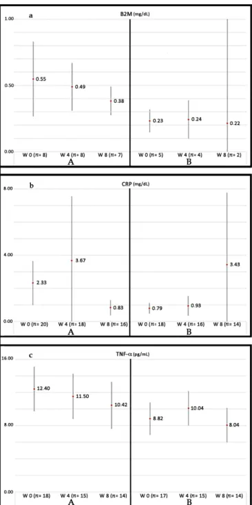
Currently, our group is conducting a phase III randomized controlled trial (RCT) in patients suffering from PAD without options for revascularization: Growth Hormone Angiogenic Study (GHAS), Eudract 2012-002228-34, approved by the Spanish Agency of Drugs and Health Products (AEMPs), and the Autonomic Committee on Research Ethics in Galicia (CAEIG, 2012/378), Spain, in which patients receive GH or placebo. Although this study is already finished and results from
it only began to be evaluated, early results show that within the markers used, TNF-αis the most frequently elevated in these patients (74%), followed byβ2-microglobulin (69%) and CRP (60%). Figure4depicts the graphic tendencies that show these patients depending on the group of treatment.
Int. J. Mol. Sci. 2018, 19, 290 14 of 40
frequently elevated in these patients (74%), followed by β2-microglobulin (69%) and CRP (60%). Figure 4 depicts the graphic tendencies that show these patients depending on the group of treatment.
Figure 4. Evolution of some biomarkers of inflammation analyzed in the GHAS study. Values are shown as the mean ± SD. (a) evolution of plasma levels of B2M (β-2-microglobulin) throughout the treatment with GH (A) or placebo (B); (b) evolution of plasma levels of CRP (C Reactive Protein) and (c) TNF-α, throughout the two groups of treatment. (A) and (B) represent different groups of treatment (GH or placebo, respectively). Note the tendency to decrease in group (A) as compared with the group (B). Patients from group (A) had significant higher basal levels of markers, indicating that patients in this group suffered from a more severe inflammatory disease as compared with group (B). Significant differences in the end of the study have not reached because of the small sample of patients still analyzed (note the differences in n). W = weeks of treatment. n= number of patients analyzed until now.
Figure 4. Evolution of some biomarkers of inflammation analyzed in the GHAS study. Values are shown as the mean±SD. (a) evolution of plasma levels of B2M (β-2-microglobulin) throughout the treatment with GH (A) or placebo (B); (b) evolution of plasma levels of CRP (C Reactive Protein) and (c) TNF-α, throughout the two groups of treatment. (A) and (B) represent different groups of treatment (GH or placebo, respectively). Note the tendency to decrease in group (A) as compared with the group (B). Patients from group (A) had significant higher basal levels of markers, indicating that patients in this group suffered from a more severe inflammatory disease as compared with group (B). Significant differences in the end of the study have not reached because of the small sample of patients still analyzed (note the differences inn). W = weeks of treatment.n= number of patients analyzed until now.
2.2. GH and Coronary Arterial Disease
The effects of GH-IGF-I in the incidence and prognosis of CAD are controversial.
As described before, GHD is associated with an increased prevalence of atherosclerosis, CAD, and stroke caused by an increased prevalence of atherosclerotic risk factors, such as alterations of body composition, lipid profile, and coagulation pattern [4,11,123], as shown in Figure5.
Int. J. Mol. Sci. 2018, 19, 290 15 of 40
2.2. GH and Coronary Arterial Disease
The effects of GH-IGF-I in the incidence and prognosis of CAD are controversial.
As described before, GHD is associated with an increased prevalence of atherosclerosis, CAD, and stroke caused by an increased prevalence of atherosclerotic risk factors, such as alterations of body composition, lipid profile, and coagulation pattern [4,11,123], as shown in Figure 5.
Figure 5. Effects of GH deficiency on atherosclerosis. GH: Growth Hormone, IGF-1: Insulin growth factor 1. NO: nitric oxide. Blue arrows indicate the effects produced by decreased GH secretion, while red arrows indicate how atherosclerosis is developed. Black arrows indicate increase or decrease, depending on the address of the arrow. Green arrow indicate that the lack of GH leads to decreased IGF-I secretion.
The changes in lipid profile observed in AGHD consist of increased in LDL and triglycerides, and decreased in high-density lipoprotein (HDL) (the latter observed only in women), with no differences in lipoprotein (a) [149]. GH replacement positively reverses this negative lipid profile. In addition, a decrease in CRP has been observed in these patients after GH therapy, while no clear changes seem to be produced in circulating triglycerides [149–151]. However, no study has determined whether GH has an additive effect that optimizes statin therapy; therefore, this remains an open question.
Regarding hypertension and peripheral resistance conflicting results have been reported in the literature [123]. Hypertension is quite frequent in GHD patients, and this condition results in impaired vasodilation responses to stress and/or exercise. As described, the GH–IGF-I axis reduces vascular tone by several mechanisms [152], although some vasoactive effects of GH may also have a central origin. In fact, GHD patients have markedly increased muscle sympathetic nerve activity, and GH replacement therapy has been shown to reduce it, suggesting an involvement of the GH-IGF-I axis in the autonomous sympathetic system regulation [153].
In some AGHD (patients with high base-line diastolic blood pressure, such as elderly GHD patients or those with previous Cushing disease), GH replacement reduces blood pressure, whereas in other patients (especially in young GHD patients), no changes in blood pressure have been shown [123,154].
Besides the cardiovascular risk factors mentioned above, GHD patients were shown to have increased blood vessel intima-media thickness (IMT) that is well known to represent one of the Figure 5.Effects of GH deficiency on atherosclerosis. GH: Growth Hormone, IGF-1: Insulin growth factor 1. NO: nitric oxide. Blue arrows indicate the effects produced by decreased GH secretion, while red arrows indicate how atherosclerosis is developed. Black arrows indicate increase or decrease, depending on the address of the arrow. Green arrow indicate that the lack of GH leads to decreased IGF-I secretion.
The changes in lipid profile observed in AGHD consist of increased in LDL and triglycerides, and decreased in high-density lipoprotein (HDL) (the latter observed only in women), with no differences in lipoprotein (a) [149]. GH replacement positively reverses this negative lipid profile. In addition, a decrease in CRP has been observed in these patients after GH therapy, while no clear changes seem to be produced in circulating triglycerides [149–151]. However, no study has determined whether GH has an additive effect that optimizes statin therapy; therefore, this remains an open question.
Regarding hypertension and peripheral resistance conflicting results have been reported in the literature [123]. Hypertension is quite frequent in GHD patients, and this condition results in impaired vasodilation responses to stress and/or exercise. As described, the GH–IGF-I axis reduces vascular tone by several mechanisms [152], although some vasoactive effects of GH may also have a central origin. In fact, GHD patients have markedly increased muscle sympathetic nerve activity, and GH replacement therapy has been shown to reduce it, suggesting an involvement of the GH-IGF-I axis in the autonomous sympathetic system regulation [153].
In some AGHD (patients with high base-line diastolic blood pressure, such as elderly GHD patients or those with previous Cushing disease), GH replacement reduces blood pressure, whereas in other patients (especially in young GHD patients), no changes in blood pressure have been shown [123,154].
Besides the cardiovascular risk factors mentioned above, GHD patients were shown to have increased blood vessel intima-media thickness (IMT) that is well known to represent one of the earliest morphological changes in the arterial wall in the process of atherogenesis [155]. It has to be highlight that femoral and carotid arteries IMT are independent predictors of CAD extent [156].
A decrease in IMT has been shown in several studies after the administration of GH to GHD patients [157]. Increases in IMT predict the development of symptomatic coronary disease, thus GH treatment may have a significant improvement in cardiovascular outcome, but this question has not been specifically analyzed in patients with GHD.
Regarding hard clinical endpoints, we previously commented on the increased risk of cardiovascular mortality in GHD patients. The worse cardiac risk profile (mainly hyperlipidemia) of these patients may explain part of the excess in CAD and mortality, but the studies do not allow for obtaining a definitive conclusion. As stated, GHD patients also has an increase in visceral fat, which decreases in response to GH therapy within six months after the initiation of the same, and it is maintained if the treatment is continued.
Despite all of these facts, there are no prospective, long-term randomized studies in AGHD patients comparing GH treatment to placebo on cardiovascular hard outcomes and mortality, and it is likely that there will never be such a study. A more recent and prospective trial found a lower mortality in GH treated hypopituitary patients when compared with a retrospective analysis of patients who had not been treated with GH [158]. However, again, the different time periods covered also included dramatic changes in the treatment of risk factors such as hypertension, diabetes mellitus, and hypercholesterolemia.
Some experimental models of CAD have shown the possible benefit of the GH-IGF-I axis on angiogenesis. For example, in rats with myocardial infarction (MI), inducing myocardial overexpression of IGF-I by delivering a human IGF-I gene by means of an adeno-associated viral vector led to angiogenesis [159]. This study demonstrated that the angiogenic process, measured by micro-SPECT-CT 16 weeks after administering the gene, persisted over time, leading to an improvement of the capillary network in rat hearts, a decreased left ventricle remodeling and an improved cardiac function. In rats with large MI, the early application of recombinant human GH (rhGH), starting three days after MI, attenuated LV remodeling without LV hypertrophy [160]. When administered a combination of GH and IGF-I, beneficial effects of early treatment have also been reported on infarct size, survival and cardiac gene expression after acute MI [161]. Curiously, these benefits were no observed when, instead of rhGH, recombinant rat GH (rrGH) was utilized [162]. This seems to be contradictory, as rhGH in rats might produce anti-GH antibodies and fail. Heterogeneous observations in experimental models can be explained by several facts, such as the early or late GH treatment, the different origin of HF, the different dosing regimens and short duration of treatment [89].
2.3. GH and Heart Failure
As stated, GH plays an important role during myocardial development that can easily be seen in untreated GHD children. They present cardiac atrophy with a reduction in the LV mass, ejection fraction, and cavity dimensions, as well as reduced cardiac output, high peripheral vascular resistance, and reduced functional capacity when compared with healthy controls of the same age, sex, and height [163]. When GHD appears in adults, it does not produce a reduction in cardiac mass, but cardiac performance and exercise capacity are impaired [164].
On the other hand, GH excess exerts different and opposite effects on the heart. In early-stage, it enhances cardiac performance, whereas it causes fibrosis and cardiac dysfunction in the intermediate-late phase. This apparent discrepancy is easily clarified: a physiological GH level or short-term excess exert positive inotropic effect; whereas long-term exposure to GH excess induces cardiac dysfunction and progression to heart failure by causing morphological and functional adaptive changes [165]. The most relevant histological abnormalities are interstitial fibrosis, reduced capillary density, increased extracellular collagen deposition, myofibril disorder, lympho-mononuclear infiltration, and myocyte death due to necrosis and apoptosis [165,166].
GH may regulate cardiac growth and metabolism by increasing protein synthesis (troponin I, myosin light chain-2, and actin), and cardiomyocyte size, increasing collagen synthesis and promoting cardiac hypertrophy [166–168]. IGF-I may reduce apoptosis of cardiomyocyte, thus preventing myocyte