Iranian Rehabilitation Journal, Vol. 11, No. 17, April 2013
Original Article
Dependency Coefficient in Computerized GALS Examination
Utilizing Motion Analysis Techniques
Hamed Shahidian
1; Ahmad Reza Arshi, PhD.
Amirkabir University of Technology, Tehran, IranFarhad Tabatabai Ghomshe, PhD.
University of Social Welfare and Rehabilitation Sciences, Tehran, Iran
Objectives: The GALS (Gait, Arms, Legs and Spine) examination is a compact version of standard procedures used by rheumatologists to determine musculoskeletal disorders in patients. Computerization of such a clinical procedure is necessary to ensure an objective evaluation. This article presents the first steps in such an approach by outlining a procedure to use motion analysis techniques as a new method for GALS examination.
Method: A 3D motion pattern was obtained from two subject groups using a six camera motion analysis system. The range of motion associated with GALS was consequently determined using a MATLAB program.
Results: The range of motion (ROM) of the two subject groups was determined, the validity of the approach was outlined, and the symmetry of movement on both sides of the body was quantified through introduction of a dependency coefficient.
Conclusion: Analysis of GALS examination and diagnosis of musculoskeletal problems could be addressed more accurately and reliably by adopting motion analysis techniques. Furthermore, introduction of a dependency coefficient offers a wide spectrum of prospective applications in neuromuscular studies .
Key words: Motion analysis, GALS examination, Musculoskeletal disorders, Dependency Coefficient
Submitted: 22 Oct 2012 Accepted: 20 Jan 2013
Introduction
Visual evaluation of joints is an integral part of human motion assessment. Implementation of cinematography in biomechanical studies using motion capture technologies made a tangible contribution to further developments of human motion analysis systems. This particular combination of software and hardware has found diverse applications in such areas as the military and computer vision. Motion analysis systems are also comfortably relied on by medical professionals in quantitative evaluation of musculoskeletal performance in rehabilitation, neurology and sports medicine. Individual disciplines, however, require tailored software for a more coherent quantitative analysis. Examples of dedicated tools for disciplinary applications are numerous. Software for 3D analysis of the musculoskeletal system has been developed by Leardini et al (1). The reliability and validity of standing balance measurements using motion analysis systems is discussed by Kejonen et al (2). Patient
positioning verification is also addressed utilizing real-time three dimensional motion analysis (3).
Einas (4) and his colleagues worked on pelvic skeletal asymmetry and its influence on trunk movement. The range of motion and effect of foot structure in musculoskeletal overuse injuries has also been studied (5). Prediction of patellar tendon reflex is another disorder which is evaluated by 3D analysis of human movements (6). The range of motion of human segments is a related parameter to musculoskeletal system and Schmidt et al (7) addressed the issue by investigating the unconstrained motion of wrist and elbow. Finger flexion and extension following a 3D video analysis has been presented by Rash (8). Other muscular parameters like belly length with a potential for the assessment of contracture has also been investigated by Fry et al (9).
disorders. However, the preliminary evaluation of patients is still subject to manual intervention by physiotherapists, rheumatologists and orthopedic surgeons. There are a number of slightly different routines for such an evaluation. The GALS examination (gait, arms, legs, and spine) has been validated as a new approach for screening musclo-skeletal disorders in primary care (10, 11, 12). Here, the sensitivity, reliability and specificity of this examination procedure have been investigated by physiotherapists to detect rheumatoid arthritis (13). This paper represents a novel approach in adopting a dedicated motion analysis system for automatic evaluation of a patient musculoskeletal condition through substitution of the visual segment of GALS examination.
Methods
A 3D motion pattern was obtained from two subject groups using a six camera motion analysis system. The visual evaluations constitute an integral and
critical part of the GALS examination. During these clinical assessments, the physician attempts to extract features associated with body segments; at the same time the whole body configuration is kept in mind. Here factors such as ROM (range of motion), swelling, deformity, smoothness and symmetry of movements, tenderness and gripping ability are assessed. The visual evaluation however, concentrates primarily on assessment of ROM for individual joints. In the following sections the development of a protocol for parameter estimation during these examinations is explained. A number of issues that define the existing tests such as GALS should also be taken into consideration. In the first instant, the objectives of the original test should be adhered to and both sides of the body should be assessed (13). Furthermore, no additional or external forces should be applied to the subject’s body during evaluation of the active range of motion. Table (1) presents the basic structure of this protocol.
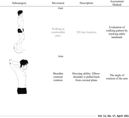
Table 1. Structure of the protocol developed for the Automatic GALS screening
Subcategory Movement Description Assessment Method
Gait
Walking at comfortable
pace 3D Gait Analysis
Evaluation of walking pattern by
tracking ankle landmark
Arm
Shoulder external rotation
Dressing ability: Elbow-shoulder is pulled back
from coronal plane
Subcategory Movement Description Assessment Method
Arm flexion Standing upright with arms hanging, the arm is then rotated upwards
The angle of rotation of the arm
Wrist flexion & extension
Arms hanging freely, hands are kept horizontally at right angles to arms, wrist
is rotated upwards and downwards
Wrist rotation
Leg
Knee flexion Lying on the couch, foreleg is free while thigh is brought up
The angle of knee flexion
Hip internal
rotation Passive internal rotation of individual hips Lateral rotation of foreleg
Ankle Dorsi & Plantar Flexion
Rotating foot from vertical position, moving back and
forth
Foot-foreleg angle
Subcategory Movement Description Assessment Method Waist Lateral
Bending
Waist Flexion
Keeping waist stationary, bending the upper extremity laterally
T10-S1 bending forward
Angle of motion of the T10-S1 line
T10-S1 forward angle The positioning of the passive or active markers
plays an important role in this screening protocol. Here the Helen-Hayse marker-set (15) is adopted for location of the markers. The other practical issue is what the patient wears during screening. Skin marking requires the male subjects to wear stretch shorts. The female subjects additionally wear a simple but specially prepared top which is similar to a kitchen apron with an open back.
The motion analysis system adopted for this study is a ‘six infrared cameras Vicon system’ with Vicon data station & workstation software. The motion was
captured at 60 fps. this speed is highly suitable for this type of movement. The results are in the form of Microsoft Excel Sheets. An M-File code is then prepared for Matlab R2007b. The code is responsible for accepting the motion analysis software output and provides the corresponding stick figures and the associated joint ROM.
For practical implementation of the protocol eight undergraduate Biomedical Engineering students at Amirkabir University of Technology (AUT) formed the two study groups. Table (2) illustrates the demographic profiles of the two subject groups. Table 2. Descriptive profiles of two study groups
Variable Male Female
Age (Yr) 20.25±0.5 19.25±0.5
Weight (Kg) 69.50±10.345 59.25±1.259
Height (Cm) 173.25±7.676 160.25±6.397
BMI (Kg/m2) 23.05452±1.894 23.14453±1.736
Results
The ROMs for the two subject groups was determined and tables 3 & 4 represent the subject data summary for male and female participants. Total average and
standard deviations were calculated for individual rows in tables (3) and (4). Here the average of female and male participants was determined separately for individual movements.
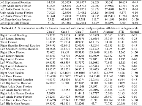
Case 1 Case 2 Case 3 Case 4 Average STD Normal Left Hip Internal Rotation 14.734 33.5029 34.6577 38.629 30.381 10.659 0-45 Right Ankle Dorsi Flexion 8.3628 41.5896 22.5732 27.309 24.9587 13.701 0-20 Left Ankle Dorsi Flexion 7.9059 47.0624 24.0755 30.878 27.4804 16.225 0-20 Right Ankle Plantar Flexion 7.3029 12.9306 17.4082 29.824 16.8665 9.577 0-50 Left Ankle Plantar Flexion 8.1441 16.4978 23.6389 34.735 20.75415 11.268 0-50 Left Knee in Gate Process 75.23 63.9687 85.781 111.7 84.1699 20.400 0-120 Left Hip in Gate Process 51.52 45.1286 62.2004 63.79 55.6597 8.886 0-80
Table 4. GALS examination results for females measured with motion analysis system. All data are in degree scale. Case 5 Case 6 Case 7 Case 8 Average STD Normal Right Lateral Bending 35.3272 27.0138 41.8696 30.0579 33.567 6.513 0-25 Left Lateral Bending 33.7315 27.3634 40.5171 31.6614 33.319 5.483 0-25
Waist Flexion 81.4195 73.2734 78.9933 63.1135 74.20 8.141 0-90
Right Shoulder External Rotation 29.9495 42.9042 52.0556 43.6244 42.135 9.123 0-45 Left Shoulder External Rotation 40.2838 34.6775 53.0795 49.1322 44.29 8.349 0-45 Right Elbow Flexion 73.062 88.854 98.3156 103.1699 90.850 13.265 0-150 Left Elbow Flexion 65.4103 74.5736 99.2266 89.4009 82.152 15.075 0-150 Right Wrist Flexion 56.7717 52.5711 61.2721 78.1051 62.18 11.195 0-60 Left Wrist Flexion 69.6553 68.8519 58.7572 86.1088 70.843 11.320 0-60 Right Wrist Extension 18.7698 58.7179 53.5452 46.5443 39.62 18.392 0-60 Left Wrist Extension 15.6217 53.1221 60.8213 59.4073 47.243 21.344 0-60 Right Knee Flexion 127.2142 126.1644 125.0407 117.1573 123.895 4.578 0-150 Left Knee Flexion 123.4088 126.6062 127.6217 114.5348 123.043 5.949 0-150 Right Hip Internal Rotation 44.6373 38.5630 35.2774 32.0348 37.316 6.544 0-45 Left Hip Internal Rotation 46.8059 36.304 42.3485 30.703 39.040 7.029 0-45 Right Ankle Dorsi Flexion - - 39.1348 29.6971 34.415 6.673 0-20 Left Ankle Dorsi Flexion 27.9981 14.4352 46.0564 27.0056 33.686 10.723 0-20 Right Ankle Plantar Flexion 7.3029 - 11.4411 18.7717 15.106 5.183 0-50 Left Ankle Plantar Flexion 24.2201 20.4623 36.9028 16.6293 25.917 10.242 0-50 Left Knee in Gate Process 113.6394 127.741 133.7182 61.98 109.269 32.630 0-120 Left Hip in Gate Process 69.8592 91.1451 78.2261 43.7 70.732 20.036 0-80
The segmental range of motion found in references is also presented in the last column of each row. Fig.
(1) represents the right and left lateral bending against one another.
Fig. 1: Right lateral bending against left lateral bending
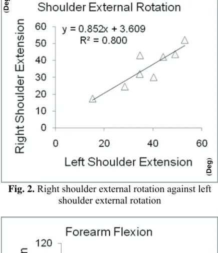
The left and right shoulder extensions are also illustrated in Fig. (2). Fig. (3) presents the forearm flexion data in both left and right sides. Figures (4 )
and (5) show wrist flexion and extension from both left and right sides. Furthermore, knee flexions in both sides are shown in Fig. (6).
(
Deg
)
Fig. 2. Right shoulder external rotation against left shoulder external rotation
Fig. 3. Right forearm flexion against left forearm flexion
Fig. 4. Right wrist flexion against left wrist flexion
Fig. 5. Right wrist extension against left wrist extension
Fig. 6. Right knee flexion against left knee flexion
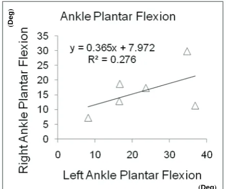
Internal rotation of the hip in both left and right sides are shown in Fig. (7). The ankle has two separate sets of data in plantar flexion and dorsi flexion as illustrated in Fig. (8) and Fig.(9). And Fig. (10) shows the left knee angle against left hip angle in gait analysis.
(Deg)
(Deg)
(Deg)
(
Deg
)
(
Deg
)
(
Deg
)
(
Deg
)
(
Deg
)
Fig. 7. Right hip internal rotation against left hip internal rotation
Fig. 8. Right ankle dorsi flexion against left ankle dorsi flexion
Fig. 9. Right ankle plantar flexion against left ankle plantar flexion
Fig. 10. Left knee angle against left hip angle in gait analysis
Discussion
In this paper a procedure based on motion analysis is presented as a new or an alternative means by which an important part of the GALS screening procedure can be performed. The current clinical procedure results in a predominantly experience-based and subjective grading arrived at by the physician. An automatic evaluation, however, could provide a far more reliable and repeatable result through objective screening. Here objectivity is obtained through motion analysis followed by an automatic comparison of the results against an accepted set of criteria (16), thus introducing a decision making platform using Matlab 2007 Rb to assist the physician a step further. The potential for addition of different algorithms to the automatic comparison stage is yet another benefit of this approach. For example, the symmetry of movement on both sides of the body could be quantified using a dependency coefficient ‘R’, which is a measure of asymmetry on individual body planes. This is exemplified by a dependency coefficient ‘R’, of waist lateral bending on both sides on the frontal plane, as shown in Fig. 10. This coefficient has values between 0 & 1, and the higher this value, the higher would be the symmetry. Higher values of R, on the other hand, are not necessarily associated with ROM. To exemplify this point, the spine and gait tests were taken to a diagnostic stage to see how the dependency coefficient (arrived at by automatic motion analysis) became clinically significant. In the case of subject 7, higher normal flexibility was encountered during waist lateral bending while the movement was quite symmetrical. Alternatively, in
(Deg)
(Deg)
(
Deg
)
(
Deg
)
(
Deg
)
(Deg)
(
Deg
)
the case of subject 4, the results of automatic assessment of GALS procedure was indicative of a lack of symmetry at the same time that higher than normal flexibility was observed. Lack of symmetry can be associated with shortening of quadratus lumborum. Alternatively, S shape scoliosis in both thoracic and lumbar areas could lead to limitations which are here manifested by a smaller than expected dependency coefficient. The torsion and shearing in pelvis, caused by sacro-iliac dysfunction, could also be considered as yet another reason for limitations in lateral bending.
In gait analysis two parameters were considered; knee angle and hip angle on sagittal plane for one complete cycle. A single side view analysis could be justified by the assumption that existence of any pathological states on one side directly affects both knee and hip angles on the other side. Cases 1, 2 and 3 in tables 3 and 4, could be considered as indications of center of mass swing deviation during the gait cycle which in turn, is an indication of knee compensation in response to weaknesses exhibited by the combination of hip and pelvis. Finally, there is a reasonable dependency between knee and hip angles in Fig. 10 that proves all the aforementioned explanations (17).
Understanding the functions affected by pathology and impairment may be critical in diagnosis. Furthermore, designing effective treatments for the prevention and cure of disabilities resulting from musculoskeletal diseases is very critical. Winter in 1990 explained that the joint mechanical power and energy reflect the underlying neuromuscular control mechanisms of human movement (18). Numerous possible solutions are required extremity kinematics (19). This flexibility in neuromuscular patterning
potentially allows one to ambulate effectively with impairments. The hip is used to compensate for weakness in knee extensor and/or ankle plantar flexor muscles of otherwise healthy (20). Gait compensations for hip muscle weakness can produce independent (i.e. successful) ambulation, although at a reduced speed as compared to normal gait (21).
Conclusions
Motion analysis provides the instrumentation necessary for an objective evaluation of GALS examination and diagnosis of musculoskeletal problems. Accuracy of medical diagnosis can be effectively altered by adopting a reliable and repeatable procedure using motion analysis techniques. Introduction of the concept of dependency coefficient could pave the way towards further neuromuscular investigations and the lack of symmetry could lead to personalized conditioning programs tailored for both healthy weaknesses and pathological states. Although implementation of such a technology might at first, seem time consuming, expensive, and require specialized technical support for medical professionals, further development of this approach will undoubtedly prove the system to be an invaluable asset. This is particularly tangible when a large group of people like the numbers encountered in health screenings for company staff is intended.
Acknowledgement:
This research has been supported by the University of Social Welfare and Rehabilitation Sciences, Department of Ergonomics, Biomechanics laboratory.
References
1. A. Leardini C. Belvedere L. Astolfi, S. Fantozzi M. Viceconti F. Taddei A. Ensini M.G. Benedetti F. Catani, A new software tool for 3D motion analyses of the musculo-skeletal system, Clinical Biomechanics 21 (2006) 870-879 2. Kejonen P, Kauranen K, Reliability and validity of standing
balance measurements with a motion analysis system, Physiotherapy, 88, 1, (2001) 25-32
3. Guido Baroni, Giancarlo Ferrigno, Roberto Orecchia, Antonio Pedotti, Real-time three-dimensional motion analysis for patient positioning verification, Radiotherapy and Oncology 54 (2000) 21-27
4. Einas Al-Eisa, David Egan, Kevin Deluzio, and Richard Wassersug, Effects of Pelvic Skeletal Asymmetry on Trunk Movement, SPINE Volume 31, Number 3, pp E71-E79 (2006)
5. Kenton R. Kaufman, Stephanie K. Brodine, Richard A. Shaffer, Chrisanna W. Johnson, Thomas R. Cullison, The
Musculoskeletal Overuse Injuries, Am J Sports Med September 1999 vol. 27 no. 5 585-593
6. L.K. Tham, N.A. Abu Osman, K.S. Lim, B. Pingguan-Murphy, W.A.B. Wan Abas, N. MohdZain, Investigation to predict patellar tendon reflex using motion analysis technique, Medical Engineering & Physics 33 (2011) 407-410
7. Joanne O. Crawford, ElpinikiLaiou, Anne Spurgeon, Grant McMillan, Musculoskeletal disorders within the telecommunications sector-A systematic review, International Journal of Industrial Ergonomics 38 (2008) 56-72
8. Ralf Schmidt, Catherine Disselhorst-Klug, Jiri Silny, Gunter Rau, marker-based measurement procedure for unconstrained wrist and elbow motions, Journal of Biomechanics 32 (1999) 615}621
measuring finger flexion and extension, Journal of Biomechanics 32 (1999) 1337-1341
10.N.R. Fry, C.R. Childs, L.C. Eve, M. Gough, R.O. Robinson, A.P. Shortland, Accurate measurement of muscle belly length in the motion analysis laboratory: potential for the assessment of contracture, Gait and Posture 17 (2003) 119-/124
11.Michael Doherty, Jane Dacre, Paul Dieppe, Michael Snaith, The 'GALS' locomotor screen, Annals of the Rheumatic Diseases 1992; 51: 1165-1169
12.MJ Plant, S Linton, E Dodd, P WJones, P T Dawes, The GALS locomotor screen and disability, Annals of the Rheumatic Diseases 1993; 52: 886-890
13.Karen A Beattie, Raja Bobba, ImaanBayoumi, David Chan, IngeSchabort, Pauline Boulos, Walter Kean, Joyce Obeid, Ruth McCallum, George Ioannidis, Alexandra Papaioannou and Alfred Cividino, Validation of the GALS musculoskeletal screening exam for use in primary care: a pilot study, BMC Musculoskeletal Disorders 2008, 9:115 14.Karen A.Beattie, NormaJ.MacIntyre, Jessica Pierobon,
Jennifer Coombs, Diana Horobetz, Alexis Petric, MaraPimm, WalterKean, MaggieJ.Larché, Alfred Cividino, The sensitivity, specificity and reliability of the GALS (gait, arms, legs and spine) examination when used by physiotherapists and physiotherapy students to detect rheumatoid arthritis, Physiotherapy (2011), doi: 10.1016/j.physio.2010.11.008
15.Tabakin D., Vaughan CL., A comparison of 3D gait models based on the Helen Hayes marker set, In: Proceedings of the sixth international symposium on the 3D analysis of human movement, 2000, p, 98-101.
16.Soucie JM, Wang C, Forsyth A, Funk S, Denney M, Roach KE, Boone D, and the Hemophilia Treatment Center Network. Range of motion measurements: reference values and a database for comparison studies. Haemophilia 2010; e-pub November 11, 2010.
17.Rodrigo S., García I., Franco M., Alonso-Vázquez A., Ambrósio, J., Energy expenditure during human gait. II - Role of muscle groups, Engineering in Medicine and Biology Society (EMBC), Aug. 31 2010-Sept. 4 2010, 4858 - 4861
18.Winter DA., Biomechanics and motor control of human movement, 2nd ed. New York: John Wiley and Sons; 1990. 19.Zajac FE., Understanding muscle coordination of the human
leg with dynamical simulations. Journal of Biomechanics 2002; 35: 1011-8.
20.Judge JO., Davis RB III, Ounpuu S., Step length reductions in advanced age: the role of ankle and hip kinetics, J Gerontol A BiolSci Med Sci 1996; 51:M303-12.