IJBC 2016; 8(4): 93-97
Relation between Estrogen and Progesterone Receptor Status with
p53, Ki67 and Her-2 Markers in Patients with Breast Cancer
Robab Sheikhpour1*, Fatemeh Poorhosseini2
1Hematology and Oncology Research Center, Shahid Sadoughi University of Medical Science, Yazd, Iran 2Department of Immunology, Kerman University of Medical Science, Kerman, Iran
A R T I C L E I N F O
Original article
Article History: Received: 11.08.2016 Accepted: 31.10.2016
Keywords: Breast cancer Estrogen receptor Her-2
p53 Ki67
Progesterone receptor
*Corresponding author: Robab Sheikhpour
Hematology and Oncology Research Center, Shahid Sadoughi University of Medical Science, Yazd, Iran
Tel: +98 913 1522462
Email: r.sheikhpour@yahoo.com
ABSTRACT
Background: Breast cancer is the most common cancer in women, containing approximately one third of all illnesses in women. Assessment of molecular markers is valuable in predicting the outcome of disease and decision making for optimal treatment. The purpose of this study was to determine the relationship between estrogen and progesterone receptors with Her-2, Ki67, P53, and clinicopathological factors in breast carcinoma.
Methods: 184 patients with breast cancer were chosen and immunohistochemistry was used for expression of p53 protein, Her-2, Estrogen receptor, Progesterone receptor and Ki67 in breast tissues. For statistical analysis, Pearson’s Chi-square tes and Spearman’s rho were used.
Results: Positive staining of estrogen receptor, progesterone receptor, Her-2, Ki67 and p53 was found in 63%, 53.8%, 54.6%, 56.2% and 42% respectively. Also there was reverse relation between estrogen receptor, progesterone receptor with Her-2 (P<0.05), but there was no relation between estrogen receptor and progesterone receptor with p53 and Ki67 (P>0.05). Also over-expression estrogen receptor was significantly associated with decreased lymph node metastasis and malignancy grade (P<0.05). Also over-expression of progesterone receptor was significantly associated with decreased malignancy grade (P<0.05).
Conclusion: Breast cancer progression is often associated with alterations in expressions of estrogen receptor, progesterone receptor, HER-2/neu, p53, and Ki67 and reverse association between hormones receptors and HER2 leads to lower or absent hormone receptors in women with HER2 positive breast cancers. Also positive estrogen receptor status can be associated with better survival in these patients.
Introduction
Breast cancer is the most common malignancy1 and
the leading cause of cancer death in women.2 It is one
of the most frequent cancers among Iranian women.3
Moreover, it is a biologically heterogeneous disease.2
The importance of several molecular markers in breast cancer are being evaluated in recent years.4 Assessment
of these biomarkers is valuable in predicting the outcome of disease and decision making for optimal treatment.5
Therefore, treatment decisions for breast cancer are commonly made based on the information derived from the immunohistochemistry (IHC) of biological markers,6 especially estrogen receptor (ER), progesterone
receptor (PR), Her2/neu, Ki67 andp53.4 Estrogen receptor
status and/or progesterone receptor status are useful as prognostic factors, although their importance lies more as predictors of response to endocrine therapy.
Patients with ER/PR positive tumors are hormone
Iranian Journal of Blood & Cancer
Journal Home Page: www.ijbc.ir
Please cite this article as: Sheikhpour R, Poorhosseini F. Relation between Estrogen and Progesterone Receptor Status with p53, Ki67 and Her-2 Markers in Patients with Breast Cancer. IJBC 2016; 8(4): 93-97.
responsive and therefore have a significantly better prognosis compared with patients whose tumors are ER/PR negative.7 Also studies showed Estrogen is an
important mitogen, exerting its activity by binding to its receptor. ER found in 50-80% of breast cancers.2
The proto-oncogene Her2-neu (c-erbB-2), localized to chromosome 17q21, encodes a transmembrane tyrosine kinase growth factor receptor. Her- 2/neu shares considerable homology with the epidermal growth factor receptor.7 It is overexpressed in 20-30% of patients with
breast cancer, with regard to its role as a prognostic and predictive factor. Although many studies have suggested that HER2 over-expression may be associated with a poor clinical outcome, other studies have not fully supported this observation.7 Also HER2 over-expression has been
associated with resistance to hormonal therapy in several studies.7 Ki-67, proliferation index,8 is a non-histone
nuclear protein that is closely linked to proliferating cells,9 and are associated with worse outcomes.10
High Ki-67 expression is associated with higher histological grade, larger tumor size, the presence of axillary lymph nodalmetastasis, shorter disease-free and overall survival in patients with breast cancer.9
Therefore, Ki67 has been suggested as a prognostic marker in patients with breast cancer. p53 as a 53 KD nuclear phosphoprotein,6,11-13 plays an important role in
many critical cellular events related to human aging and cancer, including DNA damage,14 telomere shortening
and oxidative stress.15 Also p53 is involved in regulating
cell proliferation and inducing apoptosis.16 Many studies
showed that overexpression of p53 protein in breast tumors can be associated with high cell proliferation and increased risk of progression.13 Therefore, we aimed
to evaluate the relation between expression of estrogen receptor and progesterone receptor with HER2, p53, and Ki67 in samples of breast tissue from 184 patients with breast cancer.
Patients and Methods
Breast cancer tissue was obtained from 184 patients with breast cancer after taking their consent during 2014-2015 from Hospitals in Yazd city, central Iran. Also this study was approved by the Ethics Committee and Research Committee of Yazd Research and Clinical Center for Infertility.
Following fixation, the specimens were embedded on wax paraffin and sliced to 4 µm in thickness for staining. The haematoxylin and eosin (H&E) as histological method was used to stain and analyze tissue sections. The histological grade of tumor is determined by Bloom and Richardson17 and modified by Elston.18
Immunohistochemical analysis was performed on specimens that were embedded on wax paraffin from the main tumors. 4 µm thick histological sections were mounted on poly-L-lysin coated slides. Then slides were dewaxed with xylene and rehydrated by decreasing the intensity of alcohol. For blocking endogenous peroxidase activity, sections were treated with 3% hydrogen peroxide for 15 min. Then, the slides were transferred to citrate buffer and boiled for 15 min in pH 9, but Her-2 in pH 6 in a microwave oven for antigen retrieval. Then, the sections were washed 3 times with phosphate buffered saline. For blocking non-specific binding sites, the slides were incubated in 1% BSA in phosphate buffered saline (PBS) for 20 min. Then, the sections were exposed to primary antibody.6 Table 1 shows the used primary antibodies.
Then, the secondary antibody sheep anti mouse, anti-Rabbit Horseradish peroxidase (ready to use) was used and in a later stage, sections were incubated with 3,3-diamino-benzidine tetrahydrochloride (Sigma). Then, the sections were counterstained with hematoxylinandand were washed in tap water, dehydrated, and mounted with glass cover slips. Negative control was performed by replacing the primary antibody with fetal bovine serum in each series.
Her-2 was categorized as positive or negative: 0=no staining, 1=weak, 2=moderate, and 3=strong staining. Positive staining was considered as a score of ≥1. ER and PR nuclear stains were categorized as positive or negative. The positive staining was considered as >1% of the cell nuclei stained. Ki67 staining was categorized as positive or negative. The positive staining was considered >20% of the cell nuclei. P53 staining was categorized as positive or negative. The positive staining was considered >5%.
Statistical analysis was performed using SPSS software, version 19. Initially, the correlation of each tumor marker was evaluated by Pearson and Chi-Square tests. For measuring the coefficient of concordance between two variables, Spearman’s rank correlation coefficient (Spearman’s rho) was used. Statistical significance was considered as P<0.05.
Results
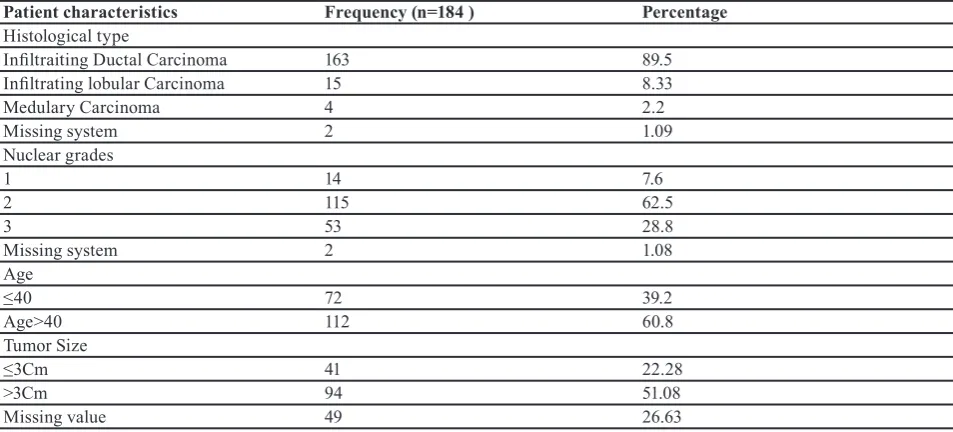
The results showed that ER and PR had the highest correlation among tumor markers. The characteristics of patients with breast cancer (histological type, nuclear grades, age and tumor size) are shown in table 2.
The result of this study showed that 68 (36.95%) patients were ER negative and 116 (63%) were positive. Frequency and percent of the immunohistochemical expression of steroid receptors, Her2/neu, Ki67 and p53 are shown in table 3.
Table 1: Antibodies used for immunohistochemical characterization of breast cancer patients
Antibody Isotype Dilution Source
ER Monoclonal mouse anti human1D5 Ready for use Dako
PR Monoclonal mouse anti human PGR 636 Ready for use Dako
Her2 Polyclonal Rabbit anti human c-erbB2 1: 400 Dako
P53 Monoclonal mouse anti human DO7 Ready for use Dako
Ki67 Monoclonal mouse anti human MIB-1 Ready for use Dako
The results of this study showed that there was reverse correlation between ER with Her-2 (P<0.01, r=-0.27). Moreover, there is a reverse relation between PR with Her-2 (P<0.01, r=-0.21). Relation between ER and PR with p53, Ki67 and Her-2 is shown in table 4.
We found a relationship between ER with grade (P<0.01, coefficient correlation=-0.28), PR with grade (P<0.05, r=-0.23) and relation between ER with Lymph nodes (P<0.05, r=-0.24). Table 5 shows relation between all biomarkers with grade and lymph nodes.
Moreover, the results showed that there is no correlation between ER with age (P=0.175) and PR with age (P=0.52).
Discussion
Tumor markers are molecules occurring in tissue that are associated with cancer and whose identification is useful in patient diagnosis, treatment or clinical management. Therefore determination of factors affecting clinicopathological features of breast cancer is important in improving insight toward this disease.5 In this study, the
immunohistochemical expression of tumor markers (ER, PR, HER- 2/neu, P53 and Ki67) in patients with breast cancer was different from other studies. This difference may be due to genetic differences, however other factors such as threshold for positivity are responsible for at least some of the differences.2 One study showed that 75% of
patients with ER+/ PR+, 40% of patients with ER+/ PR-,
25% of patients with ER-/ PR+ and 5% of patients with
ER-/PR- respond to endocrine therapy.19 Since ER+ breast
cancers are more commonly found in older women and screening mammograms are more frequently used in
older women, the detection method may have resulted in a greater relative increase in the age-adjusted rates for ER+ cancers than for ER- cancers. If mammogram
screening was positively correlated with environmental pollutants this could partially account for the correlation Table 2: Characteristics of breast cancer patients (histological type, nuclear grades, age and tumor size)
Patient characteristics Frequency (n=184 ) Percentage
Histological type
Infiltraiting Ductal Carcinoma 163 89.5
Infiltrating lobular Carcinoma 15 8.33
Medulary Carcinoma 4 2.2
Missing system 2 1.09
Nuclear grades
1 14 7.6
2 115 62.5
3 53 28.8
Missing system 2 1.08
Age
≤40 72 39.2
Age>40 112 60.8
Tumor Size
≤3Cm 41 22.28
>3Cm 94 51.08
Missing value 49 26.63
Table 3: Frequency of positive expression of steroid receptors, Her2/neu, Ki67 and p53 in 184 breast cancer patients
Biomarker Frequency Percent
ER 116 63.05
P53 71 38.5
Her2 100 54.6
Ki67 98 53.2
PR Positive 100 54.3
Table 4: Relation between ER with Her-2, PR, p53 and Ki67 in patients with breast cancer
Variables ER PR
P value P value
P53 98(-) 71(+)
0.435 0.302
Ki67 75(-) 98(+)
0.688 0.126
Her-2 83(-) 100(+)
0.007 0.004
P<0.05 is statistical significant
Table 5: Relation between biomarkers with grade and lymph nodes
Biomarker Lymph Nodes Grade
ER 0.045 0.002
PR 0.27 0.02
Ki67 0.35 0.085
Her2 0.51 0.28
P53 0.052 0.052
P<0.05 is statistical significant
observed between environmental pollutants and ER+ breast cancers.20
Also, we found a reverse association between hormones receptors and HER2 which led to lower or absent hormone receptors in women with HER2 positive breast cancers. This is one of the reasons why women who over-express HER2 may be resistant to tamoxifen.21 There are many
findings in agreement with our reports which showed that there is a reverse significant association between hormones receptors expression and HER2 over-expression.21 HER2
over expression may also correlate with resistance to hormonal therapy, sensitivity to anthracycline-based chemotherapy and resistance to CMF (cyclophosphamide, methotrexate, 5-FU).6 Moreover, HER2 over-expression is
associated with partial resistance to endocrine treatment. The complex cross-talk between ER and HER2 pathways might be an underlying cause of resistance, although the intrinsic biological mechanism is poorly understood.22
Another study did not find any association between ER expression and HER2 over-expression.23 Estrogen
receptor and PgR have an inverse relationship with Ki67 and when ER, PR decreased, Ki-67 increased.24 Ranade
and colleagues reported that ER and PR have a negative correlation with p53 protein.25 Therefore, increased ER
and PR were associated with resistance to apoptosis. Also our study showed that there is a reverse relation between ER with Lymph nodes and grade. Therefore it seems that ER-positive tumors are correlated with better survival than ER-negative tumors and decreased breast cancer mortality in afflicted patients. Rodriguez and co-workers found similar results26, while another study showed that
no correlation was found between ER/PR status and lymph node metastasis.2
Conclusion
In conclusion, these findings showed that breast cancer progression is often associated with alterations in expressions of ER, PR, HER-2/neu, p53 and Ki67. These changes might affect the treatment decision. The difference between positive expressions of these biomarkers with other studies may be due to genetic differences. However, other factors such as threshold of positivity are responsible for at least some of the differences. Reverse association between hormones receptors and Her-2 leads to lower or absent hormone receptors in women with HER2 positive breast cancers. Also ER positive correlated with better survival in breast cancer patients.
Conflict of Interest: None declared.
References
1. Sheikhpour R, Sarram MA, Sheikhpour R. Particle swarm optimization for bandwidth determination andfeature selection of kernel density estimation based classifiers indiagnosis of breast cancer. Applied Soft Computing. 2016; 40: 113–31. doi: 10.1016/j. asoc.2015.10.005.
2. Sofi GN, Sofi JN, Nadeem R, Shiekh RY, Khan FA, Sofi AA, et al. Estrogen receptor and progesterone
receptor status in breast cancer in relation to age, histological grade, size of lesion and lymph node involvement. Asian Pac J Cancer Prev. 2012; 13(10):5047-52. PMID: 23244108.
3. Mousavi SM, Montazeri A, Mohagheghi MA, Jarrahi AM, Harirchi I, Najafi M, et al. Breast cancer in Iran: an epidemiological review. Breast J. 2007; 13(4):383–91.doi: 10.1111/j.1524-4741.2007.00446.x. PMID: 17593043.
4. Ioannis M, Makovitzky J, Jeschke U, Volker, Friese K, Gerbe B. Expression of Her2/Neu, Steroid receptors (ER and PR), Ki67 and P53 in invasive mammary ductal carcinoma associated with ductal carcinoma in situ (DCIS) versus invasive breast cancer alone. Anticancer Res. 2005; 25(3A): 1719-23.
5. Moradi-Marjaneh M, Homaei-Shandiz F, S Shamsian SAA, Eftekhar-Zadeh Mashhadi I, Hedayati-Moghadam MR. Correlation of HER2/Neu over expression, P53 protein accumulation and steroid receptor status with tumor characteristics: An Iranian study of breast cancer patients. Iran J Public Health. 2008; 37(3): 19-28.
6. Sheikhpour R, Ghassemi N, Yaghmaei P, Mohiti Ardekani J, Shiryazd M. Immunohistochemical assessment of P53 protein and its correlation with clinicopathological characteristics in breast cancer patients. Indian J Sci Technol. 2014; 7(4): 472-9.
7. Cooke T, Reeves J, Lanigan A, Stanton P. HER2 as a prognostic and predictive marker for breast cancer. Ann Oncol. 2001; 12(Suppl 1): S23-S28. doi:10.1093/ annonc/12.suppl_1.S23.
8. Lebe B, Tuna B, Sis B, Yorukoglu K, Kargi A. Mdm2 and P53 expressions and Ki-67 proliferative index in fibrohistiocytic tumors. Aegean Pathol J. 2004; 1: 39–46.
9. Han JS, Cao D, Molberg KH, Sarode VR, Rao R, Sutton LM, Peng Y. Hormone receptor status rather than HER2 status is significantly associated with increased Ki-67 and p53 expression in triple-negative breast carcinomas, and high expression of Ki-67 but not p53 is significantly associated with axillary nodal metastasis in triple-negative and high-grade non-triple-negative breast carcinomas. Am J Clin Pathol. 2011; 135(2):230-7. doi: 10.1309/ AJCP9DV3EVZUATFV. PMID: 21228363.
10. Cheang MC, Chia SK, Voduc D, Gao D, Leung S, Snider J, et al. Ki67 index, HER2 status, and prognosis of patients with luminal B breast cancer. J Natl Cancer Inst. 2009; 101(10):736-50. doi: 10.1093/ jnci/djp082. PMID: 19436038. PubMed Central PMCID: PMC2684553.
11. Ageenko AI, Erkhov VS, Cherniaev LV, Volkova LIu. [A possible role of phosphoprotein p53 in the mechanism of autostimulation of tumor cell proliferation]. EkspOnkol. 1990; 12(1):35–7. PMID: 1688763.
12. Kerns BJ, Jordan PA, Moore MH, Humphrey PA, Berchuck A, Kohler ME, et al. P53 over expression in formalin-fixed, paraffin-embedded tissue detected
by immunohistochemistry. J Histochem Cytochem. 1992; 40(7):1047–51. PMID: 1607637.
13. Couture C, Raybaud-Diogène H, Têtu B, Bairati I, Murry D, Allard J, et al. P53 and Ki-67 as Markers of radioresistance in head and neck carcinoma. Cancer. 2002; 94(3): 713-22. doi: 10.1002/cncr.10232. PMID: 11857304.
14. Gu J, Spitz MR, Zhao H, Lin J, Grossman HB, Dinney CP, et al. Roles of tumor suppressor and telomere maintenance genes in cancer and aging— an epidemiological study. Carcinogenesis. 2005; 26(10):1741–7. doi: 10.1093/carcin/bgi126. PMID: 15905204.
15. Liu K, Bellam N, Lin HY, Wang B, Stockard CR, Grizzle WE, et al. Regulation of p53 by TopBP1: a potential mechanism for p53 inactivation in cancer. Mol Cell Biol. 2009; 29(10):2673–93. doi: 10.1128/ MCB.01140-08. PMID: 19289498. PubMed Central PMCID: PMC2682038.
16. Yamashita H, Nishio M, Toyama T, Sugiura H, Zhang Z, Kobayashi S, et al. Coexistence of HER2 over-expression and p53 protein accumulation is a strong prognostic molecular marker in breast cancer. Breast Cancer Res. 2004; 6(1):R24-30. doi: 10.1186/ bcr738. PMID: 14680497. PubMed Central PMCID: PMC314452.
17. Bloom HJG, Richardson WW. Histological grading and prognosis in breast cancer. Br J Cancer. 1957; 11:359–77. PMID: 13499785. PubMed Central PMCID: PMC2073885.
18. Elston CW. Grading of invasive carcinoma of the breast. In Page DL, Anderson TJ, editors. Diagnostic histopathology of the breast. Edinburgh: Churchill Livingstone; 1988
19. Shamsali A, Keyhanian SH, Gafari F. Relation between ER and PR with Her-2 and P53 protein in breast cancer patients. Infertil J. 2010; 27(1): 27. 20. St-Hilaire S, Mandal R, Commendador A, Mannel
S, Derryberry D. Estrogen receptor positive breast cancers and their association with environmental
factors. Int J Health Geogr. 2011; 10:32. doi: 10.1186/1476-072X-10-32. PMID: 21569288. PubMed Central PMCID: PMC3100231.
21. Gadelkarim Ahmed H, Al-Adhraei MA, Al-Thobhani AK. Correlations of hormone receptors (ER and PR), Her2/neu and P53 expression in breast ductal carcinoma among Yemeni women. Open Cancer Immunol J. 2011; 4(1): 1-9. doi: 10.2174/1876401001104010001. 22. Pinhel I, Hills M, Drury S, Salter J, Sumo G, A’Hern
R, et al. ER and HER2 expression are positively correlated in HER2 non-overexpressing breast cancer. Breast Cancer Res. 2012, 14(2):R46. doi: 10.1186/bcr3145. PMID: 22417870. PubMed Central PMCID: PMC3446380.
23. Anim JT, John B, AbdulSathar SA, Prasad A, Saji T, Akhtar N, et al. Relationship between the expression of various markers and prognostic factors in breast cancer. Acta Histochemica. 2005; 107(2): 87-93. doi: 10.1016/j.acthis.2005.01.002.
24. Nishimura R, Osako T, Okumura Y, Tashima R, YasuoToyozumi , Arima N. Changes in the ER, PgR, HER2, P53 and Ki-67 biological markers between primary and recurrent breast cancer: discordance rates and prognosis. World J Surg Oncol. 2011; 9:131. doi: 10.1186/1477-7819-9-131. PMID: 22004841. PubMed Central PMCID: PMC3214885.
25. Ranade KJ, Nerurkar AV, Phulpagar MD, Shirsat NV. Expression of survivin and P53 proteins and their correlation with hormone receptor status in Indian breast cancer patients. Indian J Med Sci. 2009; 63(11):481-90. doi: 10.4103/0019-5359.58877. PMID: 20075549.
26. Wang-Rodriguez J, Cross K, Gallagher S, Djahanban M, Armstrong JM, Wiedner N, et al. Male Breast Carcinoma: Correlation of ER, PR, Ki-67, Her2-Neu, and P53 with Treatment and Survival, a Study of 65 Cases. Modern Pathology. 2002; 15(8): 853-61. doi: 10.1097/01.MP.0000022251.61944.1D. PMID: 12181271.