I
IJJMMCCMM Original Article A
Auuttuummnn22001133,,VVooll22,,NNoo44
p53 and PCNA Expression in Keratocystic Odontogenic Tumors
Compared with Selected Odontogenic Cysts
Maryam Seyedmajidi1∗, Shima Nafarzadeh2, Sepideh Siadati3, Shahryar Shafaee4, Ali Bijani5,
Nazanin Keshmiri6
1. Dental Materials Research Center, Dental Faculty, Babol University of Medical Sciences, Babol, Iran.
2. Oral and Maxillofacial Pathology Department, Dental Faculty, Babol University of Medical Sciences, Babol, Iran.
3. Pathology Department, Medical Faculty, Babol University of Medical Sciences, Babol, Iran.
4. Cellular and Molecular Biology Research Center (CMBRC), Babol University of Medical Sciences, Babol, Iran.
5. Non-Communicable Pediatrics Diseases Research Center, Babol University of Medical Sciences, Babol, Iran.
6. Student Research Committee, Babol University of Medical Sciences, Babol, Iran.
p53 and PCNA expression in keratocystic odontogenic tumors compared with selected odontogenic cysts
Summary: The aim of this study was to evaluate p53 and PCNA expression in different odontogenic lesions
regarding their different clinical behaviors. Slices prepared from 94 paraffin-embedded tissue blocks (25
radicular cysts (RC), 23 dentigerous cysts (DC), 23 keratocystic odontogenic tumors (KCOT) and 23 calcifying
cystic odontogenic tumors (CCOT)) were stained with p53 and PCNA antibodies using immunohistochemistry
procedure. The highest level of p53 expression was in the basal layer of RC, and the highest level of PCNA
expression was in the suprabasal layer of KCOT. The differences of p53 expression in basal and suprabasal
layers as well as PCNA expression in the suprabasal layer were significant but there was no significant
difference in PCNA expression in the basal layer of these lesions. The expression of p53 in the basal layer of RC
was higher than in other cysts. This may be due to intensive inflammatory infiltration. Also, the high level of
PCNA expression in the suprabasal layer of KCOT may justify its neoplastic nature and tendency to recurrence.
KCOT and calcifying cystic odontogenic tumors did not show similar expression of studied biomarkers.
Key words: p53, PCNA, radicular cyst, dentigerous cyst, keratocystic odontogenic tumor, calcifying cystic
odontogenic tumor
∗
Corresponding author: P.O.Box 853, Dental Materials Research Center, Dental Faculty, Babol University of Medical Sciences, Babol, Iran. E-mail: ms_majidi79@yahoo.com
dontogenic cysts are common lesions in oral
cavity and in spite of similar
histo-pathological features have different clinical
behaviors. In rare cases, squamous cell carcinoma
in radicular cyst is reported. Also, epithelial
layer of dentigerous cyst may transform
to ameloblastoma, squamous cell carcinoma
or intraosseous mucoepidermoid carcinoma.
O
Submmited 8 Sep 2013; Accepted 20 Oct 2013
Odontogenic keratocyst (OKC) has specific
histopathological and clinical features, different
histopathogenesis and biological behavior. Many
authors believe that dentigerous and radicular cysts
enlarge because of an increase in intraluminal
osmotic pressure. However, enlargement of OKC
may be a result of unknown native epithelial factors
or enzymatic activity of fibroconnective tissue in
the cyst wall. In 2005, WHO workshop considered
parakeratinized OKC as a cystic neoplasm and
recommended the term Keratocystic Odontogenic
Tumor (KCOT). Pathogenic mechanisms that cause
growth and enlargement of KCOT includes high
proliferation rate, high expression of antiapoptotic
proteins like bcl-2 and expression of matrix
metalloproteinases (MMPs) 2 and 9. It seems that
KCOT has a neoplastic potential. Epithelial
dysplasia and squamous cell carcinoma may rarely
occur, but there are different findings about
ameloblastomatous changes (1-2).
Calcifying odontogenic cyst (COC) has a
variety of histopathological and clinical behaviors.
Most of the time, it seems like a non-neoplastic
cyst, whereas in other times it has no cystic
property which may be invasive or even malignant
and is therefore considered as a neoplasm. In
some conditions, it may also be seen with
other odontogenic tumors including odontoma,
adenomatoid odontogenic tumor and
amelo-blastoma. In WHO classification, COC and
all its subtypes are considered as odontogenic
tumor (1-2).
p53 protein is a product of Tp53 (Tumor
protein p53), a tumor suppressor gene that is
expressed in G1 phase of the cell cycle. It helps
DNA damage repairs and prevents cells to enter S
phase. If DNA is repaired, cell cycle arrest is ended.
But if DNA is not successfully repaired, p53
induces apoptosis and leads the cell to die. In the
absence or mutations of p53, DNA damage can not
be repaired and it leads to proliferation of damaged
cells and malignant transformation (3-4).
In normal non - stressed cells, p53 has a short
half-life of about 20 minutes. As a result of cellular
stress (e.g.DNA assault and damage), p53
undergoes posttranscriptional modifications. It is
protected from the effects of MDM2 (Murine
Double Minute 2) and its half-life increases (5-9).
The cellular proliferation increase has an
important role in the progression of odontogenic
cysts. p53 protein has an important role in
controlling the expression of cellular proliferation
inhibiting genes. Mutation in p53 gene can prevent
its inhibitory role, resulting in oncogenic activities
and neoplastic changes. Wild type p53 protein is
expressed in small amounts, has a short half-life
and cannot be detected using immunohistochemical
procedure. This detection can be possible in
conditions where the protein is expressed in large
amounts or is accumulated in mutant cells (10-11).
Overexpression of p53 in lesions without
mutation in the corresponding gene or even in
normal tissues was reported by Cruz et al. and Pillai
et al. In these tissues, positive results are due to the
persistence of wild type p53 protein by some
unknown reasons and consequently its
concen-tration increase leading to the possibility of its
detection by immunohistochemistry procedure
(12-13).
PCNA (Proliferating Cell Nuclear Antigen), is
a nuclear protein essential for nucleic acid
metabolism in DNA transcription and repair. It is
expressed in high amount in growing cells during
cell cycle. The expression of PCNA can be used as
a cell proliferative marker, because proliferating
cells remain for a longer time in G1/S phase (14).
Increased expression of PCNA may be stimulated
by growth factors or as a result of DNA injury in
the absence of cell cycle (15).
Considering the roles and effects of p53 and
PCNA in cells proliferation, this study was
performed to understand the behavior of epithelial
cells in different odontogenic cysts (radicular cyst
as an inflammatory cyst, dentigerous cyst as a
developmental cyst, calcifying cystic odontogenic
tumor and KCOT which are tumoral lesions with
cystic appearance) regarding their clinical behavior
differences, tendency to neoplastic transformation
and recurrence after treatment.
Materials and Methods
After the approval of the Institutional Ethics
Committee, 94 paraffin - embedded tissue blocks
including 25 radicular cysts, 23 dentigerous cysts,
23 KCOTs and 23 calcifying cystic odontogenic
tumors collected between March 2003 and February
2010, were obtained from the archives of Oral &
Maxillofacial Pathology Department of Babol
University of Medical Sciences, Babol, Iran.
Tissue blocks were cut into 5 microns slices,
stained with hematoxylin & eosin and reviewed by
Oral & Maxillofacial pathologist. Blocks containing
maximum epithelial length upon gross review were
selected and were cut into 3 microns slices. Tissue
slides were deparaffinized in xylene and rehydrated
through graded concentrations of ethanol (absolute
ethanol, 96% ethanol, 80% ethanol and 70%
ethanol).
After washing with tap water, the slides were
placed in sodium citrate buffer (PH=6.0) at 120ºC,
for 12 min. The slides were chilled at room
temperature for 15 min, rinsed in tap water and
placed in Tris Buffered Saline (TBS) for 5 min.
The slides were incubated in hydrogen peroxide for
10 min, then rinsed first in tap water then in TBS
for 5 min.
The slides were covered by monoclonal
antibody for p53 (Clone D07, Isotype; IgG2b
Kappa, DakoCytomation, Glostrup, Denmark) and
PCNA (Clone PC10, Isotype; IgG2a Kappa,
DakoCytomation, Glostrup, Denmark) for 1 h, then
rinsed in tap water and placed in TBS for 5 min.
After immunostaining, the slides were
coun-terstained with Meyer's hematoxylin, mounted with
entellan and coverslide and examined by light
microscopy (Olympus BX41, Shibuya-Ku, Tokyo,
Japan) under 400X magnification.
Breast carcinoma was used as the positive
control and omission of the primary antibody was
the negative control (11).
The brown stained nuclei of epithelial cells
were considered as positive. The percentages of
positive cells in the epithelium of cysts in basal and
suprabasal layers (100 cells in 10 high power fields)
were evaluated by two pathologists. The review of
microscopic slides was performed by using a
trinocular optic microscope and interobserver
agreement was seen in all cases.
A score index of 1, 2, 3, 4 and 5
corresponding to staining in < 1%, 1-10%, 11-33%,
34-66% and >67% of epithelial cells, was used
respectively (16).
The data were analyzed using Statistical
Package for the Social Sciences (version 17),
Kruskal Wallis test for comparing the mean
percentage of positive cells in four groups and
Wilcoxon Signed Ranks test for comparing the
mean percentage of positive cells in basal and
suprabasal layers between four groups. P<0.05 was
considered as significant.
Results
The percentage of p53 and PCNA stained cells
in basal and suprabasal layers of radicular cyst,
dentigerous cyst, KCOT and calcifying cystic
odontogenic tumor (CCOT) are shown in Table 1.
Figures 1-4 are the representatives of
immuno-histochemical stainings.
The highest level of p53 expression was in the
basal layer of radicular cyst followed by KCOT and
dentigerous cyst and the least amount belonged to
CCOT. The same order was true in suprabasal
layers. The highest level of PCNA expression was
seen in suprabasal layers of KCOT, followed by
radicular cyst, CCOT and dentigerous cyst. This
result was also correct for the basal layer (Chart 1).
The percentage of p53 and PCNA stained cells
in the basal and suprabasal layers of four types of
Chart 1. p53 and PCNA expression in basal and suprabasal
layers in different odontogenic cysts.
cysts were compared using Kruskal-Wallis test. The
differences between PCNA expression in the basal
layer of these cysts were not significant (P=0.090)
but the differences between the expression of P53
in both basal and suprabasal layers of these cysts as
well as the differences between the expression of
PCNA in the suprabasal layer of these cysts were
significant (P=0.008, P=0.031 and P= 0.009,
respectively).
The percentages of stained cells for p53 and
PCNA were compared in the basal and suprabasal
layers of each cyst, using Wilcoxon Signed Ranks
test. P53 expression in the basal layer of radicular
Table 1. The expression of p53 and PCNA in odontogenic cysts based on percentage of stained cells in basal
and suprabasal layers
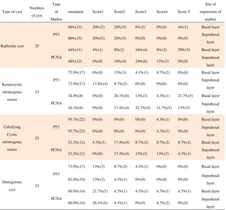
Site of expression of marker Score 5 Score4 Score3 Score2 Score1 unstained Type of Marker Numbers of cyst Type of cyst
Basal layer 4%(1) 0%(0) 8%(2) 20%(5) 20%(5) 48%(12) P53 25 Radicular cyst Suprabasal layer 0%(0) 0%(0) 0%(0) 20%(5) 20%(5) 60%(15) Basal layer 20%(5) 8%(2) 16%(4) 8%(2) 4%(1) 44%(11)
PCNA Suprabasal
layer 0%(0) 12%(3) 24%(6) 16%(4) 0%(0) 48%(12) Basal layer 0%(0) 8.7%(2) 4.3%(1) 13%(3) 0%(0) 73.9%(17) P53 23 Keratocystic odontogenic tumor Suprabasal layer 0%(0) 0%(0) 0%(0) 8.7%(2) 17.4%(4) 73.9%(17) Basal layer 21.7%(5) 4.3%(1) 13%(3) 26.1%(6) 0%(0) 34.8%(8)
PCNA Suprabasal
layer 13%(3) 21.7%(5) 21.7%(5) 17.4%(4) 0%(0) 26.1%(6) Basal layer 0%(0) 4.3%(1) 0%(0) 0%(0) 0%(0) 95.7%(22) P53 23 Calcifying Cystic odontogenic tumor Suprabasal layer 0%(0) 4.3%(1) 0%(0) 0%(0) 0%(0) 95.7%(22) Basal layer 8.7%(2) 8.7%(2) 8.7%(2) 17.4%(4) 4.3%(1) 52.2%(12)
PCNA Suprabasal
layer 4.3%(1) 13%(3) 13%(3) 17.4%(4) 0%(0) 52.2%(12) Basal layer 0%(0) 0%(0) 4.3%(1) 8.7%(2) 13%(3) 73.9%(17) P53 23 Dentigerous cyst Suprabasal layer 0%(0) 0%(0) 0%(0) 4.3%(1) 13%(3) 82.6%(19) Basal layer 4.3%(1) 4.3%(1) 4.3%(1) 4.3%(1) 21.7%(5) 60.9%(14)
PCNA Suprabasal
layer 0%(0) 8.7%(2) 0%(0) 4.3%(1) 26.1%(6) 60.9%(14)
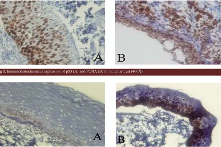
Fig 1. Immunohistochemical expression of p53 (A) and PCNA (B) in radicular cyst (400X).
Fig 2. Immunohistochemical expression of p53 (A) and PCNA (B) in keratocystic odontogenic tumor (400X).
Fig 3. Immunohistochemical expression of p53 (A) and PCNA (B) in calcifying cystic odontogenic tumor (400X).
Fig 4. Immunohistochemical expression of p53 (A) and PCNA (B) in dentigerous cyst (400X) (400X).
cyst, dentigerous cyst and KCOT was higher than
in the suprabasal layer (P=0.007, 0.024 and 0. 025,
respectively), but there was no significant
difference in CCOT (P=1.000).
In radicular cyst, PCNA expression in the
basal layer was higher than in the suprabasal layer
(P=0.003), but there was no significant difference
in other cysts (KCOT, CCOT and dentigerous cyst,
P=0.188, P=0.705 and P= 0.083, respectively).
Therefore, the highest level of p53 and PCNA
expression was found in the basal layer of radicular
cyst and the suprabasal layer of KCOT,
respectively.
Discussion
We studied the expression of p53 and PCNA
in some odontogenic cysts. p53 expression was
higher in the basal layer of radicular cyst, followed
by KCOT. PCNA expression was higher in
the suprabasal layer of KCOT, followed by
radicular cyst.
p53 expression in basal and suprabasal layers
and PCNA expression in suprabasal layers between
these cysts were significantly different but this was
not true for PCNA expression in the basal layer of
these cysts.
The results of this study showed that radicular
cyst and KCOT had a high percentage of p53
positive cells in their basal layer. In KCOT, the
suprabasal layer showed the highest expression of
PCNA. These unique findings in KCOT pointed
that proliferative components in this lesion are in
basal and suprabasal layers. This finding may
justify the neoplastic nature of KCOT and it may
also explain the clinical behavior and tendency to
recurrence of this lesion. There are some studies
with different results that evaluated the expression
of p53 and PCNA for understanding epithelial cells
behavior in different odontogenic cysts.
According to Mighell et al., the defferent
results are due to complex biology of p53 and
PCNA, histological preparation and
immuno-histochemical grading protocol used. Although, the
type of lesion, etiology and clinical behavior of
lesions should be taken into consideration (11).
Carvalhais et al. studied p53 expression in
odontogenic cysts and tumors. They did not find
any positive cells (17).
The relationship between p53 expression and
cell proliferation was showen in de Oliveira et al.'s
study. They concluded that p53 expression was
seen in proliferating cells, but it accumulated due to
several factors such as cellular stress. Also, they
showed that in radicular and dentigerous cysts, the
expression of p53 and PCNA is a response to
cellular stress resulting from inflammation, even in
the cases of developmental cyst such as
dentige-rous cyst (11).
Studies showed that growth factors and
cytokines (Interleukin 1, Interleukin 6 and tumor
necrosis factor) are released in inflammatory
processes. Inflammation may cause cell
pro-liferation and inflammatory cytokines may produce
cellular stress (18).
In dentigerous cyst, the inflammatory stimulus
may be the result of the eruption process that
caused cellular proliferation which might be
present for a short time (11). This could explain the
lower expression of p53 and PCNA in dentigerous
cyst. It was not proven that inflammatory stimuli
could cause dentigerous cyst, but usually in the
connective tissue of cyst, there was an
inflammatory infiltration that might have caused
epithelial cells proliferation.
In radicular cyst, the inflammatory stimuli are
a result of persistent bacterial contaminations of the
root canal. This has an effect on epithelial cells,
especially in the basal layer and produces the
increase and maintenance of the proliferative
activity (11).
Therefore, the high expression of p53 and
PCNA in radicular cysts is a reflection of the
cellular stress and proliferation induced by
inflammation that can inhibit the degradation of
p53 and increase the level of PCNA (11).
The relationship between inflammation and
cell proliferation in KCOT was studied by de Paula
et al. They used AgNOR staining, Ki67 and PCNA
immunohistochemistry for their analysis and
concluded that the expression of biomarkers in this
lesion showed a cellular proliferation pattern
consistent with neoplastic cells and independent of
inflammation. The high levels of p53 and PCNA
expression in the suprabasal layer, can explain why
KCOT has a proliferation and maturation pattern
that differs from other lesions. These findings may
explain its different clinical behaviors and the
tendency of recurrence (19).
Kaplan and Hirschberg studied the areas with
and without inflammation in KCOT and did not
find a significant difference in their proliferation
rate. They concluded that inflammation in KCOT
did not have any effect on its proliferative potential
(20). Also, in the present study all of KCOTs were
non-inflamed.
Gallana-Alvarez and Wagner showed that the
neoplastic cells in calcifying odontogenic cyst may
have an increasing proliferative potential.
Calcifying odontogenic cyst is considered as a
tumoral lesion and the presence of mutant proteins
should be taken into consideration. P53 expression
may be related to the proliferation rate in this
lesion (21-22).
However, in the present study, there was no
high expression of p53 and PCNA in CCOT in
comparison to other cysts. This differs from the
study of de Oliveira who found a high level of p53
and PCNA in calcifying odontogenic cysts (11).
In our study, a significant difference between the
percentages of stained cells was found for both
markers in basal and suprabasal layers of radicular
cyst, with more stained cells in basal layer,
demonstrating that in radicular cyst, basal layer is
the proliferative component. In KCOT and
dentigerous cyst, a difference between basal and
suprabasal layers was only seen for e P53
expression and no significant difference for PCNA
expression was found, indicating a similar
expression of PCNA in both basal and suprabasal
layers of dentigerous cyst and KCOT. This can be
related to the similar proliferative potential of the
two layers in these lesions.
The study of Wagner was performed on
radicular cyst, dentigerous cyst and KCOT. p53 was
just expressed in KCOT indicating different clinical
behaviors of this cyst (23).
In the present study, the high expression of PCNA
in the suprabasal layer of KCOT in comparison to
other cysts indicates that the suprabasal layer of this
cyst is a proliferative component and explains the
different growth patterns. Also, the high expression
of p53 in basal layer of radicular cyst indicates that
inflammatory stimuli produced from persistent
bacterial contamination of root canal could induce
cellular stress (11).
In KCOT, PCNA expression may show a
proliferative pattern similar to neoplastic cells.
On the other hand, the presence of mutant p53 in
KCOT must be taken in consideration. This was
explained by Gonzales-Moles using antibodies
specific to mutant p53 (24).
In KCOT, the epithelium has a little intrinsic
growth potential that is not seen in epithelial cells
of other cysts (1). Li et al. demonstrated that the
epithelium of KCOT has a suprabasal proliferative
component (15). The number of positive cells for
PCNA in the epithelium of KCOT were
significantly higher than dentigerous and radicular
cysts. This finding was described by Li et al. and
Piattelli et al. (15, 25).
p53 expression in the basal layer of radicular
cyst was more than other cysts. It might be a result
of inflammation. Also, the high expression of
PCNA protein in the suprabasal layer of KCOT
might be a result of clinical behavior and its
tendency to recurrence. On the other hand, this
lesion showed a proliferation pattern different from
those found in other lesions (11). Two tumoral
lesions, KCOT and CCOT were not similar in p53
and PCNA expression.
Conflict of interest
Authors declared no conflict of interest.
References
1. Damm DD, Bouquot JE, Neville BW, et al. Oral
and Maxillofacial Pathology. 3 ed. Missouri: WB
Saunders 2009:116-20, 590-2, 594-6.
2. Regezi JA, Sciubba JJ, Jordan RCK. Oral
pathology, clinical pathology correlations. 6 ed.
Missouri Saunders Elsevier; 2011:246-60.
3. Levine AJ. p53, the cellular gatekeeper for
growth and division. Cell 1997;88:323-31.
4. Nylander K, Dabelsteen E, Hall PA. The p53
molecule and its prognostic role in squamous cell
carcinomas of the head and neck. J Oral Pathol Med
2000;29:413-25.
5. Olson MO. Sensing cellular stress: another
new function for the nucleolus? Sci STKE
2004;2004:pe10.
6. Olson MO, Dundr M. The moving parts of the
nucleolus. Histochem Cell Biol 2005;123:203-16.
7. Stricker TP, Kumar V. Neoplasia. In: Kumar V,
Abbas AK, Fausto N, et al. (eds). Robbins and
Cotran pathologic basis of disease 8ed.
Philadelphia: Saunders Elsevier Inc; 2010:290-2.
8. Agarwala SS. Paraneoplastic syndromes. Med
Clin North Am 1996;80:173-84.
9. Chang F, Syrjanen S, Syrjanen K. Implications of
the p53 tumor-suppressor gene in clinical oncology.
J Clin Oncol 1995;13:1009-22.
10. Ozveren A, Tuskan C, Yaltirik M, et al.
Expression or the tumor suppressor gene p53 in
odontogenic cyst. Turk J Med Sci 2003;33:243-7.
11. de Oliveira MG, Lauxen Ida S, Chaves AC, et
al. Immunohistochemical analysis of the patterns of
p53 and PCNA expression in odontogenic cystic
lesions. Med Oral Patol Oral Cir Bucal
2008;13:E275-80.
12. Cruz IB, Snijders PJ, Meijer CJ, et al. p53
expression above the basal cell layer in oral mucosa
is an early event of malignant transformation and
has predictive value for developing oral squamous
cell carcinoma. J Pathol 1998;184:360-8.
13. Pillai G, Roberts H, Gatter K, et al. p53
expression in normal paraffin-embedded tissue
using different antibodies and antigen retrieval
buffer systems. Histopathology 2003;42:83-7.
14. Kelman Z. PCNA: structure, functions and
interactions. Oncogene 1997;14:629-40.
15. Li TJ, Browne RM, Matthews JB.
Quantification of PCNA+ cells within odontogenic
jaw cyst epithelium. J Oral Pathol Med
1994;23:184-9.
16. Allred DC, Harvey JM, Berardo M, et al.
Prognostic and predictive factors in breast cancer
by immunohistochemical analysis. Mod Pathol
1998;11:155-68.
17. Carvalhais J, Aguiar M, Araujo V, et al. p53
and MDM2 expression in odontogenic cysts and
tumours. Oral Dis 1999;5:218-22.
18. Hudson JD, Shoaibi MA, Maestro R, et al.
A proinflammatory cytokine inhibits p53
tumor suppressor activity. J Exp Med
1999;190:1375-82.
19. de Paula AM, Carvalhais JN, Domingues MG,
et al. Cell proliferation markers in the odontogenic
keratocyst: effect of inflammation. J Oral Pathol
Med 2000;29:477-82.
20. Kaplan I, Hirshberg A. The correlation between
epithelial cell proliferation and inflammation
in odontogenic keratocyst. Oral Oncol
2004;40:985-91.
21. Reyes D, Villanueva J, Espinosa S, et al.
Odontogenic calcificant cystic tumor: a report of
two clinical cases. Med Oral Patol Oral Cir Bucal
2007;12:E126-9.
22. Gallana-Alvarez S, Mayorga-Jimenez F,
Torres-Gomez FJ, et al. Calcifying odontogenic cyst
associated with complex odontoma: case report and
review of the literature. Med Oral Patol Oral Cir
Bucal 2005;10:243-7.
23. Wagner Y, Filippi A, Kirschner H, et al.
[Cytokeratin and p53 expression of odontogenic
cysts]. Mund Kiefer Gesichtschir 1999;3:263-9.
24. Gonzalez-Moles MA, Mosqueda-Taylor A,
Delgado-Rodriguez M, et al. Analysis of p53
protein by PAb240, Ki-67 expression and human
papillomavirus DNA detection in different types
of odontogenic keratocyst. Anticancer Res
2006;26:175-81.
25. Piattelli A, Fioroni M, Santinelli A, et al. P53
protein expression in odontogenic cysts. J Endod
2001;27:459-61.