Open Access
Research
Identification of genes down-regulated during lung cancer
progression: A cDNA array study
Mara Campioni
1, Vincenzo Ambrogi
2, Eugenio Pompeo
2, Gennaro Citro
3,
Mauro Castelli
4, Enrico P Spugnini
3, Antonio Gatti
2, Pierluigi Cardelli
3,
Laura Lorenzon
5, Alfonso Baldi*
1and Tommaso C Mineo
2Address: 1Department of Biochemistry and Biophysics "F. Cedrangolo", Section of Pathology, Second University of Naples, Naples, Italy, 2Department of Thoracic Surgery and Department of Anaesthesiology, Tor Vergata University, Rome, Italy, 3S.A.F.U. Department, CRS, "Regina
Elena" Cancer Institute, Rome, Italy, 4Experimental Oncology Department, "Regina Elena" Cancer Institute, Rome, Italy and 5Department of
Surgery "A", Second Faculty of Medicine, "La Sapienza" University, Rome, Italy
Email: Mara Campioni - [email protected]; Vincenzo Ambrogi - [email protected]; Eugenio Pompeo - [email protected]; Gennaro Citro - [email protected]; Mauro Castelli - [email protected]; Enrico P Spugnini - [email protected]; Antonio Gatti - [email protected]; Pierluigi Cardelli - [email protected]; Laura Lorenzon - [email protected]; Alfonso Baldi* - [email protected];
Tommaso C Mineo - [email protected] * Corresponding author
Abstract
Background: Lung cancer remains a major health challenge in the world. Survival for patients with stage I disease ranges between 40–70%. This suggests that a significant proportion of patients with stage I NSCLC may actually be under-staged.
Methods: In order to identify genes relevant for lung cancer development, we carried out cDNA array experiments employing 64 consecutive patients (58 men and 6 women) with a median age of 58 years and stage 1 or stage 2 non-small-cell lung cancer (NSCLC).
Results: Basic cDNA array data identified 14 genes as differentially regulated in the two groups. Quantitative RT-PCR analysis confirmed an effective different transcriptional regulation of 8 out of 14 genes analyzed. The products of these genes belong to different functional protein types, such as extra-cellular matrix proteins and proteases (Decorin and MMP11), genes involved in DNA repair (XRCC1), regulator of angiogenesis (VEGF), cell cycle regulators (Cyclin D1) and tumor-suppressor genes (Semaphorin 3B, WNT-5A and retinoblastoma-related Rb2/p130). Some previously described differences in expression patterns were confirmed by our array data. In addition, we identified and validated for the first time the reduced expression level of some genes during lung cancer progression.
Conclusion: Comparative hybridization by means of cDNA arrays assisted in identifying a series of novel progression-associated changes in gene expression, confirming, at the same time, a number of previously described results.
Published: 15 September 2008
Journal of Experimental & Clinical Cancer Research 2008, 27:38 doi:10.1186/1756-9966-27-38
Received: 28 July 2008 Accepted: 15 September 2008
This article is available from: http://www.jeccr.com/content/27/1/38
© 2008 Campioni et al; licensee BioMed Central Ltd.
Introduction
Lung cancer remains a major health challenge in the world. Despite improvements in staging and the inte-grated application of surgery, radiotherapy, and chemo-therapy, the 5-year survival rate for individuals with lung cancer is only about 15% [1]. Histologically, 80% of the lung cancers are diagnosed as non-small-cell lung cancer (NSCLC), whereas the remaining 20% of cases are diag-nosed as small-cell lung cancer (SCLC). On the basis of cell morphology, adenocarcinoma and squamous cell car-cinoma are the most common types of NSCLC. The cur-rent staging system for NSCLC is based upon the size and location of the primary tumor (T), the involvement of regional lymph nodes (N), and the presence of distant metastases (M) [1]. The standard treatment of patients with stage I NSCLC (T1-2, N0, M0) is resection of the pri-mary tumor alone (no adjuvant therapy) [2]. Survival for patients with stage I disease ranges between 40–70%, and the failure is due to distant recurrences [3]. This suggests that a significant proportion of patients with stage I NSCLC may actually be under-staged. Therefore, if cor-rectly identified, these patients may benefit from adjuvant therapy in addition to resection, with a predictable improvement in the survival rates. Indeed, to identify patients with stage I NSCLC who might benefit from adju-vant therapy, investigators have attempted to identify fac-tors predicting poor prognosis. These studies included analysis of performance status, histologic subtype, size of the primary tumor, the degree of tumor differentiation, mitotic rate, and evidence of lymphatic or vascular inva-sion [4-8]. However, all of these factors have failed, to date, to precisely identify a group of stage I patients who would benefit from adjuvant therapy. Cigarette smoking remains the main risk factor for lung cancer, accounting for about 90% of the cases in men and 70% of the cases in women [9]. Our research group has investigated in the last years the possible involvement of several molecular mech-anisms, such as cell cycle and apoptosis regulators, onco-genes and tumor suppressor onco-genes, cell adhesion molecules, in the pathogenesis and progression of lung cancer [10-19].
In this study, we utilized the cDNA array technique to identify genes differently expressed in patients at early stage of NSCLC.
Materials and methods
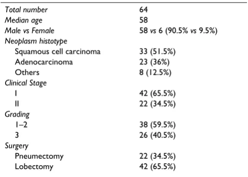
Patient selectionTable 1 summarizes the characteristics of the patients enrolled in the study. Subjects selected for the analysis were 64 patients, consecutively treated at the Department of Thoracic Surgery of the Tor Vergata University in Rome, who had radical surgery for NSCLC at pathological stage 1 or 2. The study project was submitted and approved by the Human Tissue Use Committee of the University. The
patients were staged according to operative and patholog-ical findings based on AJCC/UICC-TNM classification and stage grouping [17]. N-factor was assessed on lymph nodes removed during routine mediastinal lymphadenec-tomy. A preoperative staging computed tomography (CT) scan was performed in all patients. Histology grading and N-stage were performed on haematoxylin-eosin stained sections. In 22 patients the neoplasm was resected by pneumonectomy and in 42 by lobectomy. Patients who did not survive beyond 60 days after surgery were not included in the study to avoid bias from peri-operative death. Patients who underwent minimal resection were ruled out from the present analysis. Another mandatory prerequisite, was the lack of chemo or radiotherapy before and after surgical resection.
RNA extraction
Total RNA from frozen tumour tissues was extracted uti-lizing the Atlas™ Pure Total RNA Extraction System (CLONTECH). RNAs were quantified spectrophotometri-cally and their integrity confirmed by fractionation of 1 μg of RNA on 1% agarose gel with ethidium bromide stain-ing. Then, two populations of RNAs were prepared, including exactly 1 μg of RNA from each patients, separat-ing patients in stage 1 and stage 2.
Array hybridization
We prepared cRNA for hybridization using the Atlas™ Pure Total RNA labeling System (Clontech). The Clontech Atlas™ Human Cancer Array, composed of 588 human cDNAs, was used for hybridization according to the man-ufacturer's instructions. Arrays were scanned using a Phos-phorImager and analyzed by ImageQuant 5.0 software (Molecular Dynamics, Sunnyvale, California). The exper-iment was performed in duplicate, utilizing two different RNA preparations. Intensity values were normalized using the expression levels of the housekeeping genes spotted
Table 1: Patients' Characteristics
Total number 64
Median age 58
Male vs Female 58 vs 6 (90.5% vs 9.5%) Neoplasm histotype
Squamous cell carcinoma 33 (51.5%) Adenocarcinoma 23 (36%) Others 8 (12.5%) Clinical Stage
I 42 (65.5%)
II 22 (34.5%)
Grading
1–2 38 (59.5%)
3 26 (40.5%)
Surgery
on the arrays. A cut-off of two folds was used to select the genes differentially expressed.
Quantitative real-time reverse transcription-PCR
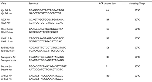
The primer sequences are shown in table 2. Primers were designed using Primer Express 2.0 software (Applied Bio-system, Foster City, CA, USA). The specificity of each tar-get amplicon was assessed by dissociation curve analysis and all amplicons were spanning over exon-exon regions to avoid genomic amplification. qPCR was performed on an ABI PRISM 7900HT Sequence Detection System (Applied Biosystems, CA, USA) in 384 well plates assem-bled by Biorobot 8000 (Qiagen, Hilden, Germany) using a final volume of 20 μl and the following cycle conditions: 50°C for 2 min., 95°C for 10 min., and then 40 cycles of 15 s at 95°C and 1 min at 60°C. All QPCR mixtures con-tained 1 μl of cDNA template (corresponding to 20 ng retro-transcribed totRNA), 1× Sybr Green PCR Master Mix (2×) (Applied Biosystems, CA, USA) and 150 μM of each target-specific primer. For each experiment a no-template reaction was included as negative control. Results have been analyzed using the Applied Biosystems analysis soft-ware and expression levels calculated from a linear regres-sion of the standard curve. Results are given as gene target expression vs GADPH expression (gene target relative expression) to correct for differences in the quantity of cDNA used in the PCR reaction. All real-time PCR reac-tions for each sample were performed in triplicate.
Results
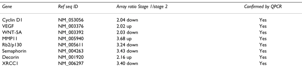
A typical representation of the results obtained hybridiz-ing two identical filter arrays with cDNA-labeled probes generated from either patients in stage 1 and patients in stage 2 is shown in Figure 1, Panels A and B. Computer-assisted analysis of these filters allowed, after normaliza-tion, to identify 14 genes, out of 588, as differentially reg-ulated in the two groups (data not shown). To confirm these changes in expression patterns, the genes were fur-ther analyzed by Quantitative RT-PCR, using two sets of independently prepared RNAs. Only 8 out of 14 of the genes analyzed were confirmed to be differentially regu-lated by quantitative RT-PCR. In table 2 are depicted the genes whose differential expression was confirmed with
quantitative RT-PCR. In table 3 the primers utilized for RT-PCR are shown. These genes code for different protein families: extra-cellular matrix proteins and proteases (Decorin and MMP11), genes involved in DNA repair (XRCC1), regulator of angiogenesis (VEGF), cell cycle reg-ulators (Cyclin D1) and tumor-suppressor genes ( Sema-phorin 3B, WNT-5A and retinoblastoma-related Rb2/ p130).
Discussion
In the present study, we analyzed two population of NSCLC patients at stage 1 and 2 to screen for tumor pro-gression-associated genes, by means of a cDNA array filter containing 588 different cDNAs. Consistent with previ-ously reported results [20], not all of the differences found by the array were effectively reproducible using another quantitative mRNA technique, such as quantitative RT-PCR. The discrepancies could be explained by methodo-logical limitations of the array technology, where thou-sands of diverse cDNAs differing exponentially in expression levels are compared by means of a single hybridization reaction, irrespective of the optimal range of reaction conditions.
The genes we found reliably differentially regulated in the two RNA populations are known to exert different func-tions, thus confirming that the biological events taking place during the malignant progression involve several molecular pathways.
The cyclin D1 gene codes for one of the cyclins involved in cell cycle regulation; specifically for the G1-S transition. The involvement of cell-cycle regulators in the progression of lung cancer is well documented [21]. This observation, therefore, further support the fact that alterations in cell cycle regulation is one of the earlier steps in the progres-sion of NSCLC.
The VEGF gene codes for a glycoprotein with potent ang-iogenic, mitogenic, and vascular permeability-enhancing activities for endothelial cells. Tumour neoangiogenesis has been recently recognized to be of importance in defin-ing subsets of patients with poor outcome in cancer
Table 2: Expression patterns obtained by array hybridization and confirmation of differential gene expression by Quantitative RT-PCR
Gene Ref seq ID Array ratio Stage 1/stage 2 Confirmed by QPCR
Cyclin D1 NM_053056 2.04 down Yes
VEGF NM_003376 2.02 up Yes
WNT-5A NM_003392 2.03 down Yes
MMP11 NM_005940 3.68 up Yes
Rb2/p130 NM_005611 3.24 down Yes
Semaphorin NM_004263 3.43 down Yes
Decorin NM_001920 2.16 up Yes
[22,23]. From that several reports [18,24] have stated that the presence of neoangiogenesis represents a significant factor in terms of overall and disease free survival in lung cancer.
The WNT-5A gene is a representative ligand that activates a beta-catenin-independent pathway in the Wnt signalling [25]. In contrast to the transforming members of the Wnt
family, shown to be up-regulated in many cancers, the role of Wnt 5a is still controversial. While it has been attributed a tumour suppressor function in some malig-nancies, there is increasing evidence of promigratory and proinvasive effects in others, mediated predominantly through the planar cell polarity pathway and activation of protein kinase C [26,27]. In our experimental setting, transcription of WNT-5A was down-regulated in stage 2
DNA Arrays Figure 1
DNA Arrays: PhosphorImager output of the two Clontech Atlas™ Human Cancer cDNa Expression Array nylon filters after hybridization with 32P-labeled cDNA derived from total RNA from either NSCLC patients at stage 1 and stage 2 (Panels A and
B, respectively).
A
B
Table 3: Primers and conditions utilized for the Quantitative RT-PCR
Gene Sequence PCR product (bp) Annealing Temp.
Cyc D1 for TGAGGCGGTAGTAGGACAGG 66 60°C
Cyc D1 rev GACCTTCGTTGCCCTCTGT
VEGF for GCAGTAGCTGCGCTGATAGA 119 60°C
VEGF rev CCTTGCTGCTCTACCTCCAC
WNT-5A for CAAAGCAACTCCTGGGCTTA 107 60°C
WNT-5A rev GCTCGGATTCCTCGGCT
MMP11 for CAGCCAAAGAAGTCAGGACC 147 60°C
MMP11 rev GGTGCCCTCTGAGATCGAC
Rb2/p130 for AGGAGTTTCTCCTGTGCGTATC 106 60°C
Rb2/p130 rev TGAAACAATGCTTTCTCCTCG
Semaphorin for TCACAGTGGCAGCATAGAGG 111 60°C
Semaphorin rev TCACAGTGGCAGCATAGAGG
Decorin for TGCAGGTCTAGCAGAGTTGTGT 91 60°C
Decorin rev AATGCCATCTTCGAGTGGTC
XRCC1 for GACACTTACCGAAAATGGCG 110 60°C
NSCLC, thus underlying a putative tumour suppressor function of its gene product in lung cancer progression.
The MMP11 gene belongs to a family of matrix metallo-proteinases, proteolytic enzymes that degrade extracellu-lar matrix and promote the local or metastatic potential of carcinoma cells, and whose action is restrained by special inhibitors (metalloproteinase inhibitors) [28,29]. The role of MMP11 protease in lung cancer progression has been clearly defined [28,29].
The Rb2/p130 gene codes for one of the retinoblastoma-related proteins. Its role as tumour-suppressor is well doc-umented [30,31], as well as its involvement in lung cancer progression [14]. Our data further suggest that alteration on Rb2/p130 gene expression is involved also in the ini-tial steps of NSCLC progression.
Loss of Semaphorin-A3F genes occurs frequently in lung cancer and correlates with advanced stage of disease [32]. Moreover, in lung cancer patients, semaphoring gene loss correlates with advanced disease and increased VEGF binding to tumour cells [33,34]. Our data confirms this phenomenon and correlates it also to the initial step of NSCLC progression.
The Decorin gene codes for one of the major extracellular matrix protein which has become the focus of various can-cer studies [35]. The exact biological role of decorin in cancer has yet to be clarified; however, several experimen-tal data suggest its involvement in cancer progression and metastatization [36].
The XRCC1 gene codes for a base excision repair. Several papers have investigated the association between lung cancer and genetic polymorphisms in XRCC1, suggesting a predisposition to lung cancer development for the patients carrying these gene alterations [37,38]. Interest-ingly, it has been recently described that XRCC1 transcript abundance levels correlate with cisplatin chemoresistance in non-small cell lung cancer cell lines [39]. To the best of our knowledge, no previous report exists about the involvement of the XRCC1 gene in the early steps of NSCLC progression.
Conclusion
This study confirms the aptitude of the cDNA array tech-nology in defining molecular pathways involved in NSCLC progression. In addition, our results corroborated previously observed expression patterns of a series of genes, and revealed new genes differentially expressed during NCSLC progression. The role of these newly iden-tified genes is being evaluated in further studies analyzing protein expression pattern and function of the proteins in vitro and in vivo in lung cancer cells.
Competing interests
The authors declare that they have no competing interests.
Authors' contributions
All authors read and approved the final manuscript. MC set up the cDNA array protocols and performed the exper-iment; VA, EP and AG collected the cancer samples; GC and MC gave advise on the work and helped in the inter-pretation of the data; EPS, PC and LL performed the RNA ectractions; AB supervised all the work and wrote the paper together with TCM.
Acknowledgements
This work was supported by grants from Ministry of Health, MIUR, Second University of Naples and FUTURA-onlus to A.B.; by FIRB/MUR
(RBIPO6LCA9-009) to G.C.; by the Italian Health Ministry (Title of the project: "Profilo genetico associato al fenotipo metastatico e alla prognosi nei tumori polmonari") to TCM.
References
1. Zochbauer-Muller S, Gazdar AF, Minna JD: Molecular pathogene-sis of lung cancer. Annu Rev Physiol 2002, 64:681-708.
2. D'Amico TA, Aloia TA, Moore MB, Conlon DH, Herndon JE 2nd, Kinch MS, Harpole DH Jr: Predicting the sites of metastases from lung cancer using molecular biologic markers. Ann Tho-rac Surg 2001, 72:1144-1148.
3. Harpole DH Jr, Herndon JE 2nd, Young WG Jr, Wolfe WG, Sabiston DC Jr: Stage I non small cell lung cancer. A multivariate anal-ysis of treatment methods and patterns of recurrence. Can-cer 1995, 76:787-796.
4. Feld R, Rubinstein LV, Weisenberger TH: Sites of recurrence in resected stage I non-small-cell lung cancer: a guide for future studies. J Clin Oncol 1984, 2:1352-1358.
5. D'Amico TA, Massey M, Herndon JE 2nd, Moore MB, Harpole DH Jr: A biologic risk model for stage I lung cancer: immunohisto-chemical analysis of 408 patients with the use of ten molec-ular markers. J Thorac Cardiovasc Surg 1999, 117:736-743. 6. Suzuki K, Nagai K, Yoshida J, Moriyama E, Nishimura M, Takahashi K,
Nishiwaki Y: Prognostic factors in clinical stage I non-small cell lung cancer. Ann Thorac Surg 1999, 67:927-932.
7. Liu D, Huang C, Kameyama K, Hayashi E, Yamauchi A, Kobayashi S, Yokomise H: E-cadherin expression associated with differenti-ation and prognosis in patients with non-small cell lung can-cer. Ann Thorac Surg 2001, 71:949-955.
8. D'Amico TA, Aloia TA, Moore MB, Herndon JE 2nd, Brooks KR, Lau CL, Harpole DH Jr: Molecular biologic substaging of stage I lung cancer according to gender and histology. Ann Thorac Surg 2000, 69:882-886.
9. Shopland DR: Tobacco use and its contribution to early cancer mortality with a special emphasis on cigarette smoking. Envi-ron Health Perspect 1995, 103:131-142.
10. Esposito V, Baldi A, De Luca A, Mazzarella G, Micheli P, Baldi F, Caputi M, Giordano A: Prognostic value of p53 in non small cell lung cancer: relationship with proliferating cell nuclear antigen and cigarette smoking. Human Path 1997, 28:233-237.
11. Esposito V, Baldi A, De Luca A, Groeger AM, Loda M, Giordano GG, Caputi M, Baldi F, Pagano M, Giordano A: Prognostic role of the cyclin-dependent kinase inhibitor p27 in non-small lung can-cer. Cancer Res 1997, 57:3381-3385.
12. Caputi M, Esposito V, Baldi A, De Luca A, Dean C, Signoriello G, Baldi F, Giordano A: p21 expression in non-small cell lung cancer: relationship to survival. Am J Respir Cell Mol Biol 1998, 18:213-217. 13. Groeger AM, Caputi M, Esposito V, Baldi A, Rossiello R, Santini D, Mancini A, Kaiser HE, Baldi F: Expression of p21 in non-small cell lung cancer: relationship with PCNA. Anticancer Res 2000, 20:3301-3306.
Publish with BioMed Central and every scientist can read your work free of charge "BioMed Central will be the most significant development for disseminating the results of biomedical researc h in our lifetime."
Sir Paul Nurse, Cancer Research UK
Your research papers will be:
available free of charge to the entire biomedical community
peer reviewed and published immediately upon acceptance
cited in PubMed and archived on PubMed Central
yours — you keep the copyright
Submit your manuscript here:
http://www.biomedcentral.com/info/publishing_adv.asp
BioMedcentral 15. Esposito V, Baldi A, Liuzzi G, Tonini G, Vincenzi B, Persichetti P,
San-tini M, Ambrogi V, Mineo TC, Montesarchio V, Wolner E, Baldi F, Groeger AM: Analysis of Fas (Apo-1/CD95) expression in non-small-cell lung cancer. Anticancer Res 2003, 23:4901-4905. 16. Groeger AM, Esposito V, De Luca A, Cassandro R, Tonini G, Ambrogi
V, Baldi F, Goldfarb R, Mineo TC, Baldi A, Wolner E: Prognostic value of immunohistochemical expression of p53, BAX, BCL-2 and BCL-Xl in resected non small cell lung cancer. Histopathology 2004, 44:54-63.
17. Esposito V, Baldi A, De Luca A, Tonini G, Vincenzi B, Santini D, Per-sichetti P, Mancini A, Citro G, Baldi F, Groeger AM, Caputi M: Cell cycle related proteins as prognostic parameters in radically resected non small cell lung cancer (NSCLC). J Clin Pathol 2005, 58:734-739.
18. Mineo TC, Ambrogi V, Baldi A, Rabitti C, Bollero P, Vincenzi B, Tonini G: Prognostic impact of VEGF, CD31, CD34, and CD105 expression and tumor vessel invasion after radical surgery for IB-IIA non-small cell lung cancer. J Clin Pathol 2004, 57:591-597.
19. Esposito V, Campioni M, De Luca A, Spugnini EP, Baldi F, Cassandro R, Mancini A, Vincenzi B, Groeger A, Caputi M, Baldi A: Analysys of HtrA1 serine protease expression in human lung cancer. Anticancer Res 2006, 26:3455-3460.
20. Baldi A, Battista T, De Luca A, Santini D, Rossiello R, Baldi F, Natali PG, Lombardi D, Picardo M, Felsani A, Paggi MG: Identification of genes down-regulated during melanoma progression: a cDNA array study. Exp Dermatol 2003, 12:213-218.
21. Vincenzi B, Schiavon G, Villetta M, Santini D, Perrone G, Di Marino M, Angeletti S, Baldi A, Tonini G: Cell cycle alterations and lung cancer. Histol Histopathol 2006, 21:423-435.
22. Macchiarini P, Fontanini G, Hardin MJ, Squartini F, Angeletti CA: Relation of neovascularization to metastasis of non-small cell lung cancer. Lancet 1992, 340:145-146.
23. Yano T, Tanikawa S, Fujie T, Masutani M, Horie T: Vascular endothelial growth factor expression and neovascularisation in non-small cell lung cancer. Eur J Cancer 2000, 36:601-609. 24. Offersen BV, Pfeiffer P, Hamilton-Dutoit S, Overgaard J: Patterns of
angiogenesis in non-small-cell lung carcinoma. Cancer 2001, 91:1500-1509.
25. Kurayoshi M, Oue N, Yamamoto H, Kishida M, Inoue A, Asahara T, Yasui W, Kikuchi A: Expression of Wnt-5a is correlated with aggressiveness of gastric cancer by stimulating cell migra-tion and invasion. Cancer Res 2006, 66:10439-10448.
26. Jönsson M, Dejmek J, Bendahl PO, Andersson T: Loss of Wnt-5a protein is associated with early relapse in invasive ductal breast carcinomas. Cancer Res 2002, 62:409-416.
27. Pukrop T, Binder C: The complex pathways of Wnt 5a in can-cer progression. J Mol Med 2008, 86:259-266.
28. Karameris A, Panagou P, Tsilalis T, Bouros D: Association of expression of metalloproteinases and their inhibitors with the metastatic potential of squamous-cell lung carcinomas. A molecular and immunohistochemical study. Am J Respir Crit Care Med 1997, 156:1930-1936.
29. Delebecq TJ, Porte H, Zerimech F, Copin MC, Gouyer V, Dacquem-bronne E, Balduyck M, Wurtz A, Huet G: Overexpression level of stromelysin 3 is related to the lymph node involvement in non-small cell lung cancer. Clin Cancer Res 2000, 6:1086-1092. 30. Paggi MG, Baldi A, Bonetto F, Giordano A: The retinoblastoma
protein family in cell cycle and cancer. J Cell Biochem 1996, 62:418-430.
31. Spugnini EP, Campioni M, D'Avino A, Caruso G, Citro G, Baldi A: Cell-cycle molecules in mesothelioma, an overview. J Exp Clin Cancer Res 2007, 26:515-521.
32. Potiron VA, Sharma G, Nasarre P, Clarhaut JA, Augustin HG, Gemmill RM, Roche J, Drabkin HA: Semaphorin SEMA3F affects multiple signaling pathways in lung cancer cells. Cancer Res 2007, 67:8708-8715.
33. Kusy S, Nasarre P, Chan D, Potiron V, Meyronet D, Gemmill RM, Constantin B, Drabkin HA, Roche J: Selective suppression of in vivo tumorigenicity by semaphorin SEMA3F in lung cancer cells. Neoplasia 2005, 7:457-465.
34. Castro-Rivera E, Ran S, Thorpe P, Minna JD: Semaphorin 3B (SEMA3B) induces apoptosis in lung and breast cancer, whereas VEGF165 antagonizes this effect. Proc Natl Acad Sci USA 2004, 101:11432-11437.
35. McDoniels-Silvers AL, Nimri CF, Stoner GD, Lubet RA, You M: Dif-ferential gene expression in human lung adenocarcinomas and squamous cell carcinomas. Clin Cancer Res 2002, 8:1127-1138.
36. Shintani K, Matsumine A, Kusuzaki K, Morikawa J, Matsubara T, Wakabayashi T, Araki K, Satonaka H, Wakabayashi H, Iino T, Uchida A: Decorin suppresses lung metastases of murine osteosar-coma. Oncol Rep 2008, 19:1533-1539.
37. Ryk C, Kumar R, Thirumaran RK, Hou SM: Polymorphisms in the DNA repair genes XRCC1, APEX1, XRCC3 and NBS1, and the risk for lung cancer in never- and ever-smokers. Lung Can-cer 2006, 54:285-292.
38. López-Cima MF, González-Arriaga P, García-Castro L, Pascual T, Marrón MG, Puente XS, Tardón A: Polymorphisms in XPC, XPD, XRCC1, and XRCC3 DNA repair genes and lung can-cer risk in a population of northern Spain. BMC Cancan-cer 2007, 7:162.