R E S E A R C H
Open Access
DHEA Attenuates Microglial Activation via
Induction of JMJD3 in Experimental
Subarachnoid Haemorrhage
Tao Tao
1†, Guang-Jie Liu
2†, Xuan Shi
3†, Yan Zhou
2, Yue Lu
1,2, Yong-Yue Gao
2, Xiang-Sheng Zhang
4, Han Wang
1,
Ling-Yun Wu
1,2, Chun-Lei Chen
1,2, Zong Zhuang
1,2, Wei Li
1,2*and Chun-Hua Hang
1,2*Abstract
Background:Microglia are resident immune cells in the central nervous system and central to the innate immune system. Excessive activation of microglia after subarachnoid haemorrhage (SAH) contributes greatly to early brain injury, which is responsible for poor outcomes. Dehydroepiandrosterone (DHEA), a steroid hormone enriched in the brain, has recently been found to regulate microglial activation. The purpose of this study was to address the role of DHEA in SAH.
Methods:We used in vivo models of endovascular perforation and in vitro models of haemoglobin exposure to illustrate the effects of DHEA on microglia in SAH.
Results:In experimental SAH mice, exogenous DHEA administration increased DHEA levels in the brain and modulated microglial activation. Ameliorated neuronal damage and improved neurological outcomes were also observed in the SAH mice pretreated with DHEA, suggesting neuronal protective effects of DHEA. In cultured microglia, DHEA elevated the mRNA and protein levels of Jumonji d3 (JMJD3, histone 3 demethylase) after
haemoglobin exposure, downregulated the H3K27me3 level, and inhibited the transcription of proinflammatory genes. The devastating proinflammatory microglia-mediated effects on primary neurons were also attenuated by DHEA; however, specific inhibition of JMJD3 abolished the protective effects of DHEA. We next verified that DHEA-induced JMJD3 expression, at least in part, through the tropomyosin-related kinase A (TrkA)/Akt signalling pathway.
Conclusions:DHEA has a neuroprotective effect after SAH. Moreover, DHEA increases microglial JMJD3 expression to regulate proinflammatory/anti-inflammatory microglial activation after haemoglobin exposure, thereby suppressing inflammation.
Keywords:Dehydroepiandrosterone, Microglia, JMJD3, Subarachnoid haemorrhage, Inflammation
Introduction
Subarachnoid haemorrhage (SAH) is a fatal neurovascu-lar disease with a simineurovascu-lar disability-adjusted life years as more common types of stroke [1]. Early brain injury (EBI), characterized by sterile inflammation and neur-onal damage, is the main cause of neurological impair-ment in SAH. Our previous studies found that remission of inflammation in EBI could improve neurological
recovery [2]. Inhibition of inflammation is a promising method to reduce EBI and improve long-term prognosis, but effective therapies are lacking.
Microglia, the resident innate immune cells in the central nervous system (CNS), play a crucial role in sterile inflammation. Upon stimulation, these cells activate and produce a variety of inflammatory factors that aggravate the inflammatory response. As inflammation progresses, microglia gradually change their functions in favour of suppressing the inflammatory response and orchestrating neuronal restorative processes [3]. Microglial activation has also been found in SAH patients [4] and animal models [5]. The paradoxical roles of microglia make the
© The Author(s). 2019Open AccessThis article is distributed under the terms of the Creative Commons Attribution 4.0 International License (http://creativecommons.org/licenses/by/4.0/), which permits unrestricted use, distribution, and reproduction in any medium, provided you give appropriate credit to the original author(s) and the source, provide a link to the Creative Commons license, and indicate if changes were made. The Creative Commons Public Domain Dedication waiver (http://creativecommons.org/publicdomain/zero/1.0/) applies to the data made available in this article, unless otherwise stated.
* Correspondence:lwxzlw@126.com;hang_neurosurgery@163.com †Tao Tao, Guang-Jie Liu and Xuan Shi contributed equally to this work.
1Department of Neurosurgery, Nanjing Drum Tower Hospital Clinical College
switch from a proinflammatory to antiinflammatory state a potential target for inhibition of inflammation in EBI.
Dehydroepiandrosterone (DHEA, 3β
-hydroxy-5-andros-ten-17-one) and its conjugate ester are the most abundant steroid hormones in circulation and are even more abun-dant in the CNS [6]. In humans, peripheral DHEA is synthesized by the adrenals and the gonads; DHEA is able to cross the blood-brain barrier (BBB) due to its lipophilic nature [7]. Moreover, in the brain, both astrocytes and neurons can synthesize DHEA [8], making the DHEA con-centration much higher in the brain (13.1~29.4 nmol/L) than in the circulation (1.83 nmol/L) [6].
DHEA has been widely used as an anti-ageing health care product and is thought to play a beneficial role in many pathological changes, including neurodegeneration [9], neuropsychiatry [10], and neuroinflammation [11]. DHEA treatment has been shown to alleviate dopamine depletion in a Parkinson’s disease (PD) mouse model [12], suggesting a neuroprotective effect of DHEA. Furthermore, DHEA reportedly inhibits activation of lipopolysaccharide (LPS)-stimulated microglia, which implies an anti-inflammatory property of DHEA [11]. By binding with many receptors, such as the N-methyl-D-aspartate (NMDA) receptor [13], G protein-coupled re-ceptors [14], and tropomyosin-related kinase A (TrkA) receptor [15], DHEA initiates different signalling path-ways to play an anti-inflammatory role. Considering that the inflammatory response is a common pathological feature of these diseases and the important role of microglia in this response, we speculate that DHEA may have a regulatory effect on microglia in EBI.
To address this hypothesis, we explored the protective effects of DHEA after SAH in vivo and discussed the specific mechanisms by which DHEA regulates micro-glial activation in vitro.
Materials and methods
All animal procedures were approved by the Ethics Re-view Committee for Animal Experimentation at Nanjing Drum Tower Hospital and were conducted in accord-ance with recommendations from the National Institutes
of Health’s Guide for the Care and Use of Laboratory
Animals.
Mice and model
A total of 91 adult male C57BL/6 mice (8–10 weeks old, 20–25 g), 5 females at 18 days of gestation, and ~ 100 pups within the first day of life from Animal Core Facility of Nanjing Medical University were used. Animals were allowed ad libitum access to food and water. Mice were kept in specific pathogen-free (SPF) and comfortable con-ditions (12-h light/dark cycle, temperature at 25 °C, and humidity at 65%) throughout the experiment. All efforts were made to minimize the number of animals killed and
their suffering in the study. Animals were randomly di-vided and assigned to experimental groups, and the mor-tality rates were 0 (0/18) in the sham group, 18.42% (7/38) in the SAH group and 11.11% (4/36) in the SAH + DHEA group (Additional file1: Figure S1).
The endovascular perforation procedure [16] was performed by the same skilled experimenter (T.T.)
(Additional file 1: Figure S2). Mice were anaesthetized
with 3% and 1.5% isoflurane inhalation (RWD Life Sci-ence, Shenzhen) for induction and maintenance. Body temperature was maintained at 37 ± 0.5 °C throughout the operation. After satisfactory anaesthesia, an ~ 1.0-cm-long median cervical incision was made. A marked 6-0 filament was inserted retrogradely from the external carotid artery into the cranium through the internal carotid artery, reaching the origin of the right middle cerebral artery. Then, the filament was progressed forward quickly to puncture the artery in the SAH group or immediately withdrawn in the sham group. All mice were given 1 ml of normal saline intraperitoneally after surgery.
The neurological score was blindly evaluated on postop-erative day (POD) 1 and POD 7 using a modified Garcia score by two independent observers (GJ.L. and CL.C.).
Animals scoring≤6 or≥15 at POD1 were excluded.
DHEA (80 mg/kg) (Yuanye Bio-Technology, Shanghai) or vehicle was injected intraperitoneally once a day [11]. The surgery was performed 1 h after the second adminis-tration, and mice were executed after the third administra-tion except the mice in neurological test (consecutive administration until POD7). Drug was administered by Y.Z., and others were blinded to the group allocation.
Enzyme-linked immunosorbent assay
The serum and the tissues from the cortex, piriform cortex, hippocampus and hypothalamus were collected after heart perfusion and homogenized (100 mg in 1 ml 1 × PBS, according to the manual). Cell supernatants from different treatment groups were collected and tested. ELISA (Cusabio, China) was conducted accord-ing to the manual.
Cell culture
For neuron culture, the cortex was harvested from em-bryonic day 18 mice after careful removal of the lepto-meninges and digested with 0.25% trypsin for 10 min in a 37 °C incubator. The digestion was stopped with 2-ml foetal bovine serum (FBS, biological industries, USA), and the medium was removed. The cortices were gently titrated in medium, and the upper single-cell medium was transferred to a new tube. The gentle titration and medium transfer were repeated 3–4 times. The resulting single-cell medium was centrifuged at 1000 rpm for 5 min, and the pellet was resuspended with Dulbecco’s
Eventually, neurons were seeded onto poly-D -lysine-pre-coated plates at a density of 7.0 × 10⁵/ml and maintained
at 37 °C and 5% CO2. Four hours later, the medium was
completely replaced with neurobasal medium containing 2% B27 and 1% GlutMax (Gibco, USA). Neurons were usually available at 7 days in vitro (DIV7). Neuronal morphology was measured by Sholl analysis with ImageJ (n= 5 neurons from each condition) [17].
For primary microglia culture, the cortex was harvested from neonatal mice and digested as described above. Mixed glial cells were cultured together with DMEM con-taining 10% FBS and 1% penicillin-streptomycin (Gibco, USA). Microglia were usually available at DIV10 to DIV14. After the plates were gently shaken, floating microglia were transferred to new plates.
Haemoglobin-induced in vitro SAH model
Hb in bovine erythrocytes (Sigma, USA) was dissolved in
complete culture medium at a concentration of 25μmol/
L. Cells were pretreated with 10−7mol/L DHEA
dis-solved in ethanol or ethanal alone 24 h before Hb expos-ure. GSK-J4 or GW 441756 was added 30 min before DHEA.
Cell viability assay by Cell Counting Kit-8
Neurons and microglia were plated in 96-well plates at a density of 1 × 10⁴cells per well. After proper treatments, the medium was replaced completely with normal
cul-ture medium and 10μl CCK8 (Dojindo Laboratories,
Japan). After a 1-h incubation at 37 °C, the absorbance was detected at 450 nm in a microplate reader (Tecan, Switzerland). The relative cell viability was calculated ac-cording to the manual.
Conditioned medium (CM) was collected from treated
microglia and filtered through a 0.22-μm filter. DIV7
neurons were cultured with the CM for 24 h and de-tected by Cell Counting Kit-8 (CCK8) as described.
Real-time PCR
Total RNA was prepared with Trizol (Invitrogen, USA) according to the manufacturer’s instructions. cDNA was reverse transcribed from mRNA with reverse transcrip-tion mix (Vazyme, Nanjing). qPCR was performed with SYBER Green mix (Roche, Switzerland) using the PCR system (Applied Biosystems, USA). The results were
analysed with the 2−ΔΔCt method, while 18S RNA was
used for normalization. Primers used in qPCR are listed in Additional file1: Table S1.
Immunofluorescence staining
Frozen sections of 10-μm thickness and cells were fixed with 4% paraformaldehyde, permeabilized with 0.3% Tri-ton X-100, and blocked with 1% BSA. Then, the sections/ cells were incubated with primary antibodies at 4 °C
overnight followed by incubation with proper second-ary antibodies. Pictures were acquired with a confocal laser scanning microscope (FluoView FV10i, Olympus, Japan). Immunofluorescence (IF) cell count and inten-sity were analysed with ImageJ software (National In-stitutes of Health). Antibodies used in IF are listed in Additional file 1: Table S2.
Western blotting
Brain tissues or cultured cells were lysed with RIPA (Thermo Scientific, USA) with protease inhibitor (Roche, Switzerland) and 1% phosphatase inhibitor (Sigma, USA). A bicinchoninic acid protein assay kit (Beyotime, China) was used for protein quantification. The same mass of protein was loaded onto SDS-PAGE gels for separation and then transferred to polyvinylidene difluoride mem-branes (Millipore, USA). Memmem-branes were blocked with 1% BSA for 2 h at room temperature and incubated with diluted primary antibody overnight at 4 °C. Bands were analysed using ImageJ. Antibodies used in WB are listed in Additional file1: Table S2.
Terminal deoxynucleotidyl transferase–mediated dUTP nick end labelling
TUNEL staining was performed on frozen brain sections with a TUNEL detection kit (Beyotime, China) according to the manufacturer’s instructions. In brief, the slides were first incubated with the primary antibody anti-NeuN (1: 200, 26975-1-AP, Proteintech) overnight, followed by in-cubation with the corresponding secondary antibody for 1 h. One microliter of TdT enzyme was diluted 5 times and mixed with 45μl of labelling solution for one section. Each section was incubated with the mixture for 30 min at room temperature. Sections were scanned by a confocal
laser scanning microscope. The whole-brain NeuN+
TUNEL+/ NeuN+ proportion was analysed with ImageJ
(n= 3 brains). NeuN+TUNEL+/NeuN+proportion = (area (NeuN+ cells) + area (TUNEL+ cells)—area (positive cells in merge figure))/Area (NeuN+cells) × 100%.
Water content
The intact brains were dissected after decapitation and divided into right and left hemispheres. Each hemisphere was weighed immediately, placed in an oven for 24 h and weighed again. Water content = ((wet weight–dry weight)/wet weight) × 100%.
Statistical analysis
All data were expressed as the mean ± standard error of the mean (SEM). All data were first tested for normality. Statistical analysis was performed using Prism 8.02
(GraphPad Software, USA). Two-tailed Student’s t test
comparisons of more than two groups. Two-way ANOVA followed Dunnett’s multiple comparisons test was used in Sholl analysis. P < 0.05 was considered sta-tistically significant.
Results
DHEA alleviates EBI after SAH in mice
We first tested the DHEA concentration in the serum and various parts of the brain by ELISA (n = 5). In the non-treated mice, the basal DHEA concentration ranged 0.20~0.49 ng/ml in the sham group and lightly higher (0.36~0.91 ng/ml) in the SAH group but no significant dif-ference. After DHEA administration, a dramatic increase in DHEA concentration was detected up to 24 h regardless of
operation (Fig. 1a). The content of DHEA in the serum
(34.28–34.18 ng/ml) was significantly higher than that in the brain (15.13–22.72 ng/ml) in both the DHEA and SAH + DHEA groups, but no obvious difference was noticed among the various brain tissues. These results confirmed that peritoneal injection was a reliable route of administra-tion and that SAH did not affect DHEA content (Fig.1a).
As shown in Fig.1b, d, severe parenchyma oedema and
massive neuronal apoptosis were observed in the ipsilat-eral hemisphere after SAH (n= 6). Corresponding to the injury, we found an apparent compromise in the neuro-logical test results, both at POD 1 and POD 7 (Fig.1c,n= 6). These results are all indicative of severe EBI after SAH. Mice pretreated with DHEA had remarkably lower
TUNEL+NeuN+proportion than those in the SAH group
(n= 3, 54.24 ± 2.24% in the SAH group versus 29.04 ±
6.66% in the SAH + DHEA group). The water content in the ipsilateral hemisphere was 80.63 ± 0.75% in the SAH + DHEA group, which was lower but not significantly
than the 83.30 ± 0.81% observed in the SAH group (n=
6). Furthermore, DHEA improved sensorimotor functions both at POD 1 and POD 7. These results indicate that DHEA plays a neuroprotective role in SAH mice.
DHEA inhibits proinflammatory microglial activation in vivo after SAH
First, we examined inflammatory status after SAH in affected brain extracts by qPCR (n= 5). mRNA levels of
cytokines such asinterleukin(IL)-1β,IL-6,tumour necro-sis factor (TNF)-α, and IL-12, chemokines such as C-C
motif chemokine ligand 2 (CCL2), and inflammasome
NLR family, pyrin domain-containing 3 (NLRP3) were
significantly higher in the SAH group than in the sham group. However, there DHEA administration remarkably attenuated these increases (Fig.2).
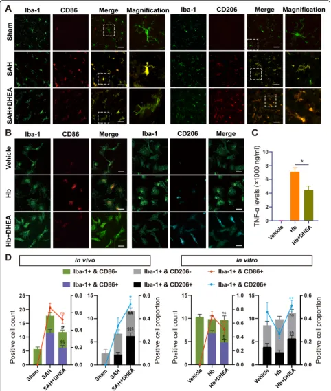
To further explore the changes and role of microglia in EBI, we performed double IF staining. In the CNS, Iba-1 is a specific microglial marker independent of microglial status, whereas CD86 is a classic marker of proinflammatory activation. When microglia are acti-vated, they transform from rod-shaped cell bodies and ramified processes to rounded amoeboid morphologies, large somas, and a short thick process.
Significantly fewer CD86+/Iba-1+microglia were accu-mulated in the affected area in the SAH + DHEA group than in the SAH group, although the proportions were not significantly different (Fig.3a, d, n= 6 fields from 3 independent experiments). Moreover, a significantly
higher number and percentage of CD206+/Iba-1+
micro-glia were detected in the SAH + DHEA group than in the SAH group (Fig.3a, d). In addition, the microglia ex-hibited an intermediate morphology between rest and
activation in the presence of DHEA (Fig. 3a). Together,
the IF images and qPCR results indicated that DHEA may inhibit SAH-induced proinflammatory microglial activation in vivo.
DHEA inhibits Hb-induced proinflammatory microglial activation and the immune response in vitro
Damage-associated molecular patterns (DAMPs), such as plasma proteins and Hb, stimulate microglial reactivity
[18]. Primary microglia were treated with 25μmol/L Hb
with or without DHEA. After 24 h, microglia transformed into a disc shape under Hb exposure, whereas the micro-glia in the vehicle group remained bipolar (Fig. 3b). Hb
also promoted the production and secretion of TNF-αin
the medium, which suggested proinflammatory microglial activation. However, the secretion of TNF-αwas restricted by DHEA (Fig.3c,n= 4).
Staining for surface markers showed results consistent with those of the in vivo experiments. Compared with the Hb group, the DHEA-treated group had a significantly higher proportion of CD206+/Iba-1+ microglia but lower proportion of CD86+/Iba-1+microglia, although the latter difference was not significant (Fig.3b, d,n= 6 fields from 3 independent experiments). Taken together, these obser-vations suggested that DHEA repressed proinflammatory
microglial activation to exert an anti-inflammatory effect in vitro.
JMJD3 is critical for the DHEA-induced mitigation of microglial inflammation after SAH
JMJD3 is a regulator of microglial activation and may play a role in the above modulation. We first located the JMJD3 protein in microglia by IF. Colocalized with Iba-1, JMJD3 was mainly located in the nucleus, which is consistent with its function (Fig.4a, b,n= 6 fields from
3 independent experiments). The JMJD3+/Iba-1+ cell
count increased dramatically after SAH, but fewer double-positive cells were counted in the SAH + DHEA
(Additional file 1: Figure S3). However, quantitative
analysis of single-cell fluorescence showed an increase in
JMJD3 fluorescence intensity within Iba-1+ cells after
DHEA exposure, which was significantly higher than
that of SAH cells (Fig. 4c). Similar results were found
in vitro (Fig.4b, d).
Upon stimulation with Hb for 24 h, the mRNA and pro-tein expression ofJmjd3increased significantly. After pre-treatment with DHEA alone in vitro, there was also an
elevation in both Jmjd3 mRNA and protein (Fig.4e, f).
In the presence of both Hb and DHEA, a more
signifi-cant increase was observed (Fig. 4e, f). Unfortunately,
we did not find this elevation in the whole-brain extract
(Additional file 1: Figure S4), potentially due to the
small proportion of microglia in the CNS.
The augmentation of JMJD3 expression after DHEA administration implies that JMJD3 might play a role in the anti-inflammatory effects of DHEA. To test this hy-pothesis, we examined the H3K27me3 levels with WB
(n= 4). DHEA eliminated the H3K27me3 increase after
Hb exposure, but this effect was blocked by a specific JMJD3 inhibitor, GSK-J4 (Fig. 5a, b), suggesting a crit-ical role of JMJD3. GSK-J4 is a specific inhibitor of JMJD3 and ubiquitously transcribed tetratricopeptide repeat, X chromosome (UTX). We determined the ex-pression of UTX after Hb and DHEA treatment and found that there was no obvious change in neurons or microglia in vitro (Additional file 1: Figure S5). There-fore, we believe that the effects of GSK-J4 on UTX can be ignored.
Inhibition of JMJD3 in microglia abolishes the anti-inflammatory and neuroprotective effects of DHEA
Various inflammatory factors are important mediators of microglial sterile inflammation. We next examined the role of JMJD3 in the production of inflammatory factors. Primary microglia were treated with a proven effective
concentration of GSK-J4 (10μmol/L) 30 min before
DHEA application [19]. After Hb exposure, the elevation
in genes IL-1β, IL-6, and TNF-α and chemokine CCL2
was validated (Fig.6a,n= 5); in contrast, the expression
of microglia homeostatic signatures, Trem2and P2RY12
[20], was downregulated (Fig. 6b, n = 5). These results
also demonstrated proinflammatory microglial activation after Hb stimulation. Supplementary DHEA suppressed IL-1β, IL-6, TNF-α, and CCL2 expression after Hb ex-posure, but this suppression was blocked by GSK-J4 (Fig. 6a). Furthermore, the upregulation ofP2RY12after DHEA was also blocked by GSK-J4, although there were no significant changes inTrem2expression (Fig.6b).
In addition to the secretion of the above proinflamma-tory factors, microglia can also secrete several neuro-trophic factors, such as brain-derived neuroneuro-trophic factor (BDNF), which act together on neurons. To ex-plore this complex influence, we cultured primary neu-rons with CM from microglia exposed to different
treatments (n= 5). The CM from Hb-exposed microglia
induced ~ 30% neuron death (Fig.6c). Importantly,
neu-rons cultured with CM from DHEA-pretreated microglia exhibited a significantly higher cell viability of ~ 95%. This protective effect of DHEA was abolished by inhib-ition of JMJD3 (~ 57%) (Fig. 6c). Finally, significant im-provement was also found in the DHEA group compared with that in the Hb group in the Sholl analysis, although no significant difference was found after JMJD3 inhibition (Fig.6d, e,n= 6).
DHEA promotes anti-inflammatory microglial activation after Hb stimulation
Some classical anti-inflammatory markers were also de-tected by both qPCR and WB. Agr1 is the direct target of JMJD3 and a classical anti-inflammatory marker
[21]. In cultured microglia, Arg1 was downregulated at
both the transcriptional and translational levels after Hb exposure and increased in the presence of DHEA,
but the increase was blocked by GSK-J4 (Fig.7a, c and
d, n = 5 in qPCR, n = 4 in WB). DHEA also restored
the mRNA level of CD206 but failed to have a
signifi-cant effect at the protein level. Inducible nitric oxide synthase (iNOS) is upregulated after stimulation and produces nitric oxide (NO) as a defence mechanism. WB showed an augmentation of iNOS protein expres-sion after Hb exposure in microglia, but this increase was mitigated by DHEA treatment. There was a weak upregulation after JMJD3 inhibition, but this effect was not significant (Fig.7c and d).
Interferon regulatory factor 4 (Irf4) and Irf7 are tar-gets of JMJD3 and play a role in the regulation of
macrophage and microglia activation [3,21–23]. DHEA
increased the mRNA expression of Irf4 and Irf7 after
Hb exposure, and upregulation of Irf7 was also
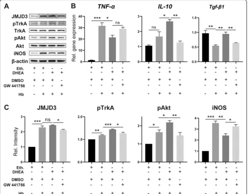
The TrkA/Akt pathway is involved in the induction of JMJD3 by DHEA
DHEA is a ligand of TrkA and sufficient to initiate the
TrkA/Akt signalling pathway [11, 15, 24]. Therefore, we
investigated whether TrkA is also involved in the regula-tion of Hb-activated microglia by DHEA. WB demon-strated increased phosphorylation of TrkA and Akt after Hb treatment, which further increased after DHEA pre-treatment (Fig. 8a,n= 3). Additionally, the protein level of JMJD3 was correlated with the phosphorylation level of TrkA and Akt. However, these changes were blocked
by the TrkA inhibitor GW 441756 (Fig.8a, c).
qPCR was further used to examine inflammatory gene expression in response to the TrkA inhibitor. The pro-tective effects of DHEA in increasing the expression of IL-10 and transforming growth factor beta 1 (Tgf-β1) were reversed by the administration of the inhibitor; however, there was no significant increase inTNF-α ex-pression with the TrkA inhibitor (Fig.8b,n= 3).
Discussion
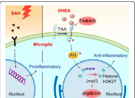
In this study, the anti-inflammatory effects of DHEA in EBI were verified in an endovascular perforation mouse model. The major findings were as follows. First, systemic administration of DHEA increased
DHEA levels in the brain and attenuated EBI after SAH. Second, DHEA administration altered microglial activation status in the ipsilateral hemisphere. Third, the expression of a H3K27 demethylase, JMJD3, was upregulated after DHEA treatment and modulated its anti-inflammatory effects. Finally, DHEA induced Jmjd3 gene expression through the TrkA/Akt signal-ling pathway in vitro (Fig.9).
More than 40% of SAH survivors experience long-term neurological impairment [25], mainly due to EBI [26, 27]. The inflammatory response is an important process in EBI. Released neuronal necrotic products, Hb, and other degradation products stimulate microglia immediately after SAH. The proinflammatory microglia initiate and amplify the inflammatory response in the acute phase, releasing a variety of inflammatory factors that further damage neurons. Our study found typical proinflammatory microglia in the neuronal damaged re-gion but no other rere-gions. The mRNA levels of a variety of cytokines were also elevated in the affected regions. The co-occurrence of massive neuronal apoptosis and proinflammatory microglial activation validates the important role of the microglia-triggered inflammatory responses in EBI, but neuronal damage may be both a cause and a consequence of microglial activation. Proin-flammatory microglial activation aggravates neuronal damage both in ischaemic stroke [28] and intraparench-ymal haemorrhage [29], whereas modulation of micro-glial activation has a beneficial effect. Our study revealed a similar result in SAH.
Dehydroepiandrosterone sulfate (DHEAS) is often dis-cussed with DHEA because it is the sulfate form and a res-ervoir of DHEA [30]. Many studies have suggested a protective profile of DHEA/DHEAS, but the detailed mechanism is not clear. DHEAS levels have been shown to be significantly lower in Alzheimer’s disease (AD) pa-tients than in controls [31]. In addition, DHEA blocked microglial nitrite production under a co-stimulation of amyloidβ-peptide and interferon-γ in vitro [32], suggest-ing a potential protective effect in AD. An observational clinical study found that high serum DHEAS was associated with a decreased IL-6 serum level and a more
favourable long-term neurological outcome after
aneurysmal SAH [33]. This is an interesting phenomenon, (See figure on previous page.)
Fig. 4Transcription and translation of JMJD3 were induced by DHEA after Hb exposure.a, bCo-localization of Iba-1 and JMJD3 demonstrated an induction of JMJD3 expression after SAH/Hb exposure that was even higher in the presence of DHEA. Magnified views are marked with dashed line boxes. Green, Iba-1; red, JMJD3; blue, DAPI; bar = 70μm.cThe relative single cell fluorescence intensity (JMJD3 in Iba-1+/mean of JMJD3 in Iba-1-) was calculated and presented in a violin plot (n= 20 in sham,n= 79 in SAH,n= 43 in SAH + DHEA from 6 fields, non-specific one-way ANOVA, ****P< 0.0001).dFluorescence intensity of JMJD3 after Hb exposure (n= 6 fields, one-way ANOVA).e, fqPCR and WB showed an induction ofJmjd3mRNA and protein expression in microglia treated with DHEA (n= 4 in qPCR and n = 3 in WB, one-way ANOVA), ns, not significant, *P< 0.05, **P< 0.01, ***P< 0.001. Eth., ethanol
but whether exogenous DHEA supplementation plays a role is unknown. Although only serum DHEA/DHEAS levels were tested in these studies, we have reasons to believe that the DHEA levels in the brain correspond with those in the periphery. These observations indicate that DHEA/DHEAS may play a protective role in the regula-tion of immune cell inflammatory responses, but the details are unclear.
Our experiments revealed that supplementary DHEA inhibited the proinflammatory response of microglia in SAH, decreased the expression of inflammatory factors and reduced neuronal damage both in vivo and in vitro. These results are generally consistent with previous reports. Regrettably, we did not perform a long-term observation and are unable to determine how DHEA affects microglia in the delayed phase of SAH to help (See figure on previous page.)
Fig. 6JMJD3 mediated the anti-inflammatory effect of DHEA. The mRNA expression levels ofaIL-1β, IL-6, TNF-α,andCCL2andbtwo
homeostasis genes were detected by qPCR (n= 5, non-parametric test forP2RY12and one-way ANOVA for the other). The gene expression levels in the vehicle group treated with dimethyl sulfoxide (DMSO) or ethanol were set as 1.cThe CM collected from microglia was added to the primary neurons for another 24 h, and neuron viability was assessed by CCK8 (n= 5, one-way ANOVA).dRepresentative fields of primary neurons cultured with CM from microglia exposed to different treatments for 24 h.eNeuronal morphology was investigated with Sholl analysis (n= 5, two-way ANOVA with Dunnett’s multiple comparisons test, the footnote* indicates the statistically significant difference between Hb and Hb + DHEA; the difference between vehicle and Hb is not shown; no significant difference between Hb + DHEA and Hb + DHEA + GSK-J4). Bar = 50μm. Eth., ethanol. *P< 0.05, **P< 0.01
brain repair and regeneration, but this will be the focus of future studies.
JMJD3 is an inducible histone demethylase [34] that pro-motes anti-inflammatory activation in macrophage/micro-glia and suppresses inflammatory responses [23]. JMJD3 in macrophages regulates the expression of molecules in-volved in anti-inflammatory activation by altering histone methylation levels of the transcription factor Irf4 [21], which decreased significantly after Hb stimulation and increased after DHEA treatment in our study (Fig.7b). The demethylase also plays a role in promoting anti-inflammatory activation in microglia. Tang’s study found that inhibition of JMJD3 blocked anti-inflammatory activa-tion and enhanced proinflammatory activaactiva-tion in N9 microglia and was associated with inflammatory responses in PD [22]. Together with our experimental results, these observations reveal that the JMJD3-Irf4 axis also plays a
regulatory role in microglia after Hb exposure and is en-hanced by DHEA treatment. Our study showed a corre-sponding change inIrf4,but further validations are needed. TrkA is phosphorylated upon ligand binding, and TrkA phosphorylation has been shown to occur as early as minutes after DHEA administration [11], indicative of direct binding. Evidence of direct binding was also found by visualization of bovine serum albumin (BSA)-bound DHEA fluorescence [15]. In our study, the specific antagonist of TrkA, GW 441786, successfully inhibited the phosphorylation of TrkA and the subsequent in-crease in JMJD3 in vitro, validating the interaction be-tween DHEA and TrkA.
main reason. We are not sure whether i.p. injection of GSK-J4/GW 441786 [35] could achieve effective concen-trations within the brain tissue in the presence of the BBB whereas intraventricular administration is less suit-able when multiple doses are required.
Second, peripheral immune cells play a role in neuroin-flammation after SAH but were not specifically studied in this study. The peripheral immune cells like T cells and neutrophil infiltrated from the 24 h after hemorrhage and peaked after 2–7 days [36], so we almost ignored the effect of DHEA on peripheral immune cells in early neuroin-flammation (24 h). However, this cannot be ignored in long-term studies.
Third, DHEA content varies with sex and age; there-fore, more experiments are needed to explore the effect of DHEA in different situations. Overall, the outstanding safety and tolerability profile of DHEA provides the pos-sibility of rapid clinical application in SAH. We are look-ing forward to more animal and clinical research of DHEA on microglial activation in SAH.
Conclusion
Our study found an anti-inflammatory effect of a widely used supplement, DHEA. In vitro experiments indicated an involvement of JMJD3 in this effect. By interacting with TrkA, DHEA induced JMJD3 expression and mod-ulated microglial activation. However, a comprehensive understanding of DHEA in vivo and its therapeutic im-plications still requires further investigation.
Supplementary information
Supplementary informationaccompanies this paper athttps://doi.org/10. 1186/s12974-019-1641-y.
Additional file 1: Table S1.Primers used in the qPCR.Table S2.
Antibodies used in the IF and WB.Figure S1.Experimental design of this study. Experiment 1, a total of 91 mice were divided into 3 groups in the in vivo experiment: 18 mice in the sham group, 38 mice in the SAH group, and 36 mice in the DHEA-treated SAH group. The mortality rates were 0 (0/18, 0) in sham, 18.42% (7/38) in SAH and 11.11% (4/36) in SAH + DHEA group. 7 and 9 mice were excluded from SAH and SAH + DHEA group respectively. Experiment 2, cultured primary microglia were treated with DHEA or GSK-J4 respectively. Experiment 3, cultured primary microglia were treated with DHEA or GW 441756 respectively.Figure S2.Experimental SAH model in C57BL/6 mouse.Figure S3.Iba-1 and JMJD3 co-stained microglia cell count after SAH (n= 6 fields, one-way ANOVA, **P <0.01, ***P< 0.001).Figure S4.Jmjd3gene expression tended to be increased but not significantly in vivo. (one-way ANOVA,n= 4, ns, not significant).
Figure S5.Jmjd3responded to haemoglobin (Hb) stimulation in microglia. Primary microglia and neurons were treated with Hb or vehicle medium for 24 h and mRNA expression was detected by qPCR. Ubiquitously transcribed tetratricopeptide repeat, X chromosome (UTX) is a homolog of Jmjd3 and both can be blocked by GSK-J4.aIn microglia, it isJmjd3, notUTX, significantly upregulated after Hb exposure. (one-way ANOVA,n= 3, ns, not significant)bIn neurons, neither expression ofJmjd3norUTX changed significantly after Hb exposure. (one-way ANOVA,n= 3, ns, not significant, ***P< 0.001)
Abbreviations
BBB:Blood-brain barrier; BDNF: Brain-derived neurotrophic factor; CCK8: Cell viability assay by Cell Counting Kit-8; CCL: C-C motif chemokine ligand; CM: Conditioned medium; CNS: Central nervous system; DAPI: 4,6-Diamidino-2-phenylindole; DHEA: Dehydroepiandrosterone;
DHEAS: Dehydroepiandrosterone sulfate; DIV: Day in vitro; DMEM: Dulbecco’s Modified Eagle Medium; EBI: Early brain injury; ELISA: Enzyme-linked immunosorbent assay; FBS: Foetal bovine serum; Hb: Haemoglobin; IF: Immunofluorescence; IL: Interleukin; iNOS: Inducible nitric oxide synthase; Irf: Interferon regulatory factor; JMJD3: Jumonji d3; LPS: Lipopolysaccharide; NLRP: NLR family, pyrin domain-containing; NMDA: N-methyl-D-aspartate; PD: Parkinson’s disease; POD: Postoperative day; qPCR: Real-time PCR; SAH: Subarachnoid haemorrhage; SEM: Standard error of the mean; SPF: Specific pathogen-free; Tgf-β1: Transforming growth factor beta 1; TNF: Tumour necrosis factor; TrkA: Tropomyosin-related kinase A;
TUNEL: Terminal deoxynucleotidyl transferase-mediated dUTP nick end label-ling; UTX: Ubiquitously transcribed tetratricopeptide repeat, X chromosome; WB: Western blotting
Acknowledgements
Not applicable.
Authors’contributions
TT is responsible for study design, performing experiments, manuscript drafting; GL and XS analyse and interpret of results; YZ is responsible for animal management and drug administration; YL is responsible for statistical analysis; YG is responsible for data analysis; XZ and HW are responsible for cell culture; LW is responsible for immunofluorescence staining; CC is responsible for neurological evaluation; ZZ is responsible for critical reversion of manuscript; WL is responsible for study design and critical reversion of manuscript; CH is responsible for study design. All authors read and approved the final manuscript.
Funding
This work was supported by National Natural Science Foundation, China [grant No. 81870922, No. 81771291, No. 81801166, No. 81901203, No. 81971127], Medical Science and Technology Development Foundation, Nanjing Department of Health [grant No. JQX18001] and the Fundamental Research Funds for the Central Universities [grant No. 021414380361]. Fig. 9As innate immune cells in the CNS, microglia are activated after
Availability of data and materials
The datasets supporting the conclusions of this article are included within the article (and its additional file).
Ethics approval and consent to participate
The study was approved by the Ethics Review Committee of Drum Tower Hospital.
Consent for publication
Not applicable.
Competing interests
The authors declare that they have no competing interests.
Author details
1Department of Neurosurgery, Nanjing Drum Tower Hospital Clinical College
of Nanjing Medical University, Nanjing 210008, Jiangsu, China.2Department of Neurosurgery, Nanjing Drum Tower Hospital, The Affiliated Hospital of Nanjing University Medical School, Nanjing 210008, Jiangsu, China.
3Department of Neurology, Jinling Hospital, Nanjing Medical University,
Nanjing, China.4Department of Neurosurgery, Beijing Friendship Hospital, Capital Medical University, Beijing 100032, China.
Received: 25 August 2019 Accepted: 14 November 2019
References
1. Macdonald RL, Schweizer TA. Spontaneous subarachnoid haemorrhage. Lancet. 2017;389:655–66.
2. Lu Y, Zhang XS, Zhang ZH, Zhou XM, Gao YY, Liu GJ, Wang H, Wu LY, Li W, Hang CH. Peroxiredoxin 2 activates microglia by interacting with Toll-like receptor 4 after subarachnoid hemorrhage. J
Neuroinflammation. 2018;15:87.
3. Hu X, Leak RK, Shi Y, Suenaga J, Gao Y, Zheng P, Chen J. Microglial and macrophage polarization—new prospects for brain repair. Nat Rev Neurol. 2014;11:56.
4. Schneider UC, Davids AM, Brandenburg S, Muller A, Elke A, Magrini S, Atangana E, Turkowski K, Finger T, Gutenberg A, et al. Microglia inflict delayed brain injury after subarachnoid hemorrhage. Acta Neuropathol. 2015;130:215–31.
5. Schallner N, Pandit R, LeBlanc R 3rd, Thomas AJ, Ogilvy CS, Zuckerbraun BS, Gallo D, Otterbein LE, Hanafy KA. Microglia regulate blood clearance in subarachnoid hemorrhage by heme oxygenase-1. J Clin Invest. 2015;125: 2609–25.
6. Maninger N, Wolkowitz OM, Reus VI, Epel ES, Mellon SH. Neurobiological and neuropsychiatric effects of dehydroepiandrosterone (DHEA) and DHEA sulfate (DHEAS). Front Neuroendocrinol. 2009;30:65–91.
7. Starka L, Duskova M, Hill M. Dehydroepiandrosterone: a neuroactive steroid. J Steroid Biochem Mol Biol. 2015;145:254–60.
8. Gottfried-Blackmore A, Sierra A, Jellinck PH, McEwen BS, Bulloch K. Brain microglia express steroid-converting enzymes in the mouse. J Steroid Biochem Mol Biol. 2008;109:96–107.
9. Hampl R, Bicikova M. Neuroimmunomodulatory steroids in Alzheimer dementia. J Steroid Biochem Mol Biol. 2010;119:97–104.
10. Souza-Teodoro LH, de Oliveira C, Walters K, Carvalho LA. Higher serum dehydroepiandrosterone sulfate protects against the onset of depression in the elderly: findings from the English Longitudinal Study of Aging (ELSA). Psychoneuroendocrinology. 2016;64:40–6.
11. Alexaki VI, Fodelianaki G, Neuwirth A, Mund C, Kourgiantaki A, Ieronimaki E, Lyroni K, Troullinaki M, Fujii C, Kanczkowski W, et al. DHEA inhibits acute microglia-mediated inflammation through activation of the TrkA-Akt1/2-CREB-Jmjd3 pathway. Mol Psychiatry. 2017;23:1410–20.
12. D'Astous M, Morissette M, Tanguay B, Callier S, Di Paolo T.
Dehydroepiandrosterone (DHEA) such as 17beta-estradiol prevents MPTP-induced dopamine depletion in mice. Synapse. 2003;47:10–4.
13. Suzuki M, Wright LS, Marwah P, Lardy HA, Svendsen CN. Mitotic and neurogenic effects of dehydroepiandrosterone (DHEA) on human neural stem cell cultures derived from the fetal cortex. Proc Natl Acad Sci U S A. 2004;101:3202–7.
14. Sicard F, Ehrhart-Bornstein M, Corbeil D, Sperber S, Krug AW, Ziegler CG, Rettori V, McCann SM, Bornstein SR. Age-dependent regulation of
chromaffin cell proliferation by growth factors, dehydroepiandrosterone (DHEA), and DHEA sulfate. Proc Natl Acad Sci U S A. 2007;104:2007–12. 15. Lazaridis I, Charalampopoulos I, Alexaki VI, Avlonitis N, Pediaditakis I,
Efstathopoulos P, Calogeropoulou T, Castanas E, Gravanis A. Neurosteroid dehydroepiandrosterone interacts with nerve growth factor (NGF) receptors, preventing neuronal apoptosis. PLoS Biol. 2011;9:e1001051.
16. Au-Schüller K, Au-Bühler D, Au-Plesnila N. A Murine Model of Subarachnoid Hemorrhage. JoVE. 2013:e50845.
17. Ferreira TA, Blackman AV, Oyrer J, Jayabal S, Chung AJ, Watt AJ, Sjostrom PJ, van Meyel DJ. Neuronal morphometry directly from bitmap images. Nat Methods. 2014;11:982–4.
18. Ransohoff RM. A polarizing question: do M1 and M2 microglia exist? Nat Neurosci. 2016;19:987–91.
19. Das A, Arifuzzaman S, Yoon T, Kim SH, Chai JC, Lee YS, Jung KH, Chai YG. RNA sequencing reveals resistance of TLR4 ligand-activated microglial cells to inflammation mediated by the selective jumonji H3K27 demethylase inhibitor. Sci Rep. 2017;7:6554.
20. Deczkowska A, Keren-Shaul H, Weiner A, Colonna M, Schwartz M, Amit I. Disease-Associated Microglia: A Universal Immune Sensor of
Neurodegeneration. Cell. 2018;173:1073–81.
21. Satoh T, Takeuchi O, Vandenbon A, Yasuda K, Tanaka Y, Kumagai Y, Miyake T, Matsushita K, Okazaki T, Saitoh T, et al. The Jmjd3-Irf4 axis regulates M2 macrophage polarization and host responses against helminth infection. Nat Immunol. 2010;11:936–44.
22. Tang Y, Li T, Li J, Yang J, Liu H, Zhang XJ, Le W. Jmjd3 is essential for the epigenetic modulation of microglia phenotypes in the immune pathogenesis of Parkinson's disease. Cell Death Differ. 2014;21:369–80. 23. Zhao SC, Ma LS, Chu ZH, Xu H, Wu WQ, Liu F. Regulation of microglial
activation in stroke. Acta Pharmacol Sin. 2017;38:445–58.
24. Lawson LJ, Perry VH, Gordon S. Turnover of resident microglia in the normal adult mouse brain. Neuroscience. 1992;48:405–15.
25. Suarez JI, Tarr RW, Selman WR. Aneurysmal subarachnoid hemorrhage. N Engl J Med. 2006;354:387–96.
26. Cahill WJ, Calvert JH, Zhang JH. Mechanisms of early brain injury after subarachnoid hemorrhage. J Cereb Blood Flow Metab. 2006;26:1341–53. 27. Sehba FA, Hou J, Pluta RM, Zhang JH. The importance of early brain injury
after subarachnoid hemorrhage. Prog Neurobiol. 2012;97:14–37. 28. Ma Y, Wang J, Wang Y, Yang GY. The biphasic function of microglia in
ischemic stroke. Prog Neurobiol. 2017;157:247–72.
29. Lan X, Han X, Li Q, Yang QW, Wang J. Modulators of microglial activation and polarization after intracerebral haemorrhage. Nat Rev Neurol. 2017;13:420–33.
30. Rutkowski K, Sowa P, Rutkowska-Talipska J, Kuryliszyn-Moskal A, Rutkowski R. Dehydroepiandrosterone (DHEA): hypes and hopes. Drugs. 2014;74:1195–207.
31. Pan X, Wu X, Kaminga AC, Wen SW, Liu A. Dehydroepiandrosterone and dehydroepiandrosterone sulfate in alzheimer’s disease: a systematic review and meta-analysis. Front Aging Neurosci. 2019;11:61.
32. Barger SW, Chavis JA, Drew PD. Dehydroepiandrosterone inhibits microglial nitric oxide production in a stimulus-specific manner. J Neurosci Res. 2000;62:503–9.
33. Hollig A, Thiel M, Stoffel-Wagner B, Coburn M, Clusmann H. Neuroprotective properties of dehydroepiandrosterone-sulfate and its relationship to interleukin 6 after aneurysmal subarachnoid hemorrhage: a prospective cohort study. Crit Care. 2015;19:231.
34. Xiang Y, Zhu Z, Han G, Lin H, Xu L, Chen CD. JMJD3 is a histone H3K27 demethylase. Cell Res. 2007;17:850.
35. Majumder S, Thieme K, Batchu SN, Alghamdi TA, Bowskill BB, Kabir MG, Liu Y, Advani SL, White KE, Geldenhuys L, et al. Shifts in podocyte histone H3K27me3 regulate mouse and human glomerular disease. J Clin Invest. 2018;128:483–99.
36. Zhou Y, Wang Y, Wang J, Anne Stetler R, Yang QW. Inflammation in intracerebral hemorrhage: from mechanisms to clinical translation. Prog Neurobiol. 2014;115:25–44.
Publisher’s Note