R E S E A R C H
Open Access
krCRISPR: an easy and efficient strategy for
generating conditional knockout of
essential genes in cells
Bei Wang
1, Zishi Wang
1, Daqi Wang
1, Baolong Zhang
2, Sang-Ging Ong
3,4, Mingqing Li
5, Wenqiang Yu
2and
Yongming Wang
1*Abstracts
Background:CRISPR/Cas9 system is a powerful tool for knocking out genes in cells. However, genes essential for cell survival cannot be directly knocked out. Traditionally, generation of conditional knockout cells requires multiple steps.
Results:In this study, we developed an easy and efficient strategy to generate conditional knockout cells by using
double episomal vectors–one which expresses gRNA and Cas9 nuclease, and the other expresses an inducible
rescue gene. Using this system which we named“krCRISPR”(knockout-rescue CRISPR), we showed that essential
genes,HDAC3andDNMT1, can be efficiently knocked out. When cells reach a desired confluency, the exogenous
rescue genes can be silenced by the addition of doxycycline. Furthermore, the krCRISPR system enabled us to study the effects of the essential gene mutations on cells. We showed that the P507L mutation inDNMT1led to
downregulation of global DNA methylation in cells, indicating that it is a disease-causing mutation.
Conclusions:The krCRISPR system offers an easy and efficient platform that facilitates the study of essential genes’ function.
Keywords:Episomal vector, CRISPR/Cas9, Essential gene, Knockout
Background
Deletion of a target gene in cells and observation of the resulting phenotype is a common strategy to determine the function of a gene in biological research [1–3]. How-ever, numerous genes that are essential for cell viability result in cell death when they are ablated [3–5]. Recent genome-wide screening has revealed that essential genes account for ~ 10% of total human genes [6]. To study es-sential genes in cells, conditional knockout strategies have been developed.
The Cre/loxP recombination system is the most com-monly used technique to knockout essential genes. This technique requires insertion of a pair of 34 bp loxP sites flanking the target genes, and expression of Cre recom-binase enzyme will cause a deletion of the genes between
the two loxP sites [7,8] (Additional file 1: Figure S1A). Other conditional strategies have also been developed. Liao et al. generated a transgenic cell line that expresses an essential gene under the control of Ptet promoter
(Doxycycline-inducible gene expression), and then knocked out the endogenous target gene [3]. Upon desirable cell density, the expression of the exogenous gene was shut down [3] (Additional file 1: Figure S1B). Matsunaga et al. inserted a tetracycline-regulated indu-cible gene promoter (tet-OFF/TRE-CMV) upstream of the endogenous target gene in cells that express tetra-cycline transactivator (tTA) [9]. The inserted promoter disrupted the endogenous promoter and controlled en-dogenous gene expression by doxycycline (Dox) [9] (Additional file 1: Figure S1C). Nevertheless, both strat-egies required multiple steps to generate stable integra-tion cell lines. The combinaintegra-tion of the CRISPR/Cas9 and episomal vector technology represent an alternative strategy to knockout essential genes.
* Correspondence:[email protected]
1MOE Key Laboratory of Contemporary Anthropology at School of Life
Sciences and Zhongshan Hospital, Fudan University, Shanghai 200438, China Full list of author information is available at the end of the article
The RNA-guided CRISPR/Cas9 system is a powerful tool for genome editing in diverse organisms and cell types [10–12]. CRISPR/Cas9 system consists of two components: a Cas9 nuclease and a 100 nucleotide guide RNA (gRNA) which directs Cas9 to cleave the target sites and generate double-strand breaks (DSBs) [13]. As an RNA-guided DNA endonuclease, Cas9 can be easily programmed to target new sites by altering its gRNA se-quence. The DSBs can be repaired by the cell’s endogen-ous DNA repair machinery through homology-directed repair (HDR) using an introduced DNA repair template, such as a double-stranded DNA donor plasmid or a single-stranded oligo DNA nucleotide (ssODN), enabling knock-in of precise mutations or reporters [14–16]. The DSBs can also be repaired by non-homologous end-joining (NHEJ), resulting in nonspecific small inser-tions and deleinser-tions (indels) useful for generating loss-of-function mutations [1,3].
We have previously used an episomal vector to express gRNA and Cas9 nuclease, and achieved high efficiency of gene knockout [17]. Episomal vector allows for long-term genome editing with the puromycin resistance gene on the episomal vector enabling enrichment of transfected cells. In this study, we developed an easy and efficient strategy to generate gene knockout-rescue sys-tems with two episomal vectors, allowing conditional knockout of essential genes. Using this system, one plas-mid encodes Cas9 and gRNA for essential gene knock-out, and the other plasmid encodes the target gene controlled by Tet-Off system. This system is designated as krCRISPR (knockout-rescue CRISPR). We showed two examples of essential gene knockout using this sys-tem. We further showed that the effects of specific mu-tation can be studied by replacing wild-type (WT) essential gene with mutant versions. Our system will fa-cilitate functional studies of essential genes.
Results
Establishment of a knockout-rescue system by using double episomal vectors
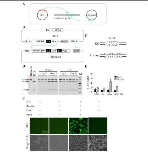
In order to knockout essential genes in cells, we de-signed a gene knockout-rescue system with double episomal vectors (Fig. 1a). Double vectors can accom-modate multiple genetic components. One plasmid which encodes Cas9 and gRNA for gene knockout was designated as KO (knockout) plasmid and the other plasmid which encodes the rescue gene was designated as Rescue plasmid. The advantage of the episomal vectors is that they can replicate in eukaryotic cells, allowing long-term Cas9 and gRNA expression [17]. The episomal vector used in this study was derived from Epstein-Barr virus (EBV) which contains two components essential for the epi-somal maintenance in cells: the latent origin oriP and
its binding protein Epstein-Barr-associated nuclear antigen 1 (EBNA1) [18]. In order to simultaneously retain two episomal plasmids in cells, the EBNA1 coding sequence was removed from the Rescue plas-mid, resulting in the episomal maintenance of the Rescue plasmid solely dependent on the KO plasmid (Fig. 1b). The Rescue plasmid encodes three genes: rescue gene, GFP and puromycin resistant gene (Puro), separated by self-cleaving T2A peptide. All three genes’ expression was controlled by the Tet-Off system. In order to reduce leaky expression, the tTA gene was encoded by the KO plasmid. Under puro-mycin selection, cells’ survival depends on the Rescue plasmid that expresses puromycin resistant gene. Therefore, cells’ survival requires the episomal main-tenance of both plasmids.
We first tested whether both plasmids can be simul-taneously maintained in cells. Both plasmids contain a common sequence that has an MfeI restriction site. To differentiate between these plasmids by restriction frag-ment length polymorphism (RFLP) assay, the MfeI re-striction site on the Rescue plasmid was destroyed by digestion and religation (Fig.1c). We co-transfected the same amount of each plasmid into different cell types and PCR-amplified the common region for the RFLP assay. Twenty-four hours post-transfection, a similar amount of both plasmids was detected (Fig. 1d, e and Additional file 1: Figure S2A). After 10 days of puro-mycin selection, higher amount of the KO plasmid was detected. A possible reason is that the KO plasmid retained the intact oriP/EBNA1 sequence which favors plasmid replication. To rule out the possibility that only one plasmid was present in a portion of cells, we trans-fected individual plasmid into cells and selected with puromycin for three days. As expected, neither KO plas-mid nor Rescue plasplas-mid could support cell’s survival (Fig. 1f ). Co-transfection of the two plasmids could support cells’survival (Fig.1f ). GFP expression increased over time with puromycin selection (Additional file 1: Figure S2B and C). We next tested the capacity of the Tet-Off system for regulation of GFP expression. Three days after addition of Dox, expression of GFP was efficiently shut down (Fig.1f, Additional file1: Figure S2B and C).
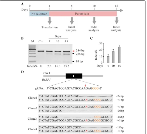
We next investigated the capacity of the knockout-rescue system for genome editing. We cloned a gRNA targeting exon1 of poly (ADP-ribose) polymerase 1 (PARP1) gene into the KO plasmid. The KO plasmid and Rescue plasmid were co-transfected into HEK293T cells with puromycin se-lection. At day 5, 10 and 15 after transfection, the indel fre-quency was analyzed by T7E1 assay (Fig.2a). As expected, the indel frequency increased over time (Fig.2b and c). We analyzed twenty single cell-derived clones by Sanger se-quencing and all of them were biallelic knockout (Fig.2d, Additional file1: Figure S3 and Table1). In summary, we
successfully established a double episomal vector system that enabled efficient genome editing. Hereafter, the double episomal vector system was designated krCRISPR (knock-out-rescue CRISPR).
The krCRISPR enabled knockout ofHDAC3gene
To investigate the capacity of the krCRISPR for essential gene knockout, we used this system to knockout histone deacetylase 3 (HDAC3) gene in human HEK293T cells.
HDAC3 is involved in apoptosis, cellular proliferation and DNA damage [19,20]. Due to the overexpression of
Fig. 2The krCRISPR system enables efficient gene knockout for thePARP1gene.aSchematic of the experimental workflow.bRepresentative gel pictures of T7E1 assay for detection of indels atPARP1sites in HEK293T cells. The indel frequency was labeled below. Ctr is the PCR band from unmodified cells with T7 enzyme digestion.cQuantification for the T7E1 assay for Fig.2b(n= 3, error bars showed mean ± SEM).dExamples of indel sequences for four single cell-derived clones. Schematic of the gRNA target site was shown above. PAM sequence is marked in orange. Cas9 cutting site is indicated by red arrow. Insertions are indicated by red letter
Table 1Efficiency of genome editing for single cell-derived clones
Heterozyote Homozygous WT Total
PARP1 5 12 3 20
DNMT1 3 16 1 20
HDAC3 3 15 2 20
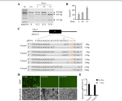
HDAC3in a variety of cancers, it is an important poten-tial target for cancer [19, 20]. It has been reported that deletion of HDAC3 is lethal for mouse embryos and mouse embryonic fibroblasts (MEFs) [21, 22]. High-throughput CRISPR/Cas9 screening revealed that deletion of HDAC3 is lethal in several human cell lines [6, 23–25]. A gRNA targeting exon7 of HDAC3 was cloned into the KO plasmid, and HDAC3 coding se-quence was cloned into the Rescue plasmid. To avoid cleavage by Cas9 nuclease, we created seven point mutations within the gRNA targeting sequence and the Protospacer Adjacent Motif (PAM) sequence that had no effects on the protein sequence (Additional file 1: Figure S4). The KO plasmid and Rescue plas-mid were co-transfected into HEK293T cells with puromycin selection. Similar to results for PARP1, the indel frequency increased over time (Fig. 3a and b). We analyzed 20 single cell-derived clones by using Sanger sequencing and 15 of them were biallelic knockout (Fig. 3c, Additional file 1: Figure S5 and Table 1). We further investigated the essentiality of the HDAC3 for cell viability by repressing exogenous
HDAC3 expression in two single cell-derived clones. After 3 days of Dox treatment, HDAC3 expression was turned off by monitoring GFP expression (Fig.
3d). The results were confirmed by qPCR with primers specifically targeting exogenous HDAC3 gene (Fig. 3e). Most cells were dead following 11 days of Dox treatment (Fig. 3d). A previous study in mouse embryonic fibroblasts (MEFs) has shown that Hdac3 knockout led to a delay in cell cycle progression, cell-cycle dependent DNA damage, and observed 20– 30% of cell death at day 5 after Hdac3 knockout [21]. In summary, these data demonstrated that the krCRISPR technology could knockout genes that are essential for cell survival.
The krCRISPR enabled knockout ofDNMT1gene
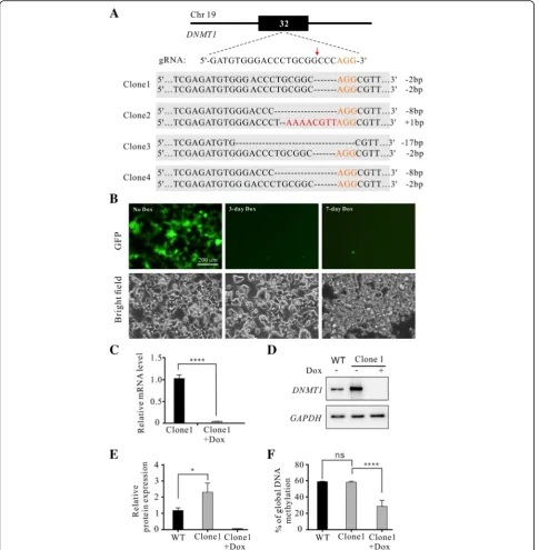
To demonstrate the capacity of the krCRISPR for essen-tial gene knockout, we showed another example of gene knockout by depleting DNMT1 which is one of the DNA methyltransferases for maintenance of DNA methylation over replication [26]. Deletion ofDNMT1is lethal for a variety of dividing somatic cells [3,27–29]. A gRNA targeting exon32 ofDNMT1 was cloned into the KO plasmid, and DNMT1 coding sequence was cloned into the Rescue plasmid. To avoid cleavage by Cas9 nu-clease, we created five point mutations within the gRNA targeting sequence and the Protospacer Adjacent Motif (PAM) sequence that have no effects on the protein se-quence (Additional file 1: Figure S4). The KO and Res-cue plasmids were co-transfected into HEK293T cells with puromycin selection for 15 days. Twenty single cell-derived clones were analyzed by Sanger sequencing
and 16 of them were biallelic knockout (Fig.4a, Additional file1: Figure S6 and Table1).
We further investigated the effects ofDNMT1 repres-sion on cell survival in two single cell-derived clones. After 3 days of Dox treatment, DNMT1 expression was shut down by indication of GFP expression (Fig.4b). At day seven after Dox treatment, the cells started undergo-ing apoptosis (Fig. 4b). A previous study in human em-bryonic stem cells has shown that all cells died within 9 days of DNMT1 knockout [3]. RT-qPCR analysis with primers specifically targeting exogenous DNMT1 gene revealed that DNMT1 expression was significantly downregulated (Fig. 4c). Western blot showed that the expression of DNMT1 in clone1 was higher than that in the WT cells at protein level, but it was un-detectable after Dox treatment at day seven (Fig. 4d and e). Luminometric methylation assay (LUMA) showed that the methylation level significantly de-creased after Dox treatment (Fig. 4f ). Altogether, these data demonstrated that we could readily obtain
DNMT1 homozygous mutant cell lines by using the krCRISPR technology.
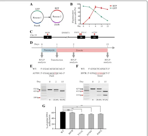
The krCRISPR enables to study effects of gene mutations In addition to gene knockout, the krCRISPR also enables us to study the effects of gene mutations. We hypothe-sized that if we established a knockout-rescue cell line with KO-Rescue1 plasmids, we could use another Res-cue plasmid (ResRes-cue2) to replace the original ResRes-cue1 plasmid. Rescue2 could encode genes that contain muta-tions of interest so that we can study them in the cells. To test whether the Rescue plasmid can be replaced by another plasmid, we designed a Rescue2 plasmid with zeocin resistance gene and RFP marker (Fig. 5a). We transfected this plasmid into cells established in Fig. 1f, where the Rescue plasmid (here we called it Rescue1) encoded GFP and puromycin resistant gene. Under zeocin selection, GFP was gradually replaced by RFP, in-dicating that the Rescue1 plasmid was replaced by Res-cue2 plasmid. At day 22, the percentage of GFP positive cells was only 17.5% and RFP was 72.5% (Fig. 5b and Additional file1: Figure S7A).
could influence DNA methylation has not been inves-tigated. Individual Rescue plasmids were transfected into the DNMT1 knockout cells with zeocin selection (Fig. 5d). To facilitate the analysis of plasmid replace-ment by RFLP, a restriction site was introduced into the DNMT1 gene without changing the protein se-quence of the Rescue2 plasmid (PmlI for A570V; XmaI for H97R) (Fig. 5e and f ). For A570V site, the ratio of the Rescue2 to total amount of plasmid DNA was 23.0% at day 2 and 93.9% at day 20, indicating that the Rescue1 plasmid was gradually replaced by Rescue2 (Fig. 5e). For H97R site, the ratio of the
Rescue2 to total amount of plasmid DNA was 29.4% at day 2 and 97.2% at day 20 (Fig. 5f ).
Next, we performed genome-wide methylation analysis for the individual mutations with LUMA. Compared to control Rescue plasmid expressing WT DNMT1, H97R variation did not influence DNA methylation level; Y511C and A570V mutation decreased DNA methylation levels, consistent with previous reports (Fig.5g) [34,36]. P507L also decreased DNA methylation level (Fig.5g), indicating that it could potentially be a disease-causing mutation. In summary, we established a platform that can be used to study the effects of mutations at cellular level.
Fig. 3Generation ofHDAC3knockout-rescue cell lines.aRepresentative gel pictures of T7E1 assay for detection of indels at HDAC3sites in HEK293T cells. Ctr is the PCR band from unmodified cells with T7 enzyme digestion.bQuantification of the T7E1 assay for Fig.3a (n= 3, error bars show mean ± SEM).c Examples of indel sequences for four single cell-derived clones. Schematic of the gRNA target site was shown above. PAM sequence was marked in orange. Cas9 cutting site is indicated by red arrow.dInhibition of exogenousHDAC3 expression inHDAC3-knockout cells caused cell death. TheHDAC3knockout-rescue cells expressed GFP. Expression of GFP was inhibited by addition of Dox for 3 days. All cells died at day 11.eRT-qPCR analysis of the exogenousHDAC3 expression with or without Dox for two clones (n= 3, error bars showed mean ± SEM)
Off-target analysis
Off-target mutations are often generated during genome editing [38, 39]. The krCRISPR system requires long-term
editing which may increase off-target effects. We used an online tool (http://www.rgenome.net/cas-offinder/) to search for potential off-target sites and selected five top
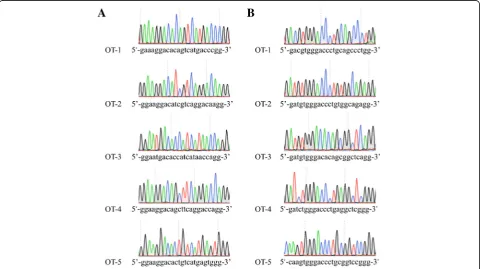
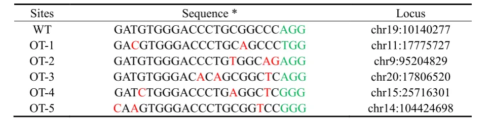
ranked potential off-target sites for gRNA-HDAC3. These potential off-targets have two or three mismatches com-pared to the targeting sequence (Table2). We tested these sites in twoHDAC3knockout-rescue clones that were de-rived from a single cell, but we did not observe off-target mutations (Fig. 6a and Additional file 1:
Figure S8A). We also analyzed five potential off-target sites for DNMT1 knockout-rescue clones, but we did not observe off-target mutations (Fig. 6b, Additional file 1: Figure S8B and Table 3). Notably, we could not exclude that off-target mutations occurred somewhere in the genome. Whole-genome sequencing will be
Fig. 5The krCRISPR system enabled analysis of the effects ofDNMT1mutation on DNA methylation.aSchematic of the Rescue1 and Rescue2 plasmid design. Rescue1 plasmid contains a puromycin resistant gene and a GFP gene, while Rescue 2 plasmid contains a zeocin resistant gene and RFP gene. Transfection of Rescue2 plasmid into the knockout-Resuce1 cells will result in replacement of Rescue1 plasmid by Rescue2 plasmid under zeocin selection.bFlow cytometry analysis showed that the GFP positive cells were gradually replaced by RFP positive cells over time (n= 3, error bars showed mean ± SEM).cDistribution of the fourDNMT1mutations.dSchematic of the experimental workflow.eandfTwo examples of Rescue plasmid replacement. The Rescue1 plasmid was gradually replaced by Rescue2 plasmid. A PmlI restriction site for A570V and an XmaI restriction site for H97R were introduced into the Rescue2 plasmids respectively. At day 0, 2 and 15, the plasmid DNA was isolated from cells and PCR-amplified for RFLP analysis. Gene mutations are labeled in red letter; the restriction sites are underlined. Black triangles indicate the Rescue1 plasmid; red triangles indicate the Rescue2 plasmid.gThe effects of individual mutations on DNA methylation were measured by LUMA. The exogenousDNMT1genes encoded by Rescue plasmids were shown below. H97R did not influence DNA methylation level. Y511C, A570V and P507L decreased DNA methylation level (n= 3, error bars showed mean ± SEM)
desirable for further detection of off-target mutations in the future.
Discussion
In this study, we demonstrated a simple and efficient method to knockout essential genes in cell lines using krCRISPR technology. This technology only requires two steps to obtain knockout-rescue cell lines: i) clone the gRNA into the KO vector and rescuing gene into the Rescue vector; ii) co-transfect both plasmids into cells and select single cell-derived clones with biallelic frame shift mutations. The expression of rescue genes can be efficiently turned off by Tet-Off technology, allowing the effects of gene knockout on cells to be studied. The krCRISPR enables efficient knockout of endogenous
genes due to the puromycin selection and long-term genome editing [17]. In contrast, previous strategies for essential gene knockout are time-consuming and labor-extensive, requiring multiple steps to generate stable integration cell lines that contain either Tet-Off elements or Cre/loxP elements [3,7–9,40,41].
In addition to gene knockout, the krCRISPR also en-ables users to study the effects of mutations on cells. Each human is estimated to carry on average ~ 60 de novo point mutations that arose in the germ line of their parents [42]. These mutations are the principal cause of heritable disease. Furthermore, genome-wide association studies (GWAS) have identified a large number of som-atic mutations that are associated with cancers [43, 44]. Although CRISPR/Cas9 technology allows efficient
Table 2Potential off-target sequences forHDAC3
*PAM sequences were marked in green and the mismatched nucleotides were labeled in red
introduction of specific mutations into the endogenous loci [45,46], it is time-consuming. The krCRISPR offers an alternative strategy to study gene mutations in cells. One can knockout endogenous genes and express rescue genes containing mutation of interest simultaneously. Once the knockout-rescue cell lines are established, the rescue plasmid can be replaced by other rescue plasmid containing different mutations. In summary, the krCRISPR technology offers a platform that facilitates the study of essential genes’functions.
Conclusions
In conclusion, we developed a double episomal vector sys-tem that allows generation of inducible knockout-rescue cell lines. In this system, one vector expresses gRNA and Cas9 nuclease, and the other vector expresses an inducible rescue gene. Users can easily knockout an essential gene by the expression of corresponding gRNA and rescue gene.
Materials and methods Cell culture and transfection
The HEK293T cell line (ATCC) was grown in Dulbecco’s Modified Eagle Medium (DMEM), supplemented with 10% fetal bovine serum (Gibco), 1x penicillin-streptomycin and passaged using 0.25% Trypsin-EDTA every other day. Cells were incubated at 37 °C with 5% CO2.
Plasmids
Knockout (KO) plasmid: this plasmid was modified from epiCRISPR plasmid [17]. The SpCas9 and tTA were co-expressed from an EF1αpromoter as a single protein separated by self-cleaving P2A peptides; gRNA was expressed from a human U6 promoter. The synthetic oligonucleotide duplexes encoding gRNAs can be cloned into BspQI restriction sites. The sequence of the plasmid is available in Additional file1: Figure S9.
The Rescue plasmid: copGFP and puromycin resist-ance genes were co-expressed from a pTRE promoter separated by two P2A peptides. The exogenous gene can be cloned into multiple cloning sites downstream of
pTRE promoter. The sequence of the plasmid is avail-able in Additional file 1: Figure S10. cDNA of DNMT1, PARP1 and HDAC3 was synthesized by GENWIZ (China) and inserted into KpnI/ AsisI restriction sites of the Rescue plasmid. Notably, synonymous mutations were introduced on gRNA targeting sequence to prevent Cas9 cleavage.
The Rescue2 plasmid: this plasmid is similar to Rescue plasmid except that copGFP-puro cassette was replaced with RFP-zeo cassette. The sequence of the plasmid is available in Additional file 1: Fig-ure S11. The DNMT1P507LDNMT1H97R, DNMT1A570v and DNMT1Y511C mutations were created on the
DNMT1WT using Gibson Assembly Cloning Kit
(#E5510S, NEB) according to the online protocol. The primers and oligonucleotides used in this study are shown in Additional file 1: Table S1.
Transfection, T7 assay and sequencing analysis for genome modification
HEK293T cells were seeded on 12-well plates in 500uL of growth medium without antibiotics. After 24 h, HEK293T cells were transfected at 60–70% confluency using Lipo-fectamine 2000 transfection regent (Invitrogen) according to the manufacturer’s protocol. For double-plasmid trans-fection, 500 ng of KO plasmid and 500 ng of Rescue plas-mid were transfected per well. From day 2, cells were selected by puromycin (1.5–2 μg/mL). At day 5, 10 and 15, genomic DNA was extracted from cells using Quick-Extract (Cat. # QE09050, Lucigen) following the manufac-turer’s instructions and T7E1 assay was performed according to a previously described method [10]. Briefly, genomic region containing the gRNA target site was PCR-amplified using Q5 High-Fidelity DNA polymerase (NEB) and the PCR products were purified using QIA-quick Gel Extraction Kit (28,706, QIAGEN). A total of 300 ng purified PCR products were re-annealed and digested with T7E1 enzyme (#M0302S, NEB) for 30 min at 37 °C. The PCR products were analyzed on 1.5% agar-ose gels. Gels were imaged with EB staining and quantified using ImageJ software according to the band intensities.
Table 3Potential off-target sequences forDNMT1
*PAM sequences were marked in green and the mismatched nucleotides were labeled in red
To analyze indel sequences for single cell-derived clones, cells were digested to single cells 10 days after transfection and seeded into 96-well plates using flow cytometer. A week later, genomic DNA of clones was extracted for PCR-amplification. The PCR products containing gRNA targeting sites were cloned into T-vector (A1410, Pro-mega) according to the manufacturer’s instructions for Sanger sequencing analysis. For plasmid replacement assay, 1μg of Rescue2 plasmid was transfected into knockout-rescue cells and selected with zeocin (300-400μg/mL) from day 2.
Protein extraction and Western blotting
Cell samples were harvested and lysed with NP-40 buffer (Beyotime) in the presence of 1 mM Phenylmethanesul-fonyl fluoride (Beyotime). After centrifugation at 12000 rpm for 10 min in a 4 °C pre-cooled centrifuge, the supernatant was collected for Western blot analysis. Pro-teins were separated by 8% SDS-PAGE gel and then transferred to a polyyinylidene fluoride (PVDF) mem-brane (Thermo). After blocking with 5% (wt/vol) BSA (Sigma) in TBS-T (0.1% Tween 20 in 1x TBS) buffer for 1 h at room temperature, the membrane was incubated with primary antibodies at 4 °C overnight. Antibodies used include: anti-DNMT1 (1:1000; ab13537 Abcam) and anti-GAPDH (1:2000, 5174S, Cell Signaling). After three washes with TBS-T of 5 min each, the membranes were incubated with secondary antibody (1:10,000; ab6721 Abcam) at room temperature for 1 h, followed by three washes and imaged.
RNA isolation and quantitative reverse transcription polymerase chain reaction
Total RNA was extracted from cells using Trizol regent (Ambion) following the manufacturer’s instructions. First-strand cDNA was synthesized from the isolated RNA using 5x All-In-One RT MasterMix kit (Cat. No. G492, abm) according to the manufacturer’s manual. 2x SYBR Green qPCR Master Mix (Cat. No. 21703, bimake) was used to quantify the expression of HDAC3 and DNMT1 mRNA. GAPDH was used as an internal con-trol for normalization. The primers were designed for amplification of exogenous gene in plasmid using Primer Premier 5.0. All primer sequences used are shown in Additional file1: Table S1. The qRT-PCR was performed using Bio-Rad Real-Time PCR. Detection System and the relative expression level was calculated using the 2-ΔΔCt method.
Flow cytometry
Cells for flow cytometry analysis were treated with 0.25% Trypsin-EDTA, washed twice and resuspended in 300uL PBS. The percentage of GFP and RFP positive cells was quantified using flow cytometer (Gallios, Beckman Coulter)
according to the manufacturer’s protocol. Data were ana-lyzed using FlowJo software.
Luminometric methylation assay (LUMA)
Firstly, total DNA was purified with phenol: chloroform. Briefly, cells were harvested and washed twice with PBS. Cells were then resuspended in 460uL of nuclear lysis buffer, 20uL of proteinase K (20 mg/mL) and 20uL of 10% SDS followed by incubation at 58 °C overnight. 5uL RNaseA (10 mg/mL) was then added, mixed by vortex-ing and incubated for 3 h at 37 °C. 500uL phenol:chloro-form was added, mixed by vortexing and incubated for 3 min at room temperature following centrifuged for 20 min at 13000 rpm. 400uL of aqueous phase containing DNA was then transferred to a fresh tube followed by the addition of 400uL isopropanol and vortexed for 30s, and finally 40uL NaAc (pH = 5.2) was added and vor-texed for 2 min. The remaining DNA precipitate was washed twice with 75% ethanol, and dissolved in 80uL ddH2O.
Subsequently, pyrosequencing was performed. 400 ng genomic DNA was cleaved with HpaII + EcoRI or MspI + EcoRI in two separate 20uL reactions containing 400 ng DNA, 1uL HpaII (or 0.5uL MspI), 0.5uL EcoRI-HF and 2uL 10x cutsmat buffer (NEB). The reactions were incubated at 37 °C for 4 h. Then pyrosequencing was performed following a previously described protocol [47]. The percentage of methylation was calculated based on the LUMA results with the following formula:
Methylation % = 100[1-(HpaII/EcoRI/MspI/EcoRI)].
Statistical analysis
In this study, statistical analysis was performed using GraphPad Prism 5. All data were presented as mean ± SEM. The unpaired Student’s t-test was adopted to de-termine the statistical differences between the samples of two groups. Significant levels: *P< 0.05, **P< 0.01, ****P< 0.001. All experiments were repeated three times independently.
Additional file
Additional file 1:Figure S1.Strategies for knockout of essential genes in cells.Figure S2.Establishment of the knockout-rescue system with double episomal vectors.Figure S3.Indel sequences atPARP1site. Figure S4.The gRNA targeting sequences on chromosome and their corresponding sequences on the rescue genes.Figure S5.Indel sequences atHDAC3site.Figure S6.Indel sequences atDNMT1site. Figure S7.Results of flow cytometry.Figure S8.Analysis of potential off-target sites.Figure S9KO plasmid sequence.Figure S10.Rsecue plasmid sequence.Figure S11Rsecue2 plasmid sequence.Table S1.Primers and oligonucleotides. (DOCX 3386 kb)
Abbreviations
PAM: Protospacer Adjacent Motif; RFLP: Restriction fragment length polymorphism; ssDNA: single-stranded DNA
Acknowledgements Not applicable.
Funding
This work was supported by grants from the National Natural Science Foundation of China (81870199), the National Basic Research Program of China (2015CB943300), the Foundation for Innovative Research Group of the National Natural Science Foundation of China (31521003) and Opening program 2018 of the State Key Laboratory of Genetic Engineering (SKLGE1809).
Availability of data and materials
All data generated or analyzed during this study are included in this published article.
Authors’contributions
Conceived and designed the experiments: BW, DW, YW Performed the experiments: BW, ZW, DW, BZ Analyzed the data: BW, BZ, ZW Contributed reagents/ materials: WY, ML Wrote the paper: YM, BW, SGO. All authors read and approved the final manuscript.
Ethics approval Not applicable.
Consent for publication Not applicable.
Competing interests
The authors declare that they have no competing interests.
Publisher’s Note
Springer Nature remains neutral with regard to jurisdictional claims in published maps and institutional affiliations.
Author details
1MOE Key Laboratory of Contemporary Anthropology at School of Life
Sciences and Zhongshan Hospital, Fudan University, Shanghai 200438, China.
2Shanghai Public Health Clinical Center & Laboratory of RNA Epigenetics,
Institute of Biomedical Sciences, Shanghai Medical College, Fudan University, Shanghai 201508, China.3Department of Pharmacology, University of Illinois
College of Medicine, Chicago, IL 60612, USA.4Division of Cardiology, Department of Medicine, University of Illinois College of Medicine, Chicago, IL 60612, USA.5The Key Lab of Reproduction Regulation of NPFPC in SIPPR,
Institute of Reproduction & Development in Obstetrics & Gynecology Hospital, Fudan University, Shanghai 200011, China.
Received: 27 December 2018 Accepted: 15 February 2019
References
1. Zhou Y, Zhu S, Cai C, Yuan P, Li C, Huang Y, Wei W. High-throughput screening of a CRISPR/Cas9 library for functional genomics in human cells. Nature. 2014;509:487–91.
2. Parnas O, Jovanovic M, Eisenhaure TM, Herbst RH, Dixit A, Ye CJ, Przybylski D, Platt RJ, Tirosh I, Sanjana NE, Shalem O, Satija R, Raychowdhury R, Mertins P, Carr SA, Zhang F, Hacohen N, Regev A. A genome-wide CRISPR screen in primary immune cells to dissect regulatory networks. Cell. 2015;162:675–86. 3. Liao J, Karnik R, Gu H, Ziller MJ, Clement K, Tsankov AM, Akopian V, Gifford
CA, Donaghey J, Galonska C, Pop R, Reyon D, Tsai SQ, Mallard W, Joung JK, Rinn JL, Gnirke A, Meissner A. Targeted disruption of DNMT1, DNMT3A and DNMT3B in human embryonic stem cells. Nat Genet. 2015;47:469–78. 4. Wang T, Wei JJ, Sabatini DM, Lander ES. Genetic screens in human cells
using the CRISPR-Cas9 system. Science. 2014;343:80–4.
5. Koike-Yusa H, Li Y, Tan EP, Velasco-Herrera Mdel C, Yusa K. Genome-wide recessive genetic screening in mammalian cells with a lentiviral CRISPR-guide RNA library. Nat Biotechnol. 2014;32:267–73.
6. Wang T, Birsoy K, Hughes NW, Krupczak KM, Post Y, Wei JJ, Lander ES, Sabatini DM. Identification and characterization of essential genes in the human genome. Science. 2015;350:1096–101.
7. Branda CS, Dymecki SM. Talking about a revolution: the impact of site-specific recombinases on genetic analyses in mice. Dev Cell. 2004;6:7–28. 8. Lewandoski M. Conditional control of gene expression in the mouse. Nat
Rev Genet. 2001;2:743–55.
9. Matsunaga T, Yamashita JK. Single-step generation of gene knockout-rescue system in pluripotent stem cells by promoter insertion with CRISPR/Cas9. Biochem Biophys Res Commun. 2014;444:158–63.
10. Cong L, Ran FA, Cox D, Lin S, Barretto R, Habib N, Hsu PD, Wu X, Jiang W, Marraffini LA, Zhang F. Multiplex genome engineering using CRISPR/Cas systems. Science. 2013;339:819–23.
11. Mali P, Yang L, Esvelt KM, Aach J, Guell M, DiCarlo JE, Norville JE, Church GM. RNA-guided human genome engineering via Cas9. Science. 2013;339:823–6. 12. Hwang WY, Fu Y, Reyon D, Maeder ML, Tsai SQ, Sander JD, Peterson RT, Yeh
JR, Joung JK. Efficient genome editing in zebrafish using a CRISPR-Cas system. Nat Biotechnol. 2013;31:227–9.
13. Jinek M, Chylinski K, Fonfara I, Hauer M, Doudna JA, Charpentier E. A programmable dual-RNA-guided DNA endonuclease in adaptive bacterial immunity. Science. 2012;337:816–21.
14. Ran FA, Hsu PD, Wright J, Agarwala V, Scott DA, Zhang F. Genome engineering using the CRISPR-Cas9 system. Nat Protoc. 2013;8:2281–308. 15. Wang Y, Zhang WY, Hu S, Lan F, Lee AS, Huber B, Lisowski L, Liang P,
Huang M, de Almeida PE, Won JH, Sun N, Robbins RC, Kay MA, Urnov FD, Wu JC. Genome editing of human embryonic stem cells and induced pluripotent stem cells with zinc finger nucleases for cellular imaging. Circ Res. 2012;111:1494–503.
16. Wang Y, Liang P, Lan F, Wu H, Lisowski L, Gu M, Hu S, Kay MA, Urnov FD, Shinnawi R, Gold JD, Gepstein L, Wu JC. Genome editing of isogenic human induced pluripotent stem cells recapitulates long QT phenotype for drug testing. J Am Coll Cardiol. 2014;64:451–9.
17. Xie Y, Wang D, Lan F, Wei G, Ni T, Chai R, Liu D, Hu S, Li M, Li D, Wang H, Wang Y. An episomal vector-based CRISPR/Cas9 system for highly efficient gene knockout in human pluripotent stem cells. Sci Rep. 2017;7:2320. 18. Van Craenenbroeck K, Vanhoenacker P, Haegeman G. Episomal vectors for
gene expression in mammalian cells. Eur J Biochem. 2000;267:5665–78. 19. Adhikari N, Amin SA, Trivedi P, Jha T, Ghosh B. HDAC3 is a potential
validated target for cancer: an overview on the benzamide-based selective HDAC3 inhibitors through comparative SAR/QSAR/QAAR approaches. Eur J Med Chem. 2018;157:1127–42.
20. West AC, Johnstone RW. New and emerging HDAC inhibitors for cancer treatment. J Clin Invest. 2014;124:30–9.
21. Bhaskara S, Chyla BJ, Amann JM, Knutson SK, Cortez D, Sun ZW, Hiebert SW. Deletion of histone deacetylase 3 reveals critical roles in S phase progression and DNA damage control. Mol Cell. 2008;30:61–72. 22. Montgomery RL, Potthoff MJ, Haberland M, Qi X, Matsuzaki S, Humphries KM,
Richardson JA, Bassel-Duby R, Olson EN. Maintenance of cardiac energy metabolism by histone deacetylase 3 in mice. J Clin Invest. 2008;118:3588–97. 23. Hart T, Chandrashekhar M, Aregger M, Steinhart Z, Brown KR, MacLeod G,
Mis M, Zimmermann M, Fradet-Turcotte A, Sun S, Mero P, Dirks P, Sidhu S, Roth FP, Rissland OS, Durocher D, Angers S, Moffat J. High-resolution CRISPR screens reveal fitness genes and genotype-specific Cancer liabilities. Cell. 2015;163:1515–26.
24. Tzelepis K, Koike-Yusa H, De Braekeleer E, Li Y, Metzakopian E, Dovey OM, Mupo A, Grinkevich V, Li M, Mazan M, Gozdecka M, Ohnishi S, Cooper J, Patel M, McKerrell T, Chen B, Domingues AF, Gallipoli P, Teichmann S, Ponstingl H, McDermott U, Saez-Rodriguez J, Huntly BJP, Iorio F, Pina C, Vassiliou GS, Yusa K. A CRISPR dropout screen identifies genetic vulnerabilities and therapeutic targets in acute myeloid leukemia. Cell Rep. 2016;17:1193–205.
25. Steinhart Z, Pavlovic Z, Chandrashekhar M, Hart T, Wang X, Zhang X, Robitaille M, Brown KR, Jaksani S, Overmeer R, Boj SF, Adams J, Pan J, Clevers H, Sidhu S, Moffat J, Angers S. Genome-wide CRISPR screens reveal a Wnt-FZD5 signaling circuit as a druggable vulnerability of RNF43-mutant pancreatic tumors. Nat Med. 2017;23:60–8.
26. Jones PA, Liang G. Rethinking how DNA methylation patterns are maintained. Nat Rev Genet. 2009;10:805–11.
27. Trowbridge JJ, Snow JW, Kim J, Orkin SH. DNA methyltransferase 1 is essential for and uniquely regulates hematopoietic stem and progenitor cells. Cell Stem Cell. 2009;5:442–9.
28. Sen GL, Reuter JA, Webster DE, Zhu L, Khavari PA. DNMT1 maintains progenitor function in self-renewing somatic tissue. Nature. 2010;463:563–7. 29. Jackson-Grusby L, Beard C, Possemato R, Tudor M, Fambrough D,
Csankovszki G, Dausman J, Lee P, Wilson C, Lander E, Jaenisch R. Loss of genomic methylation causes p53-dependent apoptosis and epigenetic deregulation. Nat Genet. 2001;27:31–9.
30. Saradalekshmi KR, Neetha NV, Sathyan S, Nair IV, Nair CM, Banerjee M. DNA methyl transferase (DNMT) gene polymorphisms could be a primary event in epigenetic susceptibility to schizophrenia. PLoS One. 2014;9:e98182. 31. Peng C, Deng Q, Li Z, Xiong C, Li C, Zheng F. Risk-association of DNMT1
gene polymorphisms with coronary artery disease in Chinese Han population. Int J Mol Sci. 2014;15:22694–705.
32. Ye C, Beeghly-Fadiel A, Lu W, Long J, Shu XO, Gao YT, Zheng W, Cai Q. Two-stage case-control study of DNMT-1 and DNMT-3B gene variants and breast cancer risk. Breast Cancer Res Treat. 2010;121:765–9.
33. Smets M, Link S, Wolf P, Schneider K, Solis V, Ryan J, Meilinger D, Qin W, Leonhardt H. DNMT1 mutations found in HSANIE patients affect interaction with UHRF1 and neuronal differentiation. Hum Mol Genet. 2017;26:1522–34. 34. Sun Z, Wu Y, Ordog T, Baheti S, Nie J, Duan X, Hojo K, Kocher JP, Dyck PJ, Klein CJ. Aberrant signature methylome by DNMT1 hot spot mutation in hereditary sensory and autonomic neuropathy 1E. Epigenetics. 2014;9:1184–93. 35. Winkelmann J, Lin L, Schormair B, Kornum BR, Faraco J, Plazzi G, Melberg A,
Cornelio F, Urban AE, Pizza F, Poli F, Grubert F, Wieland T, Graf E, Hallmayer J, Strom TM, Mignot E. Mutations in DNMT1 cause autosomal dominant cerebellar ataxia, deafness and narcolepsy. Hum Mol Genet. 2012;21:2205–10. 36. Kernohan KD, Cigana Schenkel L, Huang L, Smith A, Pare G, Ainsworth P,
Care4Rare Canada, C, Boycott KM, Warman-Chardon J, Sadikovic B. Identification of a methylation profile for DNMT1-associated autosomal dominant cerebellar ataxia, deafness, and narcolepsy. Clin Epigenetics. 2016;8:91.
37. Baets J, Duan X, Wu Y, Smith G, Seeley WW, Mademan I, McGrath NM, Beadell NC, Khoury J, Botuyan MV, Mer G, Worrell GA, Hojo K, DeLeon J, Laura M, Liu YT, Senderek J, Weis J, Van den Bergh P, Merrill SL, Reilly MM, Houlden H, Grossman M, Scherer SS, De Jonghe P, Dyck PJ, Klein CJ. Defects of mutant DNMT1 are linked to a spectrum of neurological disorders. Brain. 2015;138:845–61.
38. Ran FA, Hsu PD, Lin CY, Gootenberg JS, Konermann S, Trevino AE, Scott DA, Inoue A, Matoba S, Zhang Y, Zhang F. Double nicking by RNA-guided CRISPR Cas9 for enhanced genome editing specificity. Cell. 2013;154:1380–9. 39. Mali P, Aach J, Stranges PB, Esvelt KM, Moosburner M, Kosuri S, Yang L,
Church GM. CAS9 transcriptional activators for target specificity screening and paired nickases for cooperative genome engineering. Nat Biotechnol. 2013;31:833–8.
40. Yokoyama T, Miyazawa K, Naito M, Toyotake J, Tauchi T, Itoh M, Yuo A, Hayashi Y, Georgescu MM, Kondo Y, Kondo S, Ohyashiki K. Vitamin K2 induces autophagy and apoptosis simultaneously in leukemia cells. Autophagy. 2008;4:629–40.
41. Chen Y, Cao J, Xiong M, Petersen AJ, Dong Y, Tao Y, Huang CT, Du Z, Zhang SC. Engineering human stem cell lines with inducible gene knockout using CRISPR/Cas9. Cell Stem Cell. 2015;17:233–44.
42. Shendure J, Akey JM. The origins, determinants, and consequences of human mutations. Science. 2015;349:1478–83.
43. Sud A, Kinnersley B, Houlston RS. Genome-wide association studies of cancer: current insights and future perspectives. Nat Rev Cancer. 2017;17:692–704. 44. Tang H, Wei P, Chang P, Li Y, Yan D, Liu C, Hassan M, Li D. Genetic
polymorphisms associated with pancreatic cancer survival: a genome-wide association study. Int J Cancer. 2017;141:678–86.
45. Paquet D, Kwart D, Chen A, Sproul A, Jacob S, Teo S, Olsen KM, Gregg A, Noggle S, Tessier-Lavigne M. Efficient introduction of specific homozygous and heterozygous mutations using CRISPR/Cas9. Nature. 2016;533:125–9. 46. Salsman J, Dellaire G. Precision genome editing in the CRISPR era. Biochem
Cell biol. 2017;95:187–201.