R E V I E W
Open Access
Epigenetic assays for chemical biology and
drug discovery
Sheraz Gul
Abstract
The implication of epigenetic abnormalities in many diseases and the approval of a number of compounds that modulate specific epigenetic targets in a therapeutically relevant manner in cancer specifically confirms that some of these targets are druggable by small molecules. Furthermore, a number of compounds are currently in clinical trials for other diseases including cardiovascular, neurological and metabolic disorders. Despite these advances, the approved treatments for cancer only extend progression-free survival for a relatively short time and being
associated with significant side effects. The current clinical trials involving the next generation of epigenetic drugs may address the disadvantages of the currently approved epigenetic drugs.
The identification of chemical starting points of many drugs often makes use of screening in vitro assays against libraries of synthetic or natural products. These assays can be biochemical (using purified protein) or cell-based (using for example, genetically modified, cancer cell lines or primary cells) and performed in microtiter plates, thus enabling a large number of samples to be tested. A considerable number of such assays are available to monitor epigenetic target activity, and this review provides an overview of drug discovery and chemical biology and describes assays that monitor activities of histone deacetylase, lysine-specific demethylase, histone
methyltransferase, histone acetyltransferase and bromodomain. It is of critical importance that an appropriate assay is developed and comprehensively validated for a given drug target prior to screening in order to improve the probability of the compound progressing in the drug discovery value chain.
Keywords:Assay development, Bromodomain, Chemical biology, Chemical probe, Drug discovery, High
throughput screening, Histone acetyltransferase, Histone deacetylase, Histone methyltransferase, Demethylase
Background
Chemical biology makes use of chemistry to under-stand biological processes and this overlaps signifi-cantly with drug discovery, especially when the latter focusses on small molecules [1]. Chemical biology could also be considered to have a more basic re-search focus in that the rere-search is largely directed towards understanding fundamental biological pro-cesses with small molecules being used as tools to fa-cilitate this [2, 3]. This approach is complementary to molecular biological methods where mutations of resi-dues in proteins are utilized to determine the roles they play in biological processes. In many cases, the small molecules in chemical biology can also serve as starting points for drug discovery and this is
exemplified by the concept of “chemical probe” [4–8]. The key attributes of a “chemical probe” includes defined mechanism of action, appropriate selectivity, often being freely available (both the physical com-pound and activity data), possessing drug-like proper-ties and being associated with a reliable structure-activity relationship (SAR). These attributes are also relevant for lead compounds, clinical candidate mole-cules and drugs, but will also have additional attri-butes such as intellectual property rights, human bioavailability, and appropriate physicochemical and pharmaceutical properties.
Drug discovery is a high risk, expensive and lengthy process, typically lasting 10 years with defined phases [9]. The pre-clinical stage of drug discovery, sometimes also referred to the gene-to-candidate phase, can span a period of 5 years before the compound is suitable for human clinical trials. During this stage, a target deemed
Correspondence:Sheraz.Gul@ime.fraunhofer.de
Fraunhofer Institute for Molecular Biology and Applied Ecology -ScreeningPort, Schnackenburgallee 114, 22525 Hamburg, Germany
worthy of therapeutic intervention is identified and subsequently a biological reagent (usually purified pro-tein or cell line) is prepared that contains the target of interest. In the case of small-molecule drug discovery, this biological reagent would then be utilized to de-velop an appropriate assay for monitoring target activity and screened against libraries of small mole-cules (hundreds to millions of compounds) [10–12]. Evaluation of the active compounds from the screening campaign (hits) with freshly synthesized compounds meeting acceptable purity and integrity in a panel of relevant assays would ultimately yield a validated hit list comprising a data package pertaining to the biological activity [13]. Each validated hit series would then be an-notated with additional data such as the Lipinski rule of five [(i) molecular weight less than 500, (ii) logP, a par-tition coefficient measuring hydrophobicity less than five, (iii) no more than five hydrogen bond donors and (iv) no more than 10 hydrogen bond acceptors]. Bear-ing in mind the high attrition of drug discovery, more than one of the most promising validated hit series would be progressed to the hit-to-lead (H2L) phase [14]. Several iterative rounds of synthesis would enable the optimisation of the potency of compounds against the target of interest to the desired criteria for a lead series (typically in the sub-micromolar range) whilst retaining an appropriate selectivity profile. Additional information required when selecting the final lead series will include demonstrable and acceptable SAR, off-target selectivity profile, toxicity, physicochemical pro-file, solubility and stability in aqueous solution and human plasma, in vivo pharmacokinetics, Absorption, Distribu-tion, Metabolism and Excretion (ADME) properties, pat-entability and competitor activity. Further significant optimisation of a compound within the lead series would result in the generation of a pre-clinical candidate com-pound and, upon approval by the relevant regulatory orga-nizations, can enter human clinical trials [9].
In the post-Human Genome Project era [15], target-based drug discovery accelerated considerably and is fittingly illustrated by the kinase target class [16]. A consequence of target-based drug discovery has been the multitude of assays being available for most target classes and the remainder of this article focusses on general concepts of assay development with a specific focus on screening compatible assays for epigenetics targets, and Table 1 provides a summary of the as-says. Many of the epigenetic assays reported in the literature and referred to herein make use of com-mercial extensively validated kits. Where possible, ori-ginal references are cited that would enable an understanding of the rationale for the development of epigenetic assays and their utilization in a variety of research activities.
Assay development, high throughput and high content screening in pre-clinical drug discovery Assays that are screened against libraries of compounds to identify chemical starting points in the early stages of drug discovery can be classified as being biochemical or cell-based in nature. The exact assay that is utilized in a screen is decided upon a case-by-case basis after taking into account a number of factors such as provision of reagents, throughput, cost, and many others that have been discussed extensively in the literature [17]. The biochemical target-based (reductionist) approach was largely adopted in the post-Human Genome Project era where specific genes were identified and cloned and the corresponding proteins expressed in sufficient quantity with acceptable activity for screening [18]. This was a marked shift from earlier cell-based assays where modulation of specific targets did not occur, but in-stead relevant cellular phenotypic responses were mea-sured [19, 20]. Significant effort has been expended to mimic these physiologically relevant cell-based systems with a significantly higher throughput [21] and ad-vances have been made using a variety of these and subsequently deployed in cancer drug discovery in par-ticular [22–24] as well as being expanded to areas such as predictive toxicology [25].
Table 1Screening compatible epigenetic assays
Enzyme Assay format Key features of the assay References
Histone deacetylase (HDAC)
Chemiluminescent (AlphaLISA®) •Assay reported in literature and commercial validated assay kit
•Substrate: histone proteins •Detection: H3-K9(Ac) or H3-K27(Ac) •High sensitivity
•High throughput functional assay
122–124
Chromatin immunoprecipitation •Assay reported in literature using specific commercial reagents
•Detection: Ac-H3 •High sensitivity •Low throughput assay
125
Colorimetric (Color de Lys®) •Commercial validated assay kit
•Substrate: peptide containingε-acetylated lysine •Detection: deacetylated peptide via coupled assay •Low sensitivity
•Low/Medium throughput functional assay •Prone to optical interference with compounds
126
Fluorometric (Fluor de Lys®) •Assay reported in literature and commercial validated assay kit
•Substrate: peptide containingε-acetylated lysine •Detection: deacetylated peptide via coupled assay •Medium/High sensitivity
•Medium/High throughput functional assay
127, 128
Luminescence (HDAC-Glo™I/II) •Assay reported in literature and commercial validated assay kit
•Substrate: peptide containingε-acetylated lysine •Detection: deacetylated peptide via coupled assay •High sensitivity
•Medium/High throughput functional assay
131, 132
TR-FRET (LANCE® Ultra) •Uses specific commercial reagents
•Substrate: biotinylated Histone H3-K27(Ac) or Histone H3-K9(Ac) peptide
•Detection: H3-K9(Ac) or H3-K27(Ac) •High sensitivity
•High throughput functional assay
133
TR-FRET (LanthaScreen™) •Assay reported in literature and commercial validated assay kit
•Ligand: Alexa Fluor® 647-labelled HDAC inhibitor as a tracer •Detection: displacement of Alexa Fluor® 647-labelled
HDAC inhibitor •High sensitivity
•High throughput binding assay
134
Demethylase (LSD and Jumonji C domain-containing histone demethylase)
Colorimetric •Assay reported in literature using specific commercial reagents
•Substrates: Histone H3-K4 peptide •Detection: H2O2or H3-K4 via coupled assay •Low sensitivity
•Low throughput functional assay
•Prone to optical interference with compounds
139–142
Colorimetric (Epigenase™) •Commercial validated assay kit
•Substrates: Histone H3-K4(Me2) or dimethylated Histone H3-K4 peptide
•Detection: H2O2via coupled assay •Low sensitivity
•Low throughput functional assay •Requires wash steps
•Prone to optical interference with compounds
143
Fluorescence polarization •Assay reported in literature using methylstatfluortracer •Ligand: methylstatfluortracer
•Detection: displacement of methylstatfluortracer •High sensitivity
•High throughput binding assay
144–145
Table 1Screening compatible epigenetic assays(Continued)
•Substrate: Histone H3-K4(Me2) peptide •Detection: H2O2via coupled assay •Low sensitivity
•Low throughput functional assay •Requires wash steps
•Prone to optical interference with compounds Fluorometric •Commercial validated assay kit
•Substrate: Histone H3-K4(Me2) protein •Detection: formaldehyde via coupled assay •Low sensitivity
•Low throughput functional assay •Requires wash steps
•Prone to optical interference with compounds
148–149
High content screening •Assay reported in literature using specific commercial reagents
•Substrate: Histone H3-K27(Me)3peptide •Detection: H3-K27(Me)3
•Medium sensitivity
•Medium throughput functional assay
150
Mass spectrometry •Assay reported in literature using specific commercial reagents
•Substrate: dimethylated peptides •Detection: demethylated products •Medium/High sensitivity •Low throughput functional assay •No optical interference from compounds
151, 152
Radioactive •Assay reported in literature using specific commercial reagents
•Substrate:3H-labelled methylated histone •Detection:3H-formaldehyde via coupled
functional assay
•No optical interference from compounds •Radioactive waste
153, 154
TR-FRET (LANCE® Ultra) •Commercial validated assay kit
•Substrate: biotinylated Histone H3-K4(Me) peptide •Detection: H3-K4
•High sensitivity
•High throughput functional assay
155
Histone methyltransferase (HMT) Chemiluminescent (AlphaLISA®) •Commercial validated assay kit •Substrate: Histone H3-K79(Me2) protein •Detection: H3-K79(Me2)
•High sensitivity
•High throughput functional assay
161
Fluorescence polarization •Assay reported in literature using specific commercial reagents
•Substrate: protein or peptide
•Detection: displacement of labelled-AMP in coupled assay
•High sensitivity
•High throughput binding assay
162
Fluorometric •Assay reported in literature using specific commercial reagents
•Substrate: Histone H3 peptide
•Detection: homocysteine via coupled assay •High sensitivity
•High throughput functional assay
163–165
High content screening •Assay reported in literature using specific commercial reagents
•Substrate: Histone H3-K27(Me)3 •Detection: H3-K27(Me)3 •Medium sensitivity
•Medium throughput functional assay
166
Luminescence •Assay reported in literature using specific commercial reagents
Table 1Screening compatible epigenetic assays(Continued)
•Substrate: protein or peptide •Detection: complex coupled assay •High sensitivity
•High throughput functional assay Radiometric •Assay reported in literature using specific
commercial reagents
•Substrate: biotinylated Histone H3-K9 peptide •Detection:3H-incorporated into peptide •No optical interference from compounds •Radioactive waste
168–170
Histone acetyltransferase (HAT) Colorimetric •Commercial validated assay kit •Substrate: proprietary peptide •Detection: NADH via coupled assay •Low sensitivity
•Low/Medium throughput functional assay •Prone to optical interference with compounds
174
ELISA •Commercial validated assay kit
•Substrate: histone
•Detection: H3-K4(Ac) via coupled assay •Low/Medium sensitivity
•Requires wash steps
•Medium throughput functional assay
175
Fluorometric •Assay reported in literature using specific commercial reagents
•Substrate: Histone H3 or Histone H4 peptide •Detection: CoA-SH via coupled assay •High sensitivity
•High throughput functional assay
•Prone to optical interference with compounds
176
Fluorometric •Commercial validated assay kit •Substrate: Histone protein
•Detection: CoA-SH via coupled assay •High sensitivity
•High throughput functional assay
177
Microfluidic mobility shift •Assay reported in literature using specific commercial reagents
•Substrate: fluorolabelled Histone H3 or Histone H4 peptide
•Detection: charge difference of substrate/product •High sensitivity
•Medium throughput functional assay •No optical interference from compounds
178
Radiometric •Assay reported in literature using specific commercial reagents
•Substrate: biotinylated Histone H4 peptide or histone protein
•Detection:3H-incorporated into peptide or histone •High sensitivity
•Low throughput functional assay •No optical interference from compounds •Radioactive waste
179–181
TR-FRET(LANCE® Ultra) •Commercial validated assay kit
•Substrate: biotinylated Histone H3-K9 peptide •Detection: H3-K9(Ac)
•High sensitivity
•High throughput binding assay
182
Bromodomain Chemiluminescent (AlphaScreen™) •Assay reported in literature using specific commercial reagents
•Substrate: biotinylated Histone H4-K5(Ac) •Detection: presence of BRD4/peptide complex •High sensitivity
•High throughput binding assay
188, 189
Differential Scanning Fluorometry (BromoMELT™)
•Assay reported in literature and commercial validated assay kit
redistribution [41, 42] and multiplex assays [43, 44], and these have been successfully applied in screening against small-molecule libraries. More recent state-of-the-art screening compatible assays use three-dimensional spher-oids that offer the potential to represent the microenviron-ment of cells in the body [45].
The pre-requisites for high throughput screening (HTS) are access to a suitable assay as briefly described above and a suitable compound library. Compound libraries are usually stored in pure DMSO at concentrations between 1 mM and 10 mM as this will allow for a range of final assay concentrations of compound whilst retaining <1% DMSO (v/v) in the final assay. The extent of automation when embarking upon an HTS campaign will depend upon the numbers of compounds screened and it would be reasonable to screen a compound library composed of a few thousand compounds manually in miniaturized
formats (e.g. 384- or 1536-well microtiter plates). How-ever, where >5000 compounds are screened (in 384-well microtiter plates), it would be prudent to use some degree of automation such as stand-alone reagent dispensers or a robotic screening system [46–49]. One way to minimize the consumption of reagents when screening very large numbers of compounds is to miniaturize and parallelize an assay into 1536-well microtiter plates [50]. However, such miniaturization requires the addition of very small volumes of compound stock solutions and technologies such as the contactless acoustic dispenser from Labcyte Inc. makes this possible [51].
High content screening (HCS) is now an established technique that is routinely utilized in chemical biology and drug discovery and has made a significant impact upon understanding the output of phenotypic screening. This is a cell-based approach that can offer a
multi-Table 1Screening compatible epigenetic assays(Continued)
•Substrate: BRD4
•Detection:Tmof BRD4/SYPRO Orange complex •Low/Medium sensitivity
•Medium throughput binding assay •No optical interference from compounds Fluorescence polarization •Assay reported in literature using specific
commercial reagents
•Substrate: fluorescent BODIPY labelled tracer •Detection: BRD4/BODIPY labelled tracer complex •Medium sensitivity
•Medium throughput binding assay
190
TR-FRET •Commercial validated assay kit
•Substrate: biotinylated peptide
•Detection: BRD4/biotinylated peptide complex •High sensitivity
•High throughput binding assay
192
Streptavidin Donor beads
Histone H3-K9(Ac) or Histone H3-K27(Ac) Biotinylated-anti-H3 antibody
Acceptor beads conjugated to anti-H3-K9(Ac) or H3-K27(Ac)
antibody
680 nm 615 nm
a
b
Fig. 2aColorimetric coupled histone deacetylate assay that makes use of a chromogenic peptide substrate (proprietaryColor de Lys® Substrate) containing aε-acetylated lysine residue. When an HDAC enzyme acts upon the substrate and the sidechain of aε-acetylated lysine residue is deacetylated, it becomes susceptible to further degradation by an enzyme in the developer reagent (proprietaryColor de Lys® Developer). The action of the enzyme within the developer reagent results in the release of a chromophore detected by measuring the absorbance of the reaction at 405 nm.bFluorometric coupled histone deacetylate assay that makes use of a fluorogenic peptide substrate (proprietaryFluor de Lys® Substrate) containing aε-acetylated lysine residue. When an HDAC enzyme acts upon the substrate and the sidechain of aε-acetylated lysine residue is deacetylated, it becomes susceptible to further degradation by an enzyme in the developer reagent (proprietaryFluor de Lys® Developer) resulting in the release of 7-amino-4-methylcoumarin fluorophore which undergoes excitation at 360 nm and emits at 460 nm
HDAC
Developer
luciferase “glow-type” luminescence
Acetylated peptide substrate Deacetylated peptide
NH3+
parameter readout detecting simultaneously a multitude of cellular changes that are subsequently attributed to specific targets [52–56]. This approach is particularly relevant in epigenetics as the discovery of Romidepsin and Vorinostat as anti-cancer drugs originates from phenotypic assays [57].
General concepts underlying the common deployed screening compatible assays Amplified Luminescent Proximity Homogeneous (AlphaLISA® and AlphaScreen®) assays
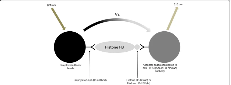
These are proximity-based assays that have successfully been used to study the activity of a wide range of targets [58–61]. The technology requires two bead types, termed donor beads and acceptor beads with the former containing the photosensitizer phthalocyanine, which converts ambient oxygen to an excited and reactive singlet oxygen upon illumination at 680 nm. This react-ive singlet oxygen can diffuse approximately 200 nm in solution and has a half-life of 4μs. If an acceptor bead is within that distance, energy is transferred from the singlet oxygen to thioxene derivatives within the ac-ceptor bead, resulting in light production at 520– 620 nm (AlphaScreen®) or at 615 nm (AlphaLISA®) [62]. These assays do not require wash steps unlike in a standard ELISA, see Fig. 1.
Colorimetric assays
These rely upon the difference in the electronic absorp-tion spectrum of the substrate and product of a reac-tion. Chromogenic substrates are composed of organic
molecules that contain a conjugated system, i.e. a deloca-lized π-bond system which is usually attributed to alter-nating single and double bonds. When chromophores absorb ultraviolet (UV) and visible radiation, their elec-trons undergo excitation from their ground state to excited state and the wavelength of UV or visible light (approximately 200–800 nm) absorbed depends largely on the extent of conjugation, such that the greater the degree of conjugation within the chromophore, the longer the wavelength of light will be absorbed [63, 64]. In some cases, both the substrate and product will absorb light and it will be necessary to monitor the formation of product where the absorption of the substrate does not change. Additionally, the optimal wavelength at which product formation can be detected should be determined after collecting the absorption of pure samples of substrate and product. When the natural substrate is itself chromogenic, this offers the potential to monitor the activity of an enzyme without the need for a synthetic chromogenic substrate. Thus obviating the effects of steric hindrance by an artificial chromophore in the molecule that can inter-fere with binding within the active centre of the enzyme and potentially confound the identification of substrate competitive compounds. Despite the successful use of colorimetric assays in screening, they are no longer the preferred option and have largely been replaced by alter-native assay formats such as fluorescence-based methods [65, 66]. This has been driven by a number of reasons such as colorimetric assays being relatively insensitive, often requiring substantial concentrations of product (typically low micromolar) to be generated for adequate
Biotinylated Histone H3-K9(Ac) or histone
H3-K27(Ac) peptide substrate streptavidin-ULight™-Acceptor
Europium chelate Donor (W1024-Eu) linked to an anti-H3-K27(Ac) or anti-H3-K9(Ac) antibody
340 nm 665 nm
NH3+
HDAC
streptavidin-ULight™-Acceptor
615 nm
detection. Colorimetric assays are also particularly prone to optical interference due to coloured compounds which are commonly found in small-molecule libraries. These optically interfering compounds are likely to result in many of these being identified as apparent hits in a small-molecule screening campaign but subsequently shown not to be genuine modulators of the activity of the target protein [34, 37, 38, 67–69]. These false positives need to be identified and removed prior to the progression of compounds for drug discovery purposes. One strategy to reduce the number of apparent hits being overrepresented with optically interfering compounds is to determine the activity of the target protein in the presence of compound in kinetic mode; however, this will reduce the throughput of the assay [70].
Differential scanning fluorimetry assays
This technique makes use of dyes that are fluorescent when present in a non-polar environment such as hydro-phobic sites of unfolded proteins relative to aqueous solution (in the case of unfolded proteins) where their fluorescence is quenched [71]. When lowMrligands bind
and stabilize proteins, the temperature at which this com-plex unfolds will be raised and this can be quantified from a fluorescence–temperature plot, with the midpoint of the protein unfolding transition defined as the Tm (melting
temperature), reflecting the potency of the lowMrligand
towards the protein [72–75].
Enzyme-linked immunosorbent assay (ELISA)
This technique is used in a variety of industries including diagnostics and quality-control checks [76]. In most cases an ELISA involves an antigen being immobilized to a sur-face that is capable of capturing a molecule that resembles the antigen. Subsequent to a series of wash steps to remove non-specifically bound proteins, a secondary antibody is ap-plied that is linked to an enzyme and the enzyme substrate is added that yields a signal, usually colorimetric or fluoro-metric [77–79]. The major drawback of ELISA from a screening perspective is their non-homogenous nature and the requirement for wash steps [80].
Fluorescence polarization assays
This technique relies upon a change in the hydro-dynamic radius of a fluorescent entity (when bound to a protein and free in solution) that alters its hydrodynamic radius [81–83]. Most of these assays are based on an indirect measurement of the size change of a protein and fluorescently labelled ligand. A requirement for this technique is the easy conjugation of a fluorophore to a relevant molecular entity. Binding of this ligand would result in a relatively high fluorescence polarization sig-nal. Its displacement from the target by a competitor
molecule would lead to a decrease in the fluorescence polarization signal [84, 85].
Fluorescence intensity assays
These have been used extensively in drug discovery and offer a number of advantages over colorimetric assays such as being significantly more sensitive and less prone to optical interference [86]. There are a large number of fluorophores available that cover most of the electromag-netic spectrum. As a result it is possible to design and synthesize molecules that contain these fluorophores in order to enable them to be employed as tools to develop assays for the investigation of difficult drug targets [87, 88]. Fluorescein has been widely used as a fluor-ophore in assays but others are available that are associ-ated with reduced compound-mediassoci-ated interference [89].
High content screening assays
These make use of a microscope-based method to image cells that can categorize multiple features when using appropriate fluorescent dyes. The image analysis requires algorithms to allow their categorization, espe-cially after exposure to compounds [90, 91]. These assays can be enhanced when working with primary cells and three-diensional cultures that are more physiologically relevant [92].
Luminescence assays
These use enzymes such as luciferases, and the comple-mentary luciferin photon-emitting substrates [93, 94]. The most widely used enzymes are firefly luciferase, Renilla luciferase, and aequorin [95–97]. In the case of firefly luciferase-based assays, beetle luciferin and ATP are combined to form luciferyl-AMP (an enzyme-bound intermediate). This reacts with O2to create oxyluciferin
in a high-energy state and a subsequent energy transi-tion to the ground state yielding light.
Mass spectrometry assays
This has been a longstanding technique and used as a secondary assay due to its relatively low throughput, or for screening modest libraries of compounds [98, 99]. It is a label-free approach as it relies upon the separation and subsequent quantification of typically a substrate and product that has undergone modification that the mass spectrometer is capable of detecting [100]. The current instrument for high throughput mass spectrom-etry is the Agilent RapidFire that has been used to screen a range of targets with improved quality of the identified hit compounds [101–103].
Microfluidic mobility shift assays
been most successfully used to investigate the kinase target class [104]. Although these assays have a low throughput, the major advantage they offer is to over-come compound-mediated optical interference, as it is separated during the electrophoretic separation of substrate and product [105]. The assay requires a fluo-rescently labelled substrate that can be used to detect both the product and any residual substrate [106].
Radioactive assays
These assays make use of radioisotopes such as3H,14C,
33
P, 35S and 125I. Filter-binding assays have historically been extensively adopted to monitor the activity of a wide range of targets [107, 108]. In the case of the neurotransmitter targets, these assays are considered to be the gold standard assay format as they are label-free, highly sensitive and are not prone to interference in a manner that the other optical methods are susceptible [109]. An advancement to these assays is the no-wash scintillation proximity assay (SPA) which makes use of beads embedded with scintillant that can bind the target of interest and give a signal [110–112].
Time-resolved-Förster resonance energy transfer assays
This is a proximity-based assay that makes use of lanthanide chelate complexes with a long-lived lumines-cence in comparison with conventional fluorophores. Therefore, enabling the short-lived background interfer-ences that are predominantly compound mediated to be removed [113]. Typically, a TR-FRET signal is generated when a molecule coupled to the Europium-labelled partner (donor) is brought into close proximity to an acceptor molecule, e.g. Allophycocyanin (APC). Upon irradiation at 340 nm, the energy from the Europium-donor is transferred to the acceptor which in turn gener-ates a signal at 665 nm, Fig. 4 [114].
The histone deacetylase (HDAC) target class and relevant screening compatible assays
The HDAC family of enzymes remove an acetyl group from acetylated lysine residues in appropriate substrates (both histone and non-histone based) [115, 116]. This protein target class has been implicated in cancer [117, 118], cardiovascular [119], inflammatory and in-fectious diseases [120] and neurodegeneration [121].
HDAC AlphaLISA®assays
A commercial assay kit is available that detects changes in the levels of Histone H3-acetylated lysine 9 (H3-K9(Ac)) and Histone H3-K27(Ac) in cellular systems [122–124]. The changes in the levels of acetylated histones is performed with histones extracted from cells, followed by addition of a biotinylated anti-H3 antibody and AlphaLISA®-acceptor beads conjugated specific to
the acetylated lysine. Streptavidin-donor beads then cap-ture the biotinylated antibody, bringing the acceptor and donor beads into proximity. Upon laser irradiation of the donor beads at 680 nm, short-lived singlet oxygen molecules produced by the donor beads can reach the acceptor beads in proximity to generate an amplified chemiluminescent signal at 615 nm (Fig. 1). Due to the nature of the assay, any changes in the observed signal may be due to reasons other than HDAC inhibition; therefore, care needs to be taken when interpreting the data. As this assay format is essentially proximity based, it can be employed to monitor the modification of a var-iety of molecules such as appropriately labelled peptide and protein substrates (e.g. biotin, FLAG, GST, His) that undergo acetylation, demethylation, methylation as well as phosphorylation (in the case of kinases) when using an antibody against a specific modulation. A chromatin immunoprecipitation (ChIP) assay has also been re-ported to extract and quantify Ac-H3 from a cellular system [125].
HDAC colorimetric assay
In contrast to the above, a commercial HDAC-specific coupled assay kit is available that makes use of a chromogenic peptide substrate (proprietaryColor de Lys® Substrate) containing aε-acetylated lysine residue [126]. When an HDAC enzyme acts upon the substrate and the sidechain of aε-acetylated lysine residue is deacety-lated, it becomes susceptible to further degradation by an enzyme in the developer reagent (proprietary Color de Lys® Developer). The action of the enzyme within the developer reagent results in the release of a chro-mophore detected by measuring the absorbance of the reaction at 405 nm (Fig. 2a). Since this is a colorimetric-based assay, it is generally of low sensitivity and prone to optical interference.
HDAC fluorometric assays
HDAC luminescence assays
This is another commercial coupled assay kit simi-lar to those described above (colorimetric and fluo-rometric) but makes use of specific amino-luciferin labelled ε-acetylated lysine peptide substrates for HDAC Class I/II enzymes (Promega Corp. HDAC-Glo™ I/II Assays and Screening Systems) [131]. When the substrate undergoes deacetylation by the HDAC enzyme, the product becomes susceptible to the developer reagent and results in the release of amino-luciferin. This amino-luciferin is the substrate for a luciferase enzyme (also in the developer re-agent) and yields a glow-type luminescence (Fig. 3). This assay has been validated and used to screen a natural product library [132].
HDAC TR-FRET assay
This is a commercial assay kit (LANCE® Ultra TR-FRET) that uses a proprietary Europium chelate donor (W1024-Eu) linked to an K27(Ac) or anti-H3-K9(Ac) antibody, together with streptavidin-ULight™ -ac-ceptor. The available substrates for this are biotinylated Histone H3-K9(Ac) and Histone H3-K27(Ac) peptides. A TR-FRET signal is generated when the unmodified peptides are captured by the Europium-labelled anti-body donor and streptavidin-ULight™ acceptor that brings the Europium-donor and ULight™-acceptor molecules into close proximity. Upon irradiation at 340 nm, the energy from the Europium-donor is trans-ferred to the ULight™-acceptor, which in turn generates a signal at 665 nm (Fig. 4) [133].
A similar assay has been reported that is based on measuring the binding affinity of inhibitors rather than enzyme activity. As catalytically functional protein is not required, there is no requirement for a substrate and instead an Alexa Fluor® 647-labelled HDAC inhibitor is used as a tracer (acceptor in the TR-FRET assay). This can bind with specific HDAC enzymes tagged with GST in the presence of Europium anti-GST tag antibody (donor in the TR-FRET assay) and if the tracer is dis-placed by an appropriate compound, a decrease in signal would be observed [134].
The demethylase target class and relevant screening compatible assays
The demethylase family of enzymes are responsible for the demethylation of lysine and arginine side chains in appropriate substrates (both histone and non-histone based) [135]. Specific examples of pro-teins in this class include lysine-specific demethylase (LSD) and the Jumonji C domain-containing histone demethylase (JHDM). This protein target class has been implicated in cancer [136], diabetes [137] and cardiovascular disease [138].
LSD colorimetric assay
In this assay, the activity of human LSD1 makes use of dimethylated Histone H3-K4 peptide. This is a coupled assay in which the oxidative demethylation reaction catalysed by LSD1 results in the production of hydrogen peroxide (H2O2) [139–141]. This, in the presence of
3,5-dichloro-2-hydroxybenzenesulfonic acid and horseradish peroxidase (HRP), results in an absorbance change at 515 nm [142].
A commercial coupled assay kit is also available (Epigenase™LSD1 Demethylase Activity/Inhibition Assay Kit) that makes use of a chromogenic peptide substrate. In the assay, microtiter plates coated with Histone H3-K4(Me2) LSD1 substrate are used, after which addition
of LSD1 results in the removal of substrate methyl groups. After a wash step, the Histone H3-K4 demethy-lated product recognition takes place using a specific antibody and subsequently the colorimetric signal gener-ated at 450 nm after addition of a proprietary detection mix (making use of the H2O2or formaldehyde released
as the by-product of LSD1 enzymatic reaction) [143].
Jumonji C domain-containing histone demethylase fluorescence polarization assay
The crystal structure of histone demethylases have used in a structure-based drug design exercise to develop a substrate-derived inhibitor for Jumonji C domain-containing histone demethylase, termed methylstat [144]. This compound was shown to be active in vitro against isolated protein in a mass spectrometry (meas-uring H3-K9(Me3)) and in a cell-based HCS assay
(measuring H3-K9(Me3)) using immunostaining with
an anti-H3-K9(Me3) antibody. Modification of this
compound with a fluorescent label has led to methyl-statfluor, which has successfully been employed as a tracer in fluorescence polarization binding assay to monitor JHDM 1A activity [145].
LSD fluorometric assay
This commercial assay kit works in a similar manner to the colorimetric kit described above but being fluores-cence based. The assay is based on the multistep enzym-atic reaction in which LSD1 first produces H2O2during
the demethylation of an Histone H3-K4(Me2) peptide. In
the presence of HRP, H2O2 reacts with
10-acetyl-3,7-dihydroxyphenoxazine (also called Amplex Red) that results in the formation of Resorufin that can be quanti-fied by fluorescence readout at excitation at 530 nm and emits at 590 nm [146]. A similar commercial kit is also available with an identical protocol but containing a proprietary Fluoro-Developer solution [147].
released as the by-product of LSD1 reaction reacts with the proprietary detection reagent to generate a fluorescent signal with excitation at 410 nm and an emission at 480 nm [148]. Although the detecting reagent in the kit is proprietary, formaldehyde can be quantitated as the fluor-escent condensation product 3,5,-diacetyl-1,4dihydroluti-dine (DDL) which is formed with acetyl-acetone and ammonia in the Hantzsch reaction [149].
LSD high content screening assay
This approach has been used to monitor the changes in H3-K27(Me3) and H3-K4(Me3) due to demethylase
activ-ity quantified in cell-based system using specific anti-H3-K27(Me3) and anti-H3-K4(Me3) antibodies. This approach
was complemented with an in vitro assay using isolated lysine demethylase 6B (KDM6B) and chromatin immuno-precipitation (ChIP) assays using the same antibodies [150]. This panel of assays could be used to screen compounds in a low throughput manner and collectively they could provide information as to whether or not the compounds are LSD inhibitors.
LSD mass spectrometry assay
This label-free approach has been used to measure LSD1 activity when using an Histone H3-K4(Me2)
peptide substrate. The detection of demethylated product (H3-K4(Me)) was quantitatively determined by HPLC-MS [151]. As this is a low throughput assay, a relatively low number of compounds were screened.
This technique has also been used to monitor LSD2 activity using an Histone H3-K4(Me2) peptide substrate.
The demethylation efficiency of LSD2 was estimated by mass spectrometry on the basis of detection of the prod-uct H3-K4(Me) peptide [152].
LSD radioactive assay
This assay measures the release of radioactive formal-dehyde from 3H-labelled methylated histone substrates when acted upon by LSD1 [153]. The radioactive formaldehyde is captured and separated from residual substrate and this assay is very sensitive and com-patible for use with tissue and cell lysates [153]. How-ever, it is limited by the method of radioactive substrate preparation and the formaldehyde detection method which requires the conversion of formalde-hyde to DDL [154].
LSD TR-FRET assay
This is a commercial assay kit (LANCE® Ultra TR-FRET) that works upon the same principle as shown above for the analogous assay for HDAC enzyme. In this case, the assay makes use of a biotinylated Histone H3-K4(Me) peptide substrate, with the unmodified peptide being captured by an Europium-labelled antibody as
donor and ULight™-streptavidin that binds the peptide substrate [155].
The detection of H3-K27(Me3) in cell-based assay
system has also been reported and the findings were fur-ther confirmed using alternative assay formats, namely AlphaLISA®and Western blot [156, 157].
The histone methyltransferase (HMT) target class and relevant screening compatible assays
Histone methyltransferases (HMTs) enzymes catalyse the transfer of methyl groups to histone proteins and consequently, this can control or regulate DNA methyla-tion through chromatin-dependent transcripmethyla-tion repres-sion or activation. Histone methylation serves in both epigenetic gene activation and silencing, thereby making it important to measure the activity or inhibition of HMTs and are implicated in cancer [158], HIV [159] and cardiovascular disease [160].
HMT AlphaLISA®assay
This is a commercial assay kit that detects changes in the levels of Histone H3-K79(Me2) protein [161]. The
changes in the levels of Histone H3-K79(Me2) were
performed by addition of anti-Histone H3 (C-ter-minal) AlphaLISA® acceptor beads and biotinylated anti-dimethyl-H3-K79(Me2) antibody and
streptavidin-donor beads.
HMT fluorescence polarization assay
This is a generic methyltransferase assay that detects S-adenosylhomocysteine (SAH) product formation. The assay uses a highly specific immunodetection of nucleo-tide reaction products with the fluorescence polarization readout. This method requires an antibody that specific-ally binds SAH in the presence of excess S-adenosyl-L-methionine (SAM) and can differentiate on the basis of a single methyl group [162]. This assay has the advan-tage of being compatible with other enzymes of the same target class.
HMT fluorometric assay
HMT high content screening assay
An ultra-high throughput screening assay (1536 wells) has been reported for determining the changes in H3-K27(Me3) in HeLa cells [166]. The assay quantifies the
reduction in total H3-K27(Me3) using a specific
anti-body. The use of this assay in conjunction with a target-based assay for Enhancer of zeste homolog 2 (EZH2) histone-lysine N-methyltransferase enzyme enabled the assignment of any cellular activity to this specific target.
HMT luminescence assay
This assay has been reported for histone methyltransfer-ases in which the enzymes catalyse the transfer of a methyl group from SAM to a lysine amino group in a histone substrate resulting in the formation of SAH. The assay is novel in that the quantification of enzyme activ-ity takes place via three coupled steps [167] and there-fore is undesirable from a screening perspective.
HMT radiometric assay
The activity of protein arginine methyltransferase 1 and 5 have been reported to make use of biotinylated pep-tides, 3H-SAM and streptavidin-coated SPA beads in a homogenous format that do not require any wash steps. Incorporation of radioactivity into the biotinylated pep-tides immobilized onto the SPA beads would lead to an increase in signal [168]. An analogous assay for Neuros-pora crassa Dim-H3-K9 methyltransferase that involves wash steps has also been reported which uses streptavi-din microtiter plates coated with biotintinylated-H3K9 peptide substrate. Subsequently, the enzyme and 3 H-SAM are added resulting in the transfer of the methyl groups to the target peptide. This brings the radioactive methyl group and scintillator in close proximity and an increase in signal [169]. This assay has also been applied to most other human methyltransferases [170].
Histone acetyltransferase (HAT) assays
Histone acetyltransferase (HAT) enzymes catalyse the transfer of acetyl group from acetyl-CoA to histone pro-teins and are implicated in cancer [171], cardiovascular disease [172] and neurodegenerative disorders [173].
Colorimetric assay
This is a commercial assay kit in which acetylation of a proprietary peptide substrate by all HAT enzymes releasing CoA-SH, which then serves as an essential coenzyme for producing NADH. The detection of NADH takes place spectrophotometrically at 440 nm upon it reacting with a soluble tetrazolium dye [174].
ELISA
This is also a commercial assay kit to detect the presence of Histone H3-K4(Ac). In this assay, histone substrates are
captured using Histone H3-coated antibody, followed by incubation with HAT enzymes allowing generation of product. Subsequent addition of a modification-specific primary antibody, anti-H3-K4(Ac) and sec-ondary antibody coupled to HRP and a proprietary developing solution results in an increase in absorb-ance at 450 nm [175].
Fluorometric assay
An assay for lysine acetyltransferase Rtt109 that upon transferring an acetyl group from acetyl-CoA to specific histone-lysine residues of its substrate results in the gen-eration of CoA. The free thiol group of CoA reacts with the sulfhydryl-sensitive probe CPM to form a fluorescent adduct that is detected [176].
Another commercial assay kit that uses Histone H3 and Histone H4 N-terminal peptides as substrates. The HAT enzyme catalyses the transfer of acetyl groups from acetyl-CoA to the histone peptide thereby generating the acetylated peptide and CoA-SH. After stopping the reac-tion and addireac-tion of a developing solureac-tion, it reacts with the free sulfhydryl groups on the CoA-SH to give a fluorescent readout [177].
Microfluidic mobility shift assay
This makes use of fluorescently labelled peptide sub-strates (derived from Histone H3 and Histone H4). Upon modification of the peptides, the substrate and product have a difference in charge and microfluidic electrophoresis allows their separation and quantifica-tion [178]. This assay was used to profile known and novel modulators of lysine acetyltransferase enzymes.
Radiometric assay
In this assay, a synthetic biotinylated Histone H4-derived peptide acts as an HAT substrate [179, 180]. The enzyme acts upon [14C]acetyl-CoA and generates a radiolabelled peptide which is retained on strep-tavidin beads and subsequently counted in a liquid scintillation counter.
This assay makes use of radiolabelled [3H]-acetyl-CoA that is coated onto microtiter plates. Upon acetylation of lysine-rich Histone from calf thymus, the reaction is stopped and the signal counted using a scintillation counter [181].
TR-FRET assay
The bromodomain target class and relevant screening compatible assays
Bromodomains are protein modules that bind to acetylated lysine residues and hence facilitate protein–protein inte-ractions. Bromodomain-mediated interactions have key roles in transcriptional regulation and their dysfunction is implicated in a large number of diseases including cancer [183–185], atherosclerosis [186] and diabetes [187].
AlphaScreen®assay
This is an assay reporting the detection of BRD4 bind-ing to Histone H4-K5(Ac) [188]. A biotinylated Histone H4-K5(Ac) substrate binds GST-bromodomain-containing protein 4 (GST-BRD4) and this complex binds a streptavi-din-donor and glutathione-acceptor enabling them to come into close proximity, thereby yielding a signal.
In addition, interactions between His-BRD4 and bi-otinylated Histone H4-K4(Ac) have been reported that made use of streptavidin-donor beads and Ni-acceptor beads to enable the formation of the detection sand-wich [189].
Differential scanning fluorimetry assay
An assay has been reported that makes use of unlabelled BRD4 and SYPRO Orange Protein Gel Stain as a fluor-escent probe [190]. It involved heat-induced protein de-naturation which exposes hydrophobic surfaces that interact with SYPRO Orange, thereby increasing its fluorescence. The fluorescence gradually increases with increasing temperature and this data yields a melting temperature (Tm) that is represented by an inflection
point on the curve. Interactions between the protein and a ligand increases protein stability, leading to an increase in Tm and used to predict the Kd of compounds that
were tested. This is also available commercially as the BromoMELT™kit [191].
Fluorescence polarization assay
A fluorescence polarization assay has been reported that makes use of a fluorescent BODIPY labelled tracer (BODIPY-BI2536) binding to BRD4 [190]. When the BRD4/BODIPY-BI2536 complex is in the presence of a compound that can displace the tracer from BRD4, a re-duction in signal is observed. This assay was validated using a number of reference compounds.
TR-FRET assay
This is a commercial assay kit that allows the characterization of BRD4/peptide interaction. The donor consists of BRD4 bromodomain 1 peptide labelled with Europium chelate. A biotinylated peptide containing the target acetylated lysine serves as the ligand for BRD4 bromodomain 1. APC-labelled avidin can bind with high affinity to the peptide substrate via the biotin moiety
and serves as the acceptor in the assay [192] and any compound that displaces the complex will result in a decrease in signal.
Conclusions
The body of evidence implicating epigenetic proteins in regulating biological processes and their dysfunction being the cause of various diseases is continuously increasing [119, 193]. This has led to significant drug discovery research efforts and the Food and Drug Administration (FDA) approval of a number of drugs and an even larger number of compounds being evalu-ated in clinical trials [119, 194, 195]. This trajectory of epigenetic drug discovery research is similar to that for the kinase protein target class after they were discovered and it is anticipated that many of the lessons learnt will apply to the epigenetic targets. For example, despite the initial concerns that selective kinase drugs may not be achievable due to their common ATP binding sites, Lapatinib has been shown to be a highly selective recep-tor tyrosine kinase inhibirecep-tor and has been approved for clinical use [196]. Another important example of a kinase inhibitor is Palbociclib which is an oral, re-versible, selective, small-molecule inhibitor of cyclin-dependent kinase (CDK) 4 and CDK6 for the treatment of cancer. Early drug discovery efforts for the CDKs did not yield selective inhibitors and as a consequence they were considered as being intractable for inhibition by small molecules [197]. The research efforts in the kinase area have also led to the development of powerful assay methodologies to monitor their activity and currently more than 25 kinase assay formats are available [26, 198].
An additional therapeutic approach is to use combi-nations of drugs, e.g. kinase and epigenetic drugs [199, 200]. However, these may be associated with side effects and the possible origins of some of these for epigenetic drugs such as panobinostat have been elucidated using thermal proteome profiling [201]. This approach made use of a cellular thermal shift assay in conjunction with mass spectrometry-based proteomics. In this assay, HepG2 cells were incubated with panobinostat and subsequently those targets that were bound to the compound were identified. The bound proteins included HDAC targets as well as phenylalanine hydroxylase. The loss of function due to phenylalanine hydroxylase inhibition would be ex-pected to increase phenylalanine levels in plasma and eventually decrease tyrosine levels. This could explain the symptoms mimicking hypothyroidism, a common panobinostat side effect.
discovery in the clinic [202, 203]. In order to identify the toxicity and efficacy problems early in the drug discovery workflow, there is an urgent need to establish more predictive and physiologically relevant assays such as those that use three-dimensional organoid cultures to study human disease processes [53, 204–206]. It can be difficult to interpret the screening output from such assays as the observations are likely to originate from compounds modulating a variety of cellular processes. This ambiguity can be reduced significantly with the cellular thermal shift assay (CETSA) that enables target engagement by compounds to be studied [207]. Other assay techniques that are now being used more com-monly include advanced mass spectrometry [208–210] and when applied in conjunction with advanced image analysis of clinical samples, exquisite detail of cellular processes can be deciphered as shown in the case of the in situ detection of topoisomerase [211].
This review provides details of the current status of the assays that are available to monitor the activity of epigenetic targets. Since there are a number of assays that can be developed for any given target, it is prudent to develop a panel of assays as these can be used to confirm the observations across different readouts. This is illustrated methodically for the lysine demethylases that make use of target-based assays, crystallography and cell-based assays and should serve as a template for epigenetic drug discovery research [212].
Abbreviations
Acetyl-CoA:Acetyl-coenzyme A; ADME: Absorption, Distribution, Metabolism and Excretion; APC: Allophycocyanin; BODIPY: 4,4-Difluoro-4-bora-3a,4a-diaza-s-indacene; BRD4: Bromodomain-containing protein 4; CDK: Cyclin-dependent kinase; CETSA: Cellular thermal shift assay; CoA-SH: Coenzyme A; CPM: 7-Diethylamino-3-(4-maleimidophenyl)-4-methylcoumarin); DLL: 3,5,-Diacetyl-1,4dihydrolutidine; ELISA: Enzyme-linked immunosorbent assay; EZH2: Enhancer of zeste homolog 2; FDA: Food and Drug Administration; GST: Glutathione S-transferase; H2L: Hit-to-lead; H2O2: Hydrogen peroxide; HAT: Histone acetyltransferase; HCS: High content screening; HDAC: Histone deacetylase; His: Histidine; HMT: Histone methyltransferase; HRP: Horseradish peroxidase; HTS: High throughput screening; JHDM: Jumonji C domain-containing histone demethylase; LSD: Lysine-specific demethylase; mM: Millimolar; NADH: Nicotinamide adenine dinucleotide; Ni: Nickel; nm: Nanometer; SAH: S-Adenosylhomocysteine; SAM: S-Adenosyl-L-methionine; SAR: Structure-activity relationship; SPA: Scintillation proximity assay;Tm: Melting temperature; TR-FRET: Time-resolved fluorescence resonance energy transfer
Acknowledgements
The author thanks the COST action EPICHEMBIO (CM1406) for support and Paul Steckelberg for production of the Figures.
Funding
The author thanks the COST action EPICHEMBIO (CM1406) for contributing any fees paid relating to article-processing charges.
Availability of data and materials Not applicable.
Author’s contributions
This review is the sole work of its author.
Competing interests
The author declares that he has no competing interests.
Consent for publication Not applicable.
Ethics approval and consent to participate Not applicable.
Publisher’s Note
Springer Nature remains neutral with regard to jurisdictional claims in published maps and institutional affiliations.
Received: 3 September 2016 Accepted: 12 April 2017
References
1. Doudna JA. Chemical biology at the crossroads of molecular structure and mechanism. Nat Chem Biol. 2005;1(6):300–3.
2. Keiser MJ, Irwin JJ, Shoichet BK. The chemical basis of pharmacology. Biochemistry. 2010;49(48):10267–76.
3. Runcie AC, Chan KH, Zengerle M, Ciulli A. Chemical genetics approaches for selective intervention in epigenetics. Curr Opin Chem Biol. 2016;33:186–94. 4. Arrowsmith CH, Audia JE, Austin C, Baell J, Bennett J, Blagg J, et al. The
promise and peril of chemical probes. Nat Chem Biol. 2015;11:536–41. 5. Schreiber SL, Kotz JD, Li M, Aubé J, Austin CP, Reed JC, et al. Advancing
Biological Understanding and Therapeutics Discovery with Small-Molecule Probes. Cell. 2015;161(6):1252–65.
6. Kaniskan HÜ, Jin J. Chemical probes of histone lysine methyltransferases. ACS Chem Biol. 2015;10(1):40–50.
7. Workman P, Collins I. Probing the probes: fitness factors for small molecule tools. Chem Biol. 2010;17(6):561–77.
8. Brown PJ, Müller S. Open access chemical probes for epigenetic targets. Future Med Chem. 2015;7(14):1901–17.
9. Blass B. Basic Principles of Drug Discovery and Development. New York: Academic Press; 2015.
10. Butler MS, Fontaine F, Cooper MA. Natural product libraries: assembly, maintenance, and screening. Planta Med. 2014;80(14):1161–70.
11. López-Vallejo F, Giulianotti MA, Houghten RA, Medina-Franco JL. Expanding the medicinally relevant chemical space with compound libraries. Drug Discov Today. 2012;17(13-14):718–26.
12. Chuprina A, Lukin O, Demoiseaux R, Buzko A, Shivanyuk A. Drug- and lead-likeness, target class, and molecular diversity analysis of 7.9 million commercially available organic compounds provided by 29 suppliers. J Chem Inf Model. 2010;50(4):470–9.
13. Baragaña B, Hallyburton I, Lee MC, Norcross NR, Grimaldi R, Otto TD, et al. A novel multiple-stage antimalarial agent that inhibits protein synthesis. Nature. 2015;522(7556):315–20.
14. Lipinski CA. Rule of five in 2015 and beyond: Target and ligand structural limitations, ligand chemistry structure and drug discovery project decisions. Adv Drug Deliv Rev. 2016;101:34–41.
15. Venter JC, Adams MD, Myers EW, Li PW, Mural RJ, Sutton GG, et al. The sequence of the human genome. Science. 2001;291(5507):1304–51. 16. Fabbro D. 25 years of small molecular weight kinase inhibitors: potentials
and limitations. Mol Pharmacol. 2015;87(5):766–75.
17. Sittampalam GS, Coussens NP, Nelson H, Arkin M, Auld D, Austin C, Bejcek B, Glicksman M, Inglese J, Iversen PW, Li Z, McGee J, McManus O, Minor L, Napper A, Peltier JM, Riss T, Trask OJ Jr., Weidner J, editors. Assay Guidance Manual. Bethesda:Eli Lilly & Company and the National Center for Advancing Translational Sciences;2004–2016.
18. Guardiola AR, Yao T. Molecular cloning and characterization of a novel histone deacetylase HDAC10. J Biol Chem. 2002;277(5):3350–6.
19. Labarge MA, Parvin B, Lorens JB. Molecular deconstruction, detection, and computational prediction of microenvironment-modulated cellular responses to cancer therapeutics. Adv Drug Deliv Rev. 2014;69-70:123–31. 20. Chan CY, Huang PH, Guo F, Ding X, Kapur V, Mai JD, Yuen PK, Huang
TJ. Accelerating drug discovery via organs-on-chips. Lab Chip. 2013; 13(24):4697–710.
22. Moffat JG, Rudolph J, Bailey D. Phenotypic screening in cancer drug discovery - past, present and future. Nat Rev Drug Discov. 2014;13(8): 588–602.
23. Prior M, Chiruta C, Currais A, Goldberg J, Ramsey J, Dargusch R, et al. Back to the future with phenotypic screening. ACS Chem Neurosci. 2014;5(7):503–13.
24. Zheng W, Thorne N, McKew JC. Phenotypic screens as a renewed approach for drug discovery. Drug Discov Today. 2013;18(21-22):1067–73.
25. Xu JJ. Cellular imaging: a key phenotypic screening strategy for predictive toxicology. Front Pharmacol. 2015;6:191.
26. Ma H, Deacon S, Horiuchi K. The challenge of selecting protein kinase assays for lead discovery optimization. Expert Opin Drug Discovery. 2008; 3(6):607–21.
27. Wang Y, Ma H. Protein kinase profiling assays: a technology review. Drug Discov Today Technol. 2015;18:1–8.
28. Zhang R, Xie X. Tools for GPCR drug discovery. Acta Pharmacol Sin. 2012; 33(3):372–84.
29. Franchet C, Dorange I. GPCR Binding Technologies: An Overview. Curr Top Med Chem. 2015;15(24):2476–83.
30. Chen L, Jin L, Zhou N. An update of novel screening methods for GPCR in drug discovery. Expert Opin Drug Discovery. 2012;7(9):791–806.
31. Prazeres DM, Martins SA. G protein-coupled receptors: an overview of signaling mechanisms and screening assays. Methods Mol Biol. 2015;1272:3–19. 32. Conn PM, Spicer TP, Scampavia L, Janovick JA. Assay strategies for
identification of therapeutic leads that target protein trafficking. Trends Pharmacol Sci. 2015;36(8):498–505.
33. Shoichet BK. Screening in a spirit haunted world. Drug Discov Today. 2006; 11(13-14):607–15.
34. Thorne N, Auld DS, Inglese J. Apparent activity in high-throughput screening: origins of compound-dependent assay interference. Curr Opin Chem Biol. 2010;14(3):315–24.
35. Johnston PA. Redox cycling compounds generate H2O2in HTS buffers containing strong reducing reagents-real hits or promiscuous artifacts? Curr Opin Chem Biol. 2011;15(1):174–82.
36. Dahlin JL, Walters MA. The essential roles of chemistry in high-throughput screening triage. Future Med Chem. 2014;6(11):1265–90.
37. Brenke JK, Salmina ES, Ringelstetter L, Dornauer S, Kuzikov M, Rothenaigner I, et al. Identification of Small-Molecule Frequent Hitters of Glutathione S-Transferase-Glutathione Interaction. J Biomol Screen. 2016;21(6):596–607. 38. Schorpp K, Rothenaigner I, Salmina E, Reinshagen J, Low T, Brenke JK, et al. Identification of Small-Molecule Frequent Hitters from AlphaScreen High-Throughput Screens. J Biomol Screen. 2014;19(5):715–26. 39. Kho D, MacDonald C, Johnson R, Unsworth CP, O’Carroll SJ, du Mez E, et al.
Application of xCELLigence RTCA Biosensor Technology for Revealing the Profile and Window of Drug Responsiveness in Real Time. Biosensors (Basel). 2015;5(2):199–222.
40. Ke N, Nguyen K, Irelan J, Abassi YA. Multidimensional GPCR profiling and screening using impedance-based label-free and real-time assay. Methods Mol Biol. 2015;1272:215–26.
41. Ferrie AM, Goral V, Wang C, Fang Y. Label-free functional selectivity assays. Methods Mol Biol. 2015;1272:227–46.
42. Pinto G, Alhaiek AA, Godovac-Zimmermann J. Proteomics reveals the importance of the dynamic redistribution of the subcellular location of proteins in breast cancer cells. Expert Rev Proteomics. 2015;12(1):61–74. 43. Tang H, Panemangalore R, Yarde M, Zhang L, Cvijic ME. 384-Well
Multiplexed Luminex Cytokine Assays for Lead Optimization. J Biomol Screen. 2016;21(6):548–55.
44. Merezhko M, Muggalla P, Nykänen NP, Yan X, Sakha P, Huttunen HJ. Multiplex assay for live-cell monitoring of cellular fates of amyloid-β precursor protein (APP). PLoS One. 2014;9(6):e98619.
45. Langer G. Implementation and Use of State-of-the-Art, Cell-Based In Vitro Assays. Handb Exp Pharmacol. 2016;232:171–90.
46. Huggins DJ, Venkitaraman AR, Spring DR. Rational methods for the selection of diverse screening compounds. ACS Chem Biol. 2011;6(3):208–17. 47. Kong F, Yuan L, Zheng YF, Chen W. Automatic liquid handling for life
science: a critical review of the current state of the art. J Lab Autom. 2012;17(3):169–85.
48. Rasmussen L, Tigabu B, White EL, Bostwick R, Tower N, Bukreyev A, Rockx B, LeDuc JW, Noah JW. Adapting high-throughput screening methods and assays for biocontainment laboratories. Assay Drug Dev Technol. 2015;13(1):44–54.
49. Gaisford W. Robotic liquid handling and automation in epigenetics. J Lab Autom. 2012;17(5):327–9.
50. Michael S, Auld D, Klumpp C, Jadhav A, Zheng W, Thorne N, Austin CP, Inglese J, Simeonov A. A robotic platform for quantitative high-throughput screening. Assay Drug Dev Technol. 2008;6(5):637–57.
51. Edwards B, Lesnick J, Wang J, Tang N, Peters C. Miniaturization of High-Throughput Epigenetic Methyltransferase Assays with Acoustic Liquid Handling. J Lab Autom. 2016;21(1):208–16.
52. Li L, Zhou Q, Voss TC, Quick KL, LaBarbera DV. High-throughput imaging: Focusing in on drug discovery in 3D. Methods. 2016;96:97–102. 53. Joshi P, Lee MY. High Content Imaging (HCI) on Miniaturized
Three-Dimensional (3D) Cell Cultures. Biosensors (Basel). 2015;5(4):768–90. 54. Kang J, Hsu CH, Wu Q, Liu S, Coster AD, Posner BA, et al. Improving drug
discovery with high-content phenotypic screens by systematic selection of reporter cell lines. Nat Biotechnol. 2016;34(1):70–7.
55. Inglese J, Johnson RL, Simeonov A, Xia M, Zheng W, Austin CP, Auld DS. High-throughput screening assays for the identification of chemical probes. Nat Chem Biol. 2007;3(8):466–79.
56. Massey AJ. Multiparametric Cell Cycle Analysis Using the Operetta High-Content Imager and Harmony Software with PhenoLOGIC. PLoS One. 2015; 10(7):e0134306.
57. Bates SE, Robey RW, Piekarz RL. CCR 20th Anniversary Commentary: Expanding the Epigenetic Therapeutic Portfolio. Clin Cancer Res. 2015; 21(10):2195–7.
58. Eglen RM, Reisine T, Roby P, Rouleau N, Illy C, Bossé R, Bielefeld M. The use of AlphaScreen technology in HTS: current status. Curr Chem Genomics. 2008;1:2–10.
59. Wang J, Fang P, Chase P, Tshori S, Razin E, Spicer TP, et al. Development of an HTS-Compatible Assay for Discovery of Melanoma-Related
Microphthalmia Transcription Factor Disruptors Using AlphaScreen Technology. J Biomol Screen. 2017;22(1):58–66.
60. Yasgar A, Jadhav A, Simeonov A, Coussens NP. AlphaScreen-Based Assays: Ultra-High-Throughput Screening for Small-Molecule Inhibitors of Challenging Enzymes and Protein-Protein Interactions. Meth Mol Biol. 2016;1439:77–98.
61. Roberts JM, Bradner JE. A Bead-Based Proximity Assay for BRD4 Ligand Discovery. Curr Protoc Chem Biol. 2015;7(4):263–78.
62. Sierecki E, Giles N, Polinkovsky M, Moustaqil M, Alexandrov K, Gambin Y. A cell-free approach to accelerate the study of protein-protein interactions in vitro. Interface Focus. 2013;3:20130018.
63. Sittampalam GS, Kahl SD, Janzen WP. High-throughput screening: advances in assay technologies. Curr Opin Chem Biol. 1997;1(3):384–91.
64. Gul S, Sreedharan SK, Brocklehurst K. Enzyme Assays. Chichester: Wiley; 1998. 65. Gribbon P, Sewing A. Fluorescence readouts in HTS: no gain without pain?
Drug Discov Today. 2003;8(22):1035–43.
66. Eggeling C, Brand L, Ullmann D, Jäger S. Highly sensitive fluorescence detection technology currently available for HTS. Drug Discov Today. 2003; 8(14):632–41.
67. Hall MD, Simeonov A, Davis MI. Avoiding Fluorescence Assay Interference-The Case for Diaphorase. Assay Drug Dev Technol. 2016;14(3):175–9. 68. Dahlin JL, Baell J, Walters MA. Assay Interference by Chemical Reactivity.
In: Sittampalam GS, Coussens NP, Brimacombe K, Grossman A, Arkin M, Auld D, Austin C, Baell J, Bejcek B, Chung TDY, Dahlin JL, Devanaryan V, Foley TL, Glicksman M, Hall MD, Hass JV, Inglese J, Iversen PW, Lal-Nag M, Li Z, McGee J, McManus O, Riss T, Trask OJ Jr., Weidner JR, Xia M, Xu X, editors. Assay Guidance Manual [Internet]. Bethesda (MD): Eli Lilly & Company and the National Center for Advancing Translational Sciences; 2004-2015.
69. Feeling BJB, Nature’s PAINS. Natural Products, Natural Product Drugs, and Pan Assay Interference Compounds (PAINS). J Nat Prod. 2016; 79(3):616–28.
70. Sotoud H, Gribbon P, Ellinger B, Reinshagen J, Boknik P, Kattner L, et al. Development of a colorimetric and a fluorescence phosphatase-inhibitor assay suitable for drug discovery approaches. J Biomol Screen. 2013;18(8):899–909.
71. Niesen FH, Berglund H, Vedadi M. The use of differential scanning fluorimetry to detect ligand inter-actions that promote protein stability. Nat Protoc. 2007;2(9):2212–21.