Open Access
Primary research
Pathway interactions between MAPKs, mTOR, PKA, and the
glucocorticoid receptor in lymphoid cells
Aaron L Miller, Anna S Garza, Betty H Johnson and E Brad Thompson*
Address: Department of Biochemistry and Molecular Biology, University of Texas Medical Branch, 301 University Boulevard, Galveston, Texas 77555-1068, USA
Email: Aaron L Miller - [email protected]; Anna S Garza - [email protected]; Betty H Johnson - [email protected]; E Brad Thompson* - [email protected]
* Corresponding author
Abstract
Background: Glucocorticoids are frequently used as a primary chemotherapeutic agent in many types of human lymphoid malignancies because they induce apoptosis through activation of the glucocorticoid receptor, with subsequent alteration of a complex network of cellular mechanisms. Despite clinical usage for over fifty years, the complete mechanism responsible for glucocorticoid-related apoptosis or resistance remains elusive. The mitogen-activated protein kinase pathway is a signal transduction network that influences a variety of cellular responses through phosphorylation of specific target substrates, including the glucocorticoid receptor. In this study we have evaluated the pharmaceutical scenarios which converge on the mitogen-activated protein kinase pathway to alter glucocorticoid sensitivity in clones of human acute lymphoblastic CEM cells sensitive and refractory to apoptosis in response to the synthetic glucocorticoid dexamethasone.
Results: The glucocorticoid-resistant clone CEM-C1-15 displays a combination of high constitutive JNK activity and dexamethasone-induced ERK activity with a weak induction of p38 upon glucocorticoid treatment. The cells become sensitive to glucocorticoid-evoked apoptosis after: (1) inhibition of JNK and ERK activity, (2) stimulation of the cAMP/PKA pathway with forskolin, or (3) inhibition of mTOR with rapamycin. Treatments 1–3 in combination with dexamethasone alter the intracellular balance of phospho-MAPKs by lowering JNK phosphorylation and increasing the level of glucocorticoid receptor phosphorylated at serine 211, a modification known to enhance receptor activity.
Conclusion: Our data support the hypothesis that mitogen-activated protein kinases influence the ability of certain malignant lymphoid cells to undergo apoptosis when treated with glucocorticoid. Activated/phosphorylated JNK and ERK appear to counteract corticoid-dependent apoptosis. Inhibiting these MAPKs restores corticoid sensitivity to a resistant clone of CEM cells. Forskolin, which activates the cAMP pathway, and rapamycin, which inhibits mTOR, also inhibit JNK. Further, the sensitizing treatments result in a largely dexamethasone-dependent increase in the total pool of glucocorticoid receptor phosphorylated at serine 211. The phospho-serine 211 receptor is known to be more potent in activating gene transcription and apoptosis. The interactive effects demonstrated here in reverting resistant cells to corticoid sensitivity could provide therapeutic clinical potential in the treatment of lymphoid malignancies.
Published: 28 March 2007
Cancer Cell International 2007, 7:3 doi:10.1186/1475-2867-7-3
Received: 26 January 2007 Accepted: 28 March 2007
This article is available from: http://www.cancerci.com/content/7/1/3
© 2007 Miller et al; licensee BioMed Central Ltd.
Background
Recent discoveries have shed light on the mechanism by which glucocorticoids (GCs) cause apoptosis of malig-nant lymphoid cells. The classical context of glucocorti-coid receptor (GR) action dictates that upon ligand binding GC, the GR sheds its cytosolic chaperones, trans-locates to the nucleus, and binds to DNA glucocorticoid response elements (GREs). There, recruitment of appro-priate accessory proteins leads to induction or repression of target genes. The GR also can alter gene expression through interactions with heterologous transcription fac-tors. In recent years, it has become clear that these GR activities are strongly affected by "crosstalk" with several major protein kinase signaling pathways. These receive signals from extracellular ligands through their cognate receptors in the plasma membrane and are affected by the redox state of the cell [1-10]. An intricate set of linked mechanisms modulate GC/GR function and help explain how GCs differentially affect various cellular processes within the body. Cell- or tissue-specific differences in the strength and composition of such crosstalk pathways may explain how some lymphoid cells with functional GRs escape apoptosis despite pharmacological treatment with GCs. By use of clones from the CEM line of childhood acute lymphoblastic leukemia (ALL) cells, we have shown that the cAMP/protein kinase A (PKA) and mitogen- acti-vated protein kinase (MAPK) signaling pathways strongly influence the response of human ALL cells to GC. These findings have recently been confirmed [11]. Activation of PKA by use of forskolin (FSK) to elevate cell cAMP levels synergizes with GC to kill inherently GC-sensitive CEM clones. More strikingly, FSK can render an inherently GC-resistant CEM clone fully sensitive to GC-evoked apopto-sis [9]. This result was confirmed and extended by others, who used a different CEM clone, CEM GH, to show that blocking cAMP phosphodiesterase activity enhanced sen-sitivity to GC [12]. Though blocking the type-4 phos-phodiesterase PDE4 did not potentiate GC's in the uncloned CCRF CEM line, treatment with FSK did. The same group found that blocking PDE4 in B-cell chronic lymphocytic leukemia was effective in enhancing GC apoptotic action. There clearly is a connection between the PKA and GC pathways, though exactly which PKA sub-strates account for the enhancement of GC apoptotic activity in lymphoid cells remains to be clarified.
The MAPKs are a second important interactive pathway that affects the GR. A tiered system of protein kinases leads from cell surface receptors to the three major classes of MAPKs: extra-cellular signal-regulated kinase (ERK), c-Jun N-terminal kinase (JNK), and p38, each of which contains several isoforms [10,13,14]. Considerable pathway redundancy and overlap exists prior to the MAP kinase kinases (MKKs), but at MKKs relative specificity of sub-strates occurs, as the activated MKKs phosphorylate and
activate particular MAPKs. Upon phosphorylation MAPK enzymatic activity increases as much as 1,000-fold to phosphorylate in turn their respective sets of target pro-teins, culminating in a biological response [14]. MAPKs are subsequently inactivated through the action of a fam-ily of dual-specificity protein phosphatases. Crosstalk between the GC and MAPK pathways has been under recent scrutiny, and several studies including our own demonstrate a direct role for MAPK regulation of GR action, including effects on the chemotherapeutic sensi-tivity of leukemic cells [1,15-17]. Our previous results in clones of GC-sensitive ALL cells showed that ERK and JNK protected against GC-dependent apoptosis, whereas p38 activation promoted such apoptosis [16]. We showed that p38 could specifically phosphorylate serine 211 (Ser 211) of the GR, resulting in enhanced transcriptional and apoptotic activity.
Herein, we focus on the GC-resistant ALL clone CEM-C1-15, comparing the effects of manipulating the PKA, mTOR, and MAPK pathways on GC sensitivity. All approaches converged on the MAPKs and GR. Direct inhi-bition of JNK and ERK, for example, allowed CEM-C1-15 cells to be killed by the synthetic GC, dexamethasone (Dex). As it did in the parental clone CEM-C1, treatment with FSK restored GC sensitivity to CEM-C1-15 cells and reduced JNK activity. Recently, the combination of GC with the immunosuppressant rapamycin has shown an ability to restore apoptotic sensitivity to CEM-c1 cells, a camptothecin-resistant CEM clone [18]. We show that rapamycin also diminished JNK activity. Thus, each GC-sensitizing treatment led to an alteration in the cellular balance of ERK and JNK vs. p38 MAPK activity. Further-more, all the GC-sensitizing treatments resulted in site-specific phosphorylation of the GR at Ser 211 accompa-nied with an increase in total GR protein. These data sup-port our hypothesis that in certain lymphoid malignancies, the balance between the anti-apoptotic activities of ERK and JNK on the one hand, and the pro-apoptotic activity of p38 on the other, are strong determi-nants of the cellular response to GC.
Results
MAPK protein levels remain unchanged after Dex treatment in CEM cells
normalization of the data for phosphorylated MAPKs over several experiments.
MAPK phosphorylation states in sensitive vs. Dex-resistant CEM clones
Figure 2A depicts the immunochemical reactions for phosphorylated MAPKs in GC-sensitive CEM-C7-14 and CEM-C1-6 cells alongside the GC-resistant CEM-C1-15 clone before and after 20–24 hour incubation in 1 μM Dex. After that interval, the sensitive cells are poised to begin recruitment into apoptosis [19,20]. Our earlier results showed that phosphorylated ERK and JNK pro-tected against GC-evoked apoptosis in sensitive clones, whereas p38 MAPK enhanced it [16]. We therefore exam-ined phosphorylated MAPK levels in the resistant clone CEM-C1-15. The pattern of basal levels of
phosphor-ylated/activated MAPKs are clearly different in clone C1-15 compared to the sensitive clones. The data also show a striking elevation of phosphorylated JNK in C1-15 cells compared to either sensitive clone; Dex treatment did not affect the quantity of phosphorylated JNK in any clone. CEM-C1-6 and CEM-C7-14 both had greatly reduced basal JNK phosphorylation relative to CEM-C1-15. JNK phosphorylation generally is believed to correspond to activation; however, to confirm differential JNK activity, we assayed cell extracts for their ability to phosphorylate c-Jun, a JNK substrate. With Dex treatment, c-Jun phos-phorylation was reduced in the sensitive clones, whereas it was increased in C1-15 cells (Fig. 2B). By this index, the cellular differential in JNK activity between sensitive and resistant seen in the basal state actually increases after Dex exposure. The results in Fig. 2A and 2B are consistent with
MAPK protein levels and proportions of phosphorylated MAPKs in GC-sensitive and resistant CEM clones
Figure 1
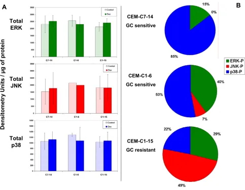
MAPK protein levels and proportions of phosphorylated MAPKs in GC-sensitive and resistant CEM clones. (A) Immunoblot of total protein of each MAPK class showed no significant differences in quantity within each class between clones or after Dex treatment, n = 4 for JNK and p38, 5 for ERK. (B) Percentages of phosphorylated MAPKs (ERK, JNK, and p38) in the presence of Dex per μg protein. (see Methods)
A
B
CEM-C7-14
GC sensitive
CEM-C1-6
GC sensitive
CEM-C1-15
GC resistant
Densit
ome
tr
y
U
n
it
s /
µg
o
f
pro
tein
Total
ERK
Total
JNK
the hypothesis that JNK has a protective effect against Dex-dependent apoptosis.
Furthermore, our earliest data demonstrated that in the sensitive clones Dex-dependent p38 phosphorylation/ activation is pro-apoptotic [16]. Analysis of p38 phospho-rylation showed higher basal levels in both sensitive clones relative to clone C1-15 (Fig. 2A). The level of p38 phosphorylation increased in response to Dex treatment in all three cell clones, but the weakest increase was seen in CEM-C1-15, in which the maximum level reached after Dex treatment was below the basal amounts in the sensi-tive clones (Fig. 2A). Basal levels of phosphorylated ERK (anti-apoptotic) were highest in C1-6 cells, intermediate in C7-14 cells, and lowest in the resistant C1-15 cells. There appeared to be an increase of phosphorylated ERK in response to Dex treatment in C1 subclones C1-6 and
C1-15, not in C7-14 cells. Blocking the anti-apoptotic activity of both ERK and JNK in the sensitive clones max-imized, and inhibition of p38 reduced, Dex-dependent apoptosis [16]. The results shown in Fig. 2 support the hypothesis that the balance between the combined anti-apoptotic activities of ERK + JNK and the pro-anti-apoptotic activity of p38 is a strong determinant of the cellular apoptotic response to Dex. Fig. 1B shows the proportions of phospho-p38 (blue), -ERK (green) and -JNK (red) after Dex treatment in the three clones. It is obvious that the relative amount of phospho-(ERK + JNK) is much greater in the resistant clone. Per unit protein, phospho-p38 rep-resents the majority of the phospho-MAPKs in the sensi-tive clones, but only 22% in the resistant clone. Again, these data are consistent with our hypothesis. A prediction of this hypothesis is that altering the balance of active
Effect of Dex on the phosphorylation state of major MAPKs and the JNK substrate c-Jun in basal and Dex treated sensitive and resistant CEM clones
Figure 2
Effect of Dex on the phosphorylation state of major MAPKs and the JNK substrate c-Jun in basal and Dex treated sensitive and resistant CEM clones. (A) Cell clones CEM-C7-14 and CEM-C1-6 (both Dex-sensitive) as well as CEM-C1-15 (Dex-resistant) were treated with ethanol vehicle (C) or 1 μM Dex (D) for 20–24 hours. Cell lysates were evalu-ated by use of phospho-specific antibodies. Shown is a typical immunoblot for phosphorylevalu-ated MAPK, from 4–5 independent experiments. (B) One of three immunoblots for phosphorylated c-Jun.
C D C D C D
ERK 2 P
ERK 1 P
C7-14 C1-6 C1-15
JNK 2 P
JNK 1 P
p38 P
c-JunP
Jun DP
β-actin
A
MAPKs should affect the Dex-dependent apoptosis of CEM-C1-15 cells. We next tested this prediction.
Inhibition of ERK and JNK activity confers a Dex-sensitive phenotype on GC-resistant CEM-C1-15 cells
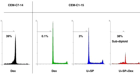
To further evaluate the roles that ERK and/or JNK play in the resistance of CEM-C1-15 cells to GCs, we pharmaco-logically blocked ERK activity with U0126 and JNK activ-ity with either the pharmacological JNK-specific inhibitor SP600125 or a cell permeable JNK inhibitory peptide (ip). Inhibition of either ERK or JNK alone partially restored apoptotic sensitivity to Dex in C1-15 cells such that Dex reduced viable cells by 30–40% compared to drug-matched controls (p = 0.03, data not shown). CEM-C1-15 cells undergo apoptosis in response to Dex in the presence of the specific pharmacological inhibitors of ERK plus JNK, whereas ERK plus JNK inhibition alone had very lit-tle effect on cell viability, though it greatly slowed cellular growth (data not shown). Fig. 3A shows results using Annexin-V to stain for membrane phosphatidlyserine eversion, a hallmark of early stage apoptosis, combined with propidium iodide (PI) uptake to evaluate cells whose membranes had been compromised. Apoptotic cells appear in the quadrants on the right. Staining with Annexin-V only indicates early or pre-apoptosis (lower right quadrant); staining with both Annexin-V and PI indicate full-blown apoptosis (upper right quadrant). Data from C7-14 cells treated with Dex alone are shown as a positive control. In C7-14 cells, Dex exposure clearly produced apoptosis, but in C1-15 cells, pharmacological inhibitors alone or Dex alone produced little apoptosis. Dex and the inhibitors in combination caused an increase in both early and late apoptotic populations in C1-15 cells. Fig. 3A also depicts an experiment in which JNK was partially inhibited by use of ip. Inhibition of both JNK and ERK again renders the cells susceptible to Dex-evoked apoptosis, but the peptide was not as effective an inhibitor of JNK activity as SP600125. Fig. 3B shows that ip is less effective at reducing phospho-c-Jun than SP and produces a corresponding lesser sensitization to Dex. Fig. 3B also demonstrates the ability of the MAPK drug combination to inhibit phospho-ERK. Fig. 4 shows flow cytometry his-tograms of the DNA in permeabilized cells stained with PI. The sub-diploid (apoptotic) fraction of CEM-C1-15 cells to the left of the G1 peak was increased by the com-bination of both inhibitors plus Dex to an extent similar to that of the sensitive CEM-C7-14 cells exposed to Dex alone. Such data shows the state of cells with membranes still intact, but not the lethal effect of the treatment, which can only be determined by quantifying viable cell num-bers as well.
Since the restored sensitivity of CEM-C1-15 to Dex did not result in a complete loss of viable cells in a single round of treatment, we evaluated the sensitivity of the residual cell
population. These cells could represent a completely resistant subpopulation or a population that survived due to several factors, e.g. inadequate block of ERK and JNK or metabolism of the blocking drugs. Cells that survived the initial treatment of double MAPK block plus Dex were washed, grown for 24 hours in the absence of the drugs and immediately retreated. The outgrown cell population responded similarly as did naïve cells (Fig. 5). In two experiments, this cycle could be serially repeated four times, after which the experiments were halted. We con-cluded that the incomplete cell kill by the combination of Dex and MAPK blocking drugs was not due to selection of a permanently resistant subpopulation.
FSK treatment of CEM-C1-15 cells sensitizes to Dex and suppresses phosphorylated JNK
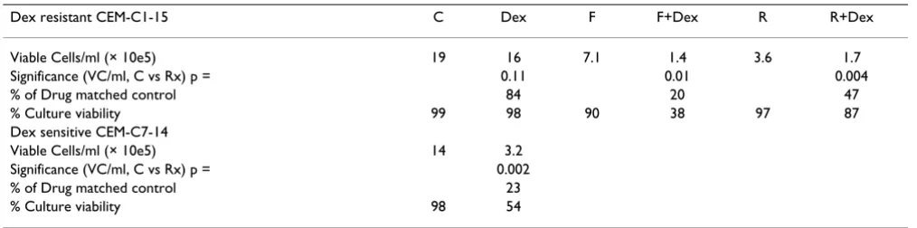
We have shown earlier that FSK alone slowed growth of CEM-C1 cell numbers with little kill and that addition of Dex caused apoptosis [9]. The same effects were seen in C1 subclone 15 (Table 1). The table shows that FSK reduced cell numbers, but caused little change in viability. Adding Dex dramatically lowered total viable cells.
To determine whether there was a convergence of path-ways, we examined the state of JNK and ERK phosphoryla-tion in these cells. FSK with or without Dex lowered JNK (Fig. 6A), but had little effect on phospho-ERK when compared to Dex alone (data not shown). FSK alone had little effect on total JNK protein (6A, lower panel), but the addition of Dex caused an alteration in the migration of the JNK2 isoform. Further investigation of this phenomenon will be required to identify the nature of the new, faster-running band.
Rapamycin lowers phosphorylated JNK and converts CEM-C1-15 cells to a Dex-sensitive state
Site-specific phosphorylation and auto-induction of the GR correlates with sensitization of CEM-C1-15 cells to Dex-dependent apoptosis
In Dex-sensitive CEM clones, treatment with Dex results in phosphorylation of the GR at Ser 211, an effect impor-tant for enhanced transcriptional and apoptotic potency of the GR and in part dependent on p38 MAPK [16]. It has been shown by others that the Dex-dependent auto-induction of GR correlates with later apoptosis in these cells [22]. Consequently we evaluated the status of the GR in CEM-C1-15 cells after various treatments by using
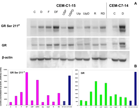
immunoblotting for phopho-Ser 211 GR and total GR with subsequent densitometry analysis (Fig. 7A and 7B). GR protein increased after treatment with FSK and more so after FSK plus Dex, by pharmacologic inhibitors of JNK and ERK alone and also plus Dex, by rapamycin alone, without further increase when Dex was added, by Uip, and similarly by Uip plus Dex. (Fig. 7B, GR).
Immunoblotting with antibodies specific for GR phos-pho-Ser 211 indicated a weak increase in phosphorylation state with no increase in GR protein in response to Dex
Inhibition of ERK plus JNK sensitizes CEM-C1-15 cells to Dex-dependent apoptosis
Figure 3
Inhibition of ERK plus JNK sensitizes CEM-C1-15 cells to Dex-dependent apoptosis. Flow cytometric analysis of 20,000 collected events for each experiment. (A) Evaluation by Annexin-V staining and PI exclusion assay. CEM-C1-15 cells were pretreated for 24 hours with U0126 (U) plus SP600125 (SP) or cell permeable JNK inhibitory peptide (ip). Dex was then added and after a further 24 hours (for treatments with U+SP or U+SP+Dex) or 72 hours (Dex or U+ip or U+ip+Dex), cells were stained with Annexin-V-FITC/PI and examined by flow cytometry. Cells of sensitive clone CEM-C7-14 treated with Dex only are shown as a positive control. Abscissa: histogram of cells positive for Annexin-V; ordinate: cells positive for PI uptake. Note log scales, lower left quadrant shows viable cells; lower right, Annexin-V positive cells (early apoptosis); upper right, cell positive for both Annexin-V and PI (late apoptosis). Inset B is an assay for the block of ERK and JNK activity. Extracts of CEM-C1-15 cells treated with vehicle (C), ip, or SP were immunochemically tested for phosphorylation of c-Jun, n = 2. Extracts tested for ERK phosphorylation were treated with vehicle (C), Dex (D), U0126 plus SP600125 (USP), or the combination (USPD), n = 1.
CEM-C7-14
Dex
CEM-C1-15
U+SP+Dex CEM-C1-15
U+SP CEM-C1-15
Dex
A
CEM-C1-15
U+ip
CEM-C1-15
U+ip+Dex
B
C ip SPc-Jun P
c-Jun
C D USP USPD
ERK P
ERK
treatment alone in CEM-C1-15 cells (Fig. 7A CEM-C1-15 and B, lanes/bars C and D) compared to the effect of Dex in the sensitive C7-14 clone (Fig. 7A and 7B, CEM-C7-14, lanes/bars C and D). The Dex-dependent phos-phorylation of GR Ser 211 in C1-15 cells was distinctly enhanced when JNK and ERK were blocked pharmacolog-ically. The inhibitors alone had minimal effect on basal low levels of GR phospho-Ser 211 (Fig. 7A and 7B, com-pare lanes/bars C, USP, and USPD). Use of the weaker peptide inhibitor ip to inhibit JNK produced qualitatively similar but lesser changes. Treatment with rapamycin, while increasing GR protein, did not increase its phospho-rylation at Ser 211, but Ser 211 did increase with the addi-tion of Dex.
FSK alone increased GR protein and GR Ser 211 phospho-rylation; Dex enhanced both effects. The net effect of Dex in combination with the various drugs resulted in every case in more total phospho-Ser 211 GR in the Dex-treated sensitized cells.
We evaluated intracellular transcriptional activity of the GR by use of a transfected promoter-reporter plasmid encoding GREs fused to a secreted alkaline phosphatase (SEAP) reporter. Upon treatment with U0126 and SP600125 in combination with Dex, transcriptional
activ-ity of the GR was significantly increased over treatment with Dex alone (Fig. 8). Substitution of the ip to JNK still supported increased GR transcriptional activity, but to a lesser extent; again consistent with the fact that the pep-tide fails to fully inhibit JNK. Thus, inhibition of JNK and ERK, which renders otherwise resistant C1-15 cells sensi-tive to dependent apoptosis, also supported Dex-dependent increases in GR phosphorylation at Ser 211, total GR protein, and the activity of the GR. The combina-tion of FSK and Dex resulted in as great an increase in SEAP induction as did blocking ERK and JNK in combina-tion with Dex. Cotreatment with rapamycin plus Dex, however, though enhancing apoptosis, decreased steroid-dependent induction of SEAP activity from the GRE-SEAP construct (data not shown). This is no doubt due to inhi-bition of SEAP mRNA translation by rapamycin; the drug does not inhibit induction of reporter mRNA [23].
Discussion
In the search for the GC-driven pathway to malignant lymphoid cell apoptosis, clones from the CEM line of ALL cells have proven extremely useful. We compared basal and Dex-induced levels of genes in three closely related clones: one inherently sensitive to Dex-induced apopto-sis; a sister clone that is inherently resistant, and a third revertant to sensitive from their resistant parental clone
Sub-diploid DNA content increases upon restoration of Dex-sensitivity through inhibition of ERK and JNK in CEM-C1-15 cells
Figure 4
Sub-diploid DNA content increases upon restoration of Dex-sensitivity through inhibition of ERK and JNK in CEM-C1-15 cells. CEM-C1-15 cells were treated simultaneously with Dex ± U+SP. Dex-treated CEM-C7-14 cells serve as a positive control for apoptotic response. After 72 hours, nuclear suspensions were evaluated for distribution of DNA content by PI staining. Histograms of cellular DNA content as well as percentages of sub-diploid DNA are presented, example of n = 4.
Dex Dex U+SP U+SP+Dex 39%
CEM-C7-14 CEM-C1-15
0.1% 3% 38%
[19]. Earlier data revealed that activation of the MAPK p38 contributed to the apoptotic outcome, whereas MAPKs, JNK and ERK acted to prevent or ameliorate Dex-depend-ent apoptosis [16]. ConsistDex-depend-ent with this finding, we show that basal levels of phosphorylated JNK were strikingly elevated in the resistant CEM-C1-15 clone compared to the sensitive clones. In addition, phospho-ERK was increased by Dex in these cells. Its sister clone CEM-C1-6, a revertant to sensitive, had greatly lowered phospho-JNK although phospho-ERK remained the highest of the three tested CEM clones. This suggested that combined contri-butions from JNK and ERK favored Dex resistance. The anti-apoptotic effect of ERK in relation to GCs in a differ-ent clone of CEM cells has recdiffer-ently been reported [11]. We hypothesized that the elevated levels of phospho-(JNK + ERK) were at least partly responsible for the resistance to Dex of CEM-C1-15 cells. We tested that hypothesis by
blocking JNK and ERK activity in clone C1-15. Single blocks produced partial Dex sensitivity, but a better con-version to the Dex-sensitive phenotype was achieved by blocking both JNK and ERK. We noted that Dex did not directly cause changes in the protein levels of the MAPKs; yet in lymphoid cells, altered gene transcription is required for corticoid-dependent apoptosis. Thus, post-translational alterations in phosphorylation/activation of the MAPKs depend on Dex-dependent changes in tran-scription of genes other than those of the MAPKs them-selves.
Recent time-course analysis of Dex-dependent changes in mRNA levels in two Dex-sensitive clones suggested that a network of induced and repressed genes coordinately affect MAPK phosphorylation (submitted). We also observed a marked difference in the c-Jun
phosphoryla-CEM-C1-15 cells that survive treatment with MAPK inhibitors plus Dex remain sensitive upon retreatment
Figure 5
CEM-C1-15 cells that survive treatment with MAPK inhibitors plus Dex remain sensitive upon retreatment. CEM-C1-15 cells were treated with vehicle (Control), Dex, U0126 (U) and SP600125 (SP) in the combinations shown (Initial). After 96 hours, the drugs were diluted by centrifugation, washed, and the cells immediately reincubated in fresh growth medium. The drug combinations were then reintroduced (Retreat). At the time points shown, viable cells were counted by trypan blue dye exclusion assay, n = 2. Ordinate, note log scale.
Hours
0
20
40
60
80
100
120
140
160
180
Viable cells/ml
10
510
610
7Control
Dex
U+SP
U+SP+Dex
Initial
Retreat
43%
64%
tion pattern in response to Dex between the sensitive and resistant subclones. CEM-C7-14 had undetectable and CEM-C1-6 had greatly reduced phospho-JNK levels while still showing c-Jun phosphorylation. It has been previ-ously reported that c-Jun can be phosphorylated by sev-eral other protein kinases besides JNK [24]. We suggest that the actions of other kinases and/or phosphatases influenced by Dex in the sensitive CEM clones are inter-vening at the level of c-Jun phosphorylation. Though JNK and ERK both must be blocked to maximize the conver-sion of CEM-C1-15 cells to Dex sensitivity, of the two, JNK seemed the more important. This is consistent with the numerous recent reports that emphasize the reliance of several types of malignant lymphoid cells on JNK and/or ERK for viability [25-31].
We had previously observed that activation of the PKA system by use of FSK transformed CEM-C1 cells – the ini-tial resistant clone – into cells Dex-sensitive for apoptosis [9]. After confirming that the same held true for subclone C1-15, we tested for a point of convergence at JNK. The data show that treatment with FSK lowers the levels of phosphorylated JNK and this effect may be enhanced by the addition of Dex. FSK is known to activate phos-phatases that dephosphorylate MAPKs [32-34]. Since FSK alone lowers phospho-JNK and inhibits growth, but does not kill either Dex-sensitive or Dex-resistant cells, the low-ered phospho-JNK must provide a background state in which Dex can produce additional alterations necessary to initiate cell death. FSK treatment consistently renders C1-15 cells more sensitive to Dex than other treatments that reduce phospho-JNK equally well. Thus, activation of PKA contributes additional factors to Dex-dependent cell death.
That JNK is a nexus for pathway interactions in Dex-dependent apoptosis is further suggested by use of
rapamycin. This drug blocks cap-dependent mRNA trans-lation through the mTOR pathway [21,23] and recently has been shown to render CEM-c1 cells partially sensitive to Dex [18]. We confirmed this result in CEM-C1-15 and now find that rapamycin inhibits JNK phosphorylation in the CEM system. Rapamycin inhibits translation of some, not all mRNAs, and it has been suggested that Dex induc-tion of those not blocked by rapamycin is the key to apop-tosis [23]. Our results suggest that reduction of the levels of anti-apoptotic phospho-JNK is a component of the sen-sitization to Dex caused by rapamycin. Undoubtedly, other rapamycin effects are involved as well.
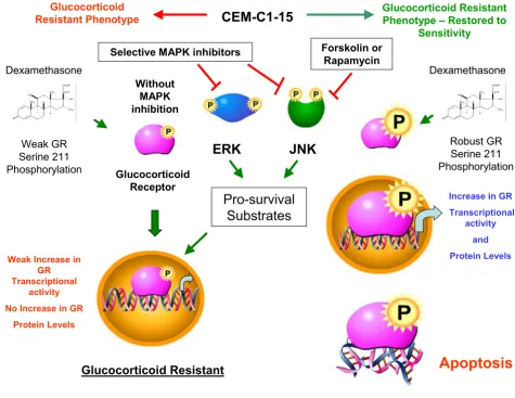
The GR is a second point of convergence between the drugs employed herein. Phosphorylation of Ser 211 in the human GR increases the transcriptional and apoptotic potency of the receptor, and mutation of Ser 211 to a non-phosphorylatable amino acid reduces GR-based apoptosis and gene transcription [16]. The p38 MAPK is capable of carrying out this phosphorylation, and other kinases may do so as well [16,35]. All of the sensitizing treatments lead to an increase in GR protein and in some cases adding Dex further increases GR. When Dex is added, the proportion of phospho-Ser 211 GR is increased. The result is that the total amount of phospho-Ser 211 is increased in cells that the drugs have rendered sensitive to Dex. This condition therefore correlates with diminished JNK activity and increased GR apoptotic/transcriptional potency. A scheme outlining these effects is presented in Fig. 9.
Conclusion
GCs appear to have a generalized metabolic effect upon most if not all of the cells in the human body. Synthetic GCs, such as Dex, in pharmacological doses have been used successfully in the treatment of lymphoid malignan-cies for many years. However, not all lymphoid cells are responsive to GCs. While it is well documented that GCs Table 1: Forskolin and Rapamycin sensitize CEM-C1-15 cells to Dex.
Dex resistant CEM-C1-15 C Dex F F+Dex R R+Dex
Viable Cells/ml (× 10e5) 19 16 7.1 1.4 3.6 1.7
Significance (VC/ml, C vs Rx) p = 0.11 0.01 0.004
% of Drug matched control 84 20 47
% Culture viability 99 98 90 38 97 87
Dex sensitive CEM-C7-14
Viable Cells/ml (× 10e5) 14 3.2
Significance (VC/ml, C vs Rx) p = 0.002
% of Drug matched control 23
% Culture viability 98 54
act through the GR to produce its multitude of responses, the complete pathway by which response or no response is accomplished is not clearly understood. We present in this paper information clarifying the relationship between the GR and the MAPK pathways in lymphoid cells. In Dex-sensitive cells the intracellular balance of MAPKs ERK and JNK which are anti-apoptotic in their active phosphor-ylated form must be maintained in a reduced state while the MAPK p38 which is pro-apoptotic must be enhanced. GC-resistant lymphoid cells may be rendered sensitive by: (1) inhibition of JNK and ERK activity, (2) stimulation of the cAMP/PKA pathway with FSK, or (3) inhibition of mTOR with rapamycin. All three treatments in combina-tion with Dex alter the balance of cellular phospho-MAPKs by lowering JNK phosphorylation, stimulating site-specific, activity-enhancing phosphorylation of the GR at Ser 211, and increasing total GR protein levels cul-minating in an apoptotic response. Thus JNK serves as the convergence point between the GR and MAPK pathways. We propose that the interactive effects found in CEM cells may apply to other hematological malignancies as well. Tests of this hypothesis are underway in our laboratory.
Methods
Cell culture and drug concentrations
The CCRF-CEM human cell line was isolated from a patient with ALL [36]. Early sensitive (C7) and Dex-resistant (C1) clones were subcloned to give CEM-C7-14, sensitive to Dex-evoked apoptosis; CEM-C1-6, a Dex-sen-sitive spontaneous revertant; and Dex-resistant CEM-C1-15 [19]. Cells were grown in RPMI 1640 (Cellgro Media Tech, Herndon, VA) pH 7.4, supplemented with 5% fetal bovine serum (FBS) (Atlanta Biologicals, Norcross, GA). Cells were cultured at 37°C in a humidified atmosphere of 95% air, 5% CO2 and subcultured regularly to ensure logarithmic growth. Cell viability was determined by trypan blue dye (Sigma-Aldrich, St. Louis, MO) exclusion using a manual hemacytometer or Vi-cell automated cell (Beckman Coulter, Miami, FL) counting. Cells were treated as appropriate for each experiment with vehicle (ethanol/DMSO/and/or sterile HPLC-grade water < 0.1% final concentration), 1 μM Dex, 4.5 μM JNK inhibitor (SP600125), 2.6 μM MEK inhibitor (U0126), 10 μM FSK, 1 μM ip, 10 nM rapamycin, or various combinations of each. All chemicals were from Sigma-Aldrich, Burdick and Jackson, Muskegon, MI or Calbiochem, San Diego, CA. For all experiments (n) equals the range of subsets of inde-pendent experiments.
Inhibition of JNK phosphorylation correlates with treatments that restore Dex-sensitivity to CEM-C1-15 cells
Figure 6
Inhibition of JNK phosphorylation correlates with treatments that restore Dex-sensitivity to CEM-C1-15 cells. (A) CEM-C1-15 cells were at once treated with vehicle control (C), Dex (D), FSK (F), or the combination of FSK plus Dex (DF) for 24 hours. JNK was evaluated by immunoblot for phosphorylation of Threonine183/Tyrosine185 and total protein,
exam-ple of n = 4. (B) CEM-C1-15 cells were pretreated with vehicle control (C), U0126 (U) plus SP600125 (SP), rapamycin (R), or combinations of these drugs for 6 hours before adding Dex (D) for an additional 18 hours. JNK was evaluated as in panel A, example of n = 2–4 for various combinations of treatments.
C D F DF
JNK 2 P
JNK 1 P
JNK 2
JNK 1
R RD Pos. USP USPD
C D
JNK 1 JNK 1 P
JNK 2 P
JNK 2
Immunochemical analysis
Cells in mid-logarithmic growth were treated with the var-ious drugs as appropriate for each experiment. To ensure accurate protein identification, positive controls were gen-erated by known strong inducers of MAPKs. We treated the cells with 50 ng/ml phorbol 12-myristate 13-acetate plus 1 μg/ml phytohemagglutinin for phospho-ERK, ultraviolet light for phospho-c-Jun (Cell Signaling Tech-nology, Beverly MA), and 100 nM anisomycin for phos-pho-JNK and p38. These controls were run in independent lanes on each blot. After the various experi-mental or positive control treatments, cells were harvested
at various time-points thereafter by centrifuging at 1,000 rpm for 5 minutes in a Beckman Allegra 6R centrifuge at 22°C. The cells were resuspended in 10 ml/22°C phos-phate buffered isotonic saline pH 7.4 (PBS), and recentri-fuged. The washed cells were transferred to 1.5 ml centrifuge tubes and lysed on ice using 4°C M-per cell lysis buffer (Pierce, Rockford, IL) supplemented with 1 × general protease inhibitor cocktail, 10 mM sodium fluo-ride, and 1 mM sodium orthovanadate (Sigma-Aldrich). Cellular debris was pelleted at 13,000 rpm for 10 minutes at 4°C in a Beckman microfuge. The clarified samples were transferred to fresh, pre-chilled microfuge tubes, and
Evaluation of GR protein and phosphorylation state in CEM-C1-15 cells rendered sensitive to Dex by several treatments
Figure 7
Evaluation of GR protein and phosphorylation state in CEM-C1-15 cells rendered sensitive to Dex by several treatments. (A)CEM-C1-15 cells were pretreated with vehicle control (C), FSK (F), U0126 (U) plus SP600125 (SP), U0126 plus (ip), rapamycin (R), or combinations of these treatments for 5.5 hours before adding Dex (D) for an additional 17 hours. Dex-sensitive CEM-C7-14 cells were treated with vehicle control or Dex for 17 hours, and results are included as a positive control. Equal amounts of protein were added to each lane. The results were evaluated by immunoblot with antibodies against phospho-GR (Ser 211), total GR, and β-actin, n = 2–4 for various combinations of treatments. (B) Densitometric analysis of immunoblot from (A). All bands were normalized to β-actin; bars = arbitrary units of variable/densitometry units of β-actin for each corresponding lane.
A
C D F DF
USP USPD Uip UipD R RD C D
CEM-C7-14
CEM-C1-15
GR Ser 211
PGR
β-actin
GR Ser 211 P
GR
Lev
e
ls (norm
a
li
zed to
ββββ
-a
cti
n
, arbitrar
y
units)
the protein concentration was estimated using BCA (Pierce). The lysate was mixed with 5 × SDS-PAGE sample buffer supplemented with 2% 2-mercaptoethanol and heated to 100°C for 5 minutes. Equally loaded proteins were separated by electrophoresis on 8% SDS-PAGE gels and transferred to a PVDF membrane (Bio-Rad, Hercules, CA or Amersham Pharmacia Biotech, Piscataway, NJ) using a semi-dry electroblotter (Hoefer, San Francisco, CA). Membranes were washed with Tris-buffered saline/ Tween-20 (TBST) (140 mM sodium chloride, 20 mM Tris, 0.1% Tween-20, pH 7.6) and blocked for 1 hour in TBST
supplemented with 5% non-fat dry milk. Membranes were rewashed and placed in a solution of TBST plus 5% bovine serum albumin containing either an antibody to phospho-(Threonine202 and Tyrosine204) -specific to ERK
MAPK, or phospho-(Threonine183 and Tyrosine185)
-spe-cific to JNK MAPK, or phospho-(Threonine180 and
Tyrosine182) -specific to p38 MAPK (Cell Signaling
Tech-nology), or to phospho-c-Jun (Serine73) (Calbiochem), or
phospho-GR (Serine211) (Cell Signaling Technology), or
to phosphorylation state independent ERK MAPK (Calbi-ochem), or JNK MAPK (Cell Signaling Technology), or
Transcriptional potency of the GR is enhanced upon restoration of Dex-sensitivity to CEM-C1-15 cells
Figure 8
Transcriptional potency of the GR is enhanced upon restoration of Dex-sensitivity to C1-15 cells. CEM-C1-15 cells were electroporated with a plasmid expressing three GREs in tandem fused to a SEAP reporter. Cells were subse-quently pretreated with vehicle control, FSK (F), U0126 (U) plus SP600125 (SP), U0126 plus (ip), or combinations of these treatments for 6 hours before adding Dex for an additional 24 hours with subsequent evaluation of SEAP activity. Drug treat-ment alone was subtracted as background and shown is the Dex effect on each treattreat-ment. Error bars: 1 standard deviation, average of 3 independent replicates; p-value is based on a t-test for matched drug treatments ± Dex, n = 2–3 for various com-binations of treatments.
GRE-SE
A
P
A
c
tiv
it
y
–
R
elat
iv
e L
igh
t
Units
(RLU)
p38 MAPK (Calbiochem), or GR (Affinity Bioreagents, Golden, CO), or β-actin (Santa Cruz Biotechnology, Santa Cruz, CA) and incubated for 16 hours at 4°C with gentle agitation. Membranes were subsequently washed with TBST and probed with horseradish peroxidase goat/anti-rabbit secondary antibody for 1 hour at 22°C. After rewashing, the membranes were saturated with horserad-ish peroxide substrate ECL (Amersham Pharmacia Bio-tech) and exposed to Blue Lite Autorad Film (ISC BioExpress, Kaysville, UT) for various times to ensure lin-earity. To analyze both phospho- and total proteins on the
same filter, after the initial reaction for phosphoprotein, the membranes were stripped of antibody by incubation in Restore™ buffer (Pierce) for 1 hour and reprobed with antibody for the appropriate protein. Densiometric anal-ysis was accomplished using AlphaEase FC™ software (Alpha Innotech Corporation, San Leandro, CA).
Calculation of proportional MAPK activity
Preliminary experiments established the linear range for directions of immunochemical reaction for ERK, JNK, and p38. Working within this range, total and phosphorylated
Agents that restore GC-sensitivity to CEM-C1-15 cells converge on the GR and MAPK pathways
Figure 9
Agents that restore GC-sensitivity to CEM-C1-15 cells converge on the GR and MAPK pathways. Dex-resistant CEM-C1-15 cells in their natural state harbor high levels of pro-survival, anti-apoptotic active JNK (green symbol) and low lev-els of active ERK which is Dex-inducible (blue symbol). The cells also contain GR (pink symbol). The sequence on the left side shows the result in CEM cells which resist Dex-dependent apoptosis. In this case, added Dex mediates a weak increase in GR phospho-Ser 211 as well as GRE reporter driven activity, but no increase in GR protein levels, and the cells remain resistant. The sequence on the right depicts the results when CEM-C1-15 cells are treated with combinations of Dex and MAPK inhibi-tors, FSK, or rapamycin. These treated cells convert to a GC-sensitive phenotype. All treatments converge at inhibition of the JNK MAPK pathway. Upon restoration of the Dex-sensitive phenotype, a robust increase in GR phospho-Ser 211, GR protein, and transcriptional activity is observed. These effects culminate in an apoptotic response.
CEM-C1-15
Glucocorticoid Resistant Phenotype
Glucocorticoid Resistant Phenotype – Restored to
Sensitivity
Selective MAPK inhibitors Forskolin or Rapamycin Dexamethasone
Weak GR Serine 211 Phosphorylation
Without MAPK inhibition
Glucocorticoid Receptor
Weak Increase in GR Transcriptional
activity
No Increase in GR
Protein Levels
Increase in GR Transcriptional
activity
and Protein Levels
Dexamethasone
Robust GR Serine 211 Phosphorylation
Glucocorticoid Resistant
Apoptosis
ERK
JNK
ERK, JNK, and p38 were estimated quantitatively by image analysis. In 4- or for ERK 5-independent experi-ments, none of the MAPK's showed variation in the basal state or after Dex treatment. Therefore, the amount of each immunochemically detected MAPK could be expressed in terms of total extract protein (adjusted to X/ml in every experiment). The relative phosphorylated forms of each MAPK, estimated immunochemically, could therefore be calculated:
Phosphatidylserine membrane eversion, DNA lysis, and cell cycle evaluation by flow cytometry
Cells were diluted to 5 × 10⁴ viable cells/ml for
CEM-C1-15 and 1 × 10⁵ viable cells/ml for CEM-C7-14 in 5 ml
aliq-uots in 6-well cell culture dishes. CEM-C1-15 cells were pre-treated with either U0126 plus SP600125 or U0126 plus ip for 24 hours before adding Dex. Cells were har-vested at various time-points thereafter by centrifuging at 1,000 rpm for 5 minutes, washed twice with ice-cold PBS, pelleted, and resuspended in 1 × ice-cold binding buffer (10 mM HEPES/NaOH pH 7.4, 140 mM NaCl, 2.5 mM CaCl2 from BD Pharmagin (San Jose, CA). 100 μl cell sus-pension was combined with 5 μl Annexin V-FITC and 10
μl PI (BD Pharmagin) for 15 minutes at 22°C in the dark. 400 μl binding buffer was then added to each sample, and 20,000 cells were processed by flow cytometer (BD FACS-canto, BD Biosciences, San Jose, CA) using filters for FITC (488 nm) and PI (> 600 nm).
Cell samples with DNA stained by PI for cell cycle exami-nation were prepared and analyzed as described [16] after treatment for 72 hours with vehicle (ethanol/DMSO), Dex, U0126 plus SP600125, or a combination of the drugs.
GR activity by GRE-reporter assay
Logarithmically growing CEM-C1-15 cells were collected by centrifugation and washed with 10 ml of sterile 37°C PBS and recollected. The cells were resuspended to a den-sity of 1 × 10⁷ viable cells/ml in serum-free 37°C RPMI
1640 containing 1.25% DMSO. 400 μl aliquots of the sus-pension were placed into 0.4 cm gap electroporation cuvettes (Bio-Rad) containing 15 μg of pGRE-SEAP reporter vector (BD Clontech, Mountain View, CA) pre-pared using a Qiagen maxi-prep kit (Qiagen, Valencia, CA). Cuvettes were electroporated using 975 μF and 270 V with a Gene Pulser II (Bio-Rad). Electroporated cells were diluted in 4 ml (per cuvette) of RPMI 1640 supple-mented with 5% FBS and 1.25% DMSO and recultured. Twenty-four hours after electroporation, cells were pel-leted, resuspended in fresh RPMI 1640 containing 5% FBS, and viable cells were quantified using trypan blue exclusion. The cultures were diluted to 4 × 10⁵ viable cells/
ml in RPMI 1640 supplemented with 5% FBS, and 500 μl triplicate aliquots per treatment were placed in a 48-well tissue culture plate (Costar, Cambridge, MA). Cells were treated for 24 hours with vehicle (ethanol/DMSO), Dex, U0126, SP600125, ip, rapamycin, or combinations thereof. Samples were subsequently tested for the pres-ence of SEAP using the Great EscAPe SEAP Chemilumines-cence Detection Kit (BD Clontech) according to the manufacturer's instructions.
List of abbreviations
ALL – acute lymphoblastic leukemia
cAMP – cyclic adenosine-3',5'-monophosphate
Dex – dexamethasone
ERK – extra-cellular signal-regulated kinase
FBS – fetal bovine serum
FSK – forskolin
GC – glucocorticoid
GR – glucocorticoid receptor
GRE – glucocorticoid response element
ip – cell permeable JNK inhibitory peptide
JNK – c-Jun N-terminal kinase
MAPK – mitogen-activated protein kinase
MKK – MAP kinase kinase
mTOR – mammalian target of rapamycin
PBS – phosphate-buffered isotonic saline
PKA – protein kinase A
PI – propidium iodide
SEAP – secreted placental alkaline phosphatase
Ser 211 – glucocorticoid receptor serine 211
TBST – Tris-buffered saline containing Tween-20
Competing interests
The author(s) declare that they have no competing inter-ests.
Phosphorylated MAPK X
Authors' contributions
ALM, ASG, and EBT were involved in experimental design and data analysis. ALM conducted the experiments and wrote the first draft of the manuscript. BHJ and EBT wrote sections and edited the manuscript. EBT served as the principal investigator.
Acknowledgements
This work was supported by grant NCI 2RO1 CA41407 to Dr. E. Brad Thompson.
The authors express their sincere appreciation to Mr. Mark Griffin in the Flow Cytometry Laboratory at the University of Texas Medical Branch, Galveston, Texas and Ms. Rhoda Thompson for assistance in preparing this paper for submission.
References
1. Adcock IM, Caramori G: Cross-talk between pro-inflammatory transcription factors and glucocorticoids. Immunol Cell Biol 2001, 79:376-384.
2. Kyriakis JM: MAP kinases and the regulation of nuclear recep-tors. Sci STKE 2000, 48:1-4.
3. McManus R: Mechanisms of steroid action and resistance in inflammation and disease. J Endocrinol 2003, 178:1-4.
4. Clark AR: MAP kinase phosphatase 1: a novel mediator of bio-logical effects of glucocorticoids? J Endocrinol 2003, 178:5-12. 5. Bruna A, Nicolas M, Munoz A, Kyriakis JM, Caelles C:
Glucocorti-coid receptor-JNK interaction mediates inhibition of the JNK pathway by glucocorticoids. EMBO J 2003, 22:6035-6044. 6. Itoh M, Adachi M, Yasui H, Takekawa M, Tanaka H, Imai K: Nuclear
export of glucocorticoid receptor is enhanced by c-Jun N-terminal kinase-mediated phosphorylation. Mol Endocrinol 2002, 16:2382-2392.
7. Rogatsky I, Logan SK, Garabedian MJ: Antagonism of glucocorti-coid receptor transcriptional activation by the c-Jun N-ter-minal kinase. Proc Natl Acad Sci USA 1998, 95:2050-2055. 8. Krstic MD, Rogatsky I, Yamamoto KR, Garabedian MJ:
Mitogen-activated and cyclin-dependent protein kinases selectively and differentially modulate transcriptional enhancement by the glucocorticoid receptor. Mol Cell Biol 1997, 17:3947-3954. 9. Medh RD, Saeed MF, Johnson BH, Thompson EB: Resistance of
human leukemic CEM-C1 cells is overcome by synergism between glucocorticoid and protein kinase A pathways: cor-relation with c-Myc suppression. Cancer Res 1998,
58:3684-3693.
10. Wada T, Penninger JM: Mitogen-activated protein kinases in apoptosis regulation. Oncogene 2004, 23:2838-2849.
11. Tanaka T, Okabe T, Gondo S, Fukuda M, Yamamoto M, Umemura T, Tani K, Nomura M, Goto K, Yanase T, Nawata H: Modification of glucocorticoid sensitivity by MAP kinase signaling pathways in glucocorticoid-induced T-cell apoptosis. Exp Hematol 2006,
34:1542-1552.
12. Ogawa R, Streiff MB, Bugayenko A, Kato GJ: Inhibition of PDE4 phosphodiesterase activity induces growth suppression, apoptosis, glucocorticoid sensitivity, p53, and p21(WAF1/ CIP1) proteins in human acute lymphoblastic leukemia cells.
Blood 2002, 99:3390-3397.
13. Kyriakis JM, Avruch J: Mammalian mitogen-activated protein kinase signal transduction pathways activated by stress and inflammation. Physiol Rev 2001, 81:807-869.
14. Cowan KJ, Storey KB: Mitogen-activated protein kinases: new signaling pathways functioning in cellular responses to envi-ronmental stress. J Exp Biol 2003, 206:1107-1115.
15. Ishizuka T, Sakata N, Johnson GL, Gelfand EW, Terada N: Rapamy-cin potentiates dexamethasone-induced apoptosis and inhib-its JNK activity in lymphoblastoid cells. Biochem Biophys Res Commun 1997, 230:386-391.
16. Miller AL, Webb MS, Copik AJ, Wang Y, Johnson BH, Kumar R, Thompson EB: p38 MAP kinase is a key mediator in glucocor-ticoid-induced apoptosis of lymphoid cells: correlation between p38 MAPK activation and site-specific
phosphoryla-tion of the human glucocorticoid receptor at Serine 211. Mol Endocrinol 2005, 19:1569-1583.
17. Wang X, Wu H, Lakdawala VS, Hu F, Hanson ND, Miller AH: Inhibi-tion of Jun N-terminal kinase (JNK) enhances glucocorticoid receptor-mediated function in mouse hippocampal HT22 cells. Neuropsychopharmacology 2005, 30:242-249.
18. Wei G, Twomey D, Lamb J, Schlis K, Agarwal J, Stam RW, Opferman JT, Sallan SE, den Boer ML, Pieters R, Golub TR, Armstrong SA: Gene expression-based chemical genomics identifies rapamycin as a modulator of MCL1 and glucocorticoid resistance. Cancer Cell 2006, 10:331-342.
19. Medh RD, Webb MS, Miller AL, Johnson BH, Fofanov Y, Li T, Wood TG, Luxon BA, Thompson EB: Gene expression profile of human lymphoid CEM cells sensitive and resistant to glucocorticoid-evoked apoptosis. Genomics 2003, 81:543-555.
20. Zhou F, Thompson EB: Role of c-jun induction in the glucocor-ticoid-evoked apoptotic pathway in human leukemic lym-phoblasts. Mol Endocrinol 1996, 10:306-316.
21. Dancey JE: mTOR inhibitors in hematologic malignancies. Clin Adv in Hematol and Oncol 2003, 1:419-423.
22. Ramdas J, Liu W, Harmon JM: Glucocorticoid-induced cell death requires autoinduction of glucocorticoid receptor expres-sion in human leukemic T cells. Cancer Res 1999, 59:1378-1385. 23. Yan H, Frost P, Shi Y, Hoang B, Sharma S, Fisher M, Gera J, Lichten-stein A: Mechanism by which mammalian target of rapamycin inhibitors sensitize multiple myeloma cells to dexametha-sone-induced apoptosis. Cancer Res 2006, 66:2305-2313. 24. Baker SJ, Kerppola TK, Luk D, Vandenberg MT, Marshak DR, Curran
T, Abate C: Jun is phosphorylated by several protein kinases at the same sites that are modified in serum-stimulated fibrob-lasts. Mol Cell Biol 1992, 12:4694-4705.
25. Gururajan M, Chui R, Karuppannan AK, Ke J, Jennings CD, Bondada S: c-Jun N-terminal kinase (JNK) is required for survival and proliferation of B-lymphoma cells. Blood 2005, 106:1382-1391. 26. Hideshima T, Hayashi T, Chauhan D, Akiyama M, Richardson P, Anderson K: Biologic sequelae of c-Jun NH(2)-terminal kinase (JNK) activation in multiple myeloma cell lines. Oncogene 2003, 22:8797-8801.
27. Fan M, Chambers TC: Role of mitogen-activated protein kinases in the response of tumor cells to chemotherapy. Drug Resist Updat 2001, 4:253-267.
28. James JA, Smith MA, Court EL, Ching CYY, Willson C, Smith JG: An investigation of the effects of the MEK inhibitor U0126 on apoptosis in acute leukemia. Hematol J 2003, 4:427-432. 29. Milella M, Precupanu CM, Gregorj C, Ricciardi MR, Petrucci MT,
Kornblau SM, Tafuri A, Andreeff M: Beyond single pathway inhi-bition: MEK inhibitors as a platform for the development of pharmacological combinations with synergistic anti-leuke-mic effects. Curr Pharm Des 2005, 11:2779-2795.
30. Lee JT Jr, McCubrey JA: The Raf/MEK/ERK signal transduction cascade as a target for chemotherapeutic intervention in leukemia. Leukemia 2002, 16:486-507.
31. Platanias LC: MAP kinase signaling pathways and hematologic malignancies. Blood 2003, 101:4667-4679.
32. Wu JJ, Bennett AM: Essential role for mitogen-activated pro-tein (MAP) kinase phosphatase-1 in stress-responsive MAP kinase and cell survival signaling. J Biol Chem 2005,
280:16461-16466.
33. Sommer A, Burkhardt H, Keyse SM, Luscher B: Synergistic activa-tion of the mkp-1 gene by protein kinase A signaling and USF, but not c-Myc. FEBS Lett 2000, 474:146-150.
34. Tamir A, Granot Y, Isakov N: Inhibition of T lymphocyte activa-tion by cAMP is associated with down-regulaactiva-tion of two par-allel mitogen-activated protein kinase pathways, the extracellular signal-related kinase and c-Jun N-terminal kinase. J Immunol 1996, 157:1514-1522.
35. Ismaili N, Garabedian MJ: Modulation of glucocorticoid receptor function via phosphorylation. Ann NY Acad Sci 2004,
1024:86-101.
36. Foley GE, Lazarus H, Farber S, Uzman BG, Boone BA, McCarthy RE: