Dunne et al. Clin Proteom (2015) 12:24
DOI 10.1186/s12014-015-9096-3
Proteins from formalin-fixed paraffin-embedded
prostate cancer sections that predict the risk
of metastatic disease
RESEARCH
Proteins from formalin-fixed
paraffin-embedded prostate cancer sections
that predict the risk of metastatic disease
Jonathan C. Dunne
1, David S. Lamb
2, Brett Delahunt
2, Judith Murray
2, Peter Bethwaite
2, Peter Ferguson
2,
John N. Nacey
3, Sven Sondhauss
1and T. William Jordan
1*Abstract
Background: Prostate cancer is the most frequently diagnosed cancer in men and the third leading cause of cancer related deaths among men living in developed countries. Biomarkers that predict disease outcome at the time of initial diagnosis would substantially aid disease management.
Results: Proteins extracted from formalin-fixed paraffin-embedded tissue were identified using nanoflow liquid chro-matography-MALDI MS/MS or after separation by one- or two-dimensional electrophoresis. The proteomics data have been deposited to the ProteomeXchange with identifier PXD000963. A list of potential biomarker candidates, based on proposed associations with prostate cancer, was derived from the 320 identified proteins. Candidate biomarkers were then examined by multiplexed Western blotting of archival specimens from men with premetastatic disease and subsequent disease outcome data. Annexin A2 provided the best prediction of risk of metastatic disease (log-rank Chi squared p = 0. 025). A tumor/control tissue >2-fold relative abundance increase predicted early biochemical failure, while <2-fold change predicted late or no biochemical failure.
Conclusions: This study confirms the potential for use of archival FFPE specimens in the search for prognostic bio-markers for prostate cancer and suggests that annexin A2 abundance in diagnostic biopsies is predictive for meta-static potential. Protein profiling each cancer may lead to an overall reduction in mortality from metameta-static prostate cancer as well as reduced treatment associated morbidity.
Keywords: Formalin-fixed paraffin embedded, Prognostic biomarkers, Prostate cancer, Proteomics
© 2015 Dunne et al. This article is distributed under the terms of the Creative Commons Attribution 4.0 International License (http://creativecommons.org/licenses/by/4.0/), which permits unrestricted use, distribution, and reproduction in any medium, provided you give appropriate credit to the original author(s) and the source, provide a link to the Creative Commons license, and indicate if changes were made. The Creative Commons Public Domain Dedication waiver (http://creativecommons.org/ publicdomain/zero/1.0/) applies to the data made available in this article, unless otherwise stated.
Background
Prostate cancer (PCa) is the most frequently diagnosed cancer in men and the third leading cause of cancer related deaths among men living in developed countries [1]. Serum prostate-specific antigen (PSA) testing used to assist in making the diagnosis of PCa is a useful tool, but it has some limitations. It suffers from poor specific-ity, and elevation of the serum PSA is often due to non-malignant causes. The biopsies performed as a result of an elevated serum PSA result in the detection of some clinically irrelevant tumors (over-diagnosis) which will
not trouble affected men during their lifetimes. Finally, the degree of elevation of the serum PSA level at the time of diagnosis does not assist in predicting how aggres-sively the cancer will behave.
Trial 96.01 was the first of two large randomized tri-als run by the Trans-Tasman Radiation Oncology Group (TROG) investigating new treatment strategies for locally advanced PCa. The trial end points at 10 years were reported in 2011 [2]. In trial patients, there appeared to be three distinct patterns of behaviour when the can-cer relapsed: one with a short PSA doubling time of less than 4 months, one with an intermediate duration PSA doubling time of 4–9 months, and the other with a PSA doubling time greater than 9 months [3]. The polariza-tion of biological behaviour on relapse suggests there are
Open Access
*Correspondence: [email protected]
1 Centre for Biodiscovery, School of Biological Sciences, Victoria University
of Wellington, PO Box 600, Wellington, New Zealand
Page 3 of 10 Dunne et al. Clin Proteom (2015) 12:24
inherent differences between cancers responsible for the speed at which they grow and their biological behaviour. If this hypothesis is correct, then PCa could have a num-ber of characteristic patterns of protein expression capa-ble of informing the patient and clinician how the cancer is likely to behave. Identification of men at the time of diagnosis that are at high risk of developing biochemi-cal relapse (PSA failure) is fundamental to improving the treatment of locally advanced PCa, and to reducing the over-treatment of men that currently occurs. Trial 96.01 showed that PSA failure was predominantly due to the development of metastatic disease [2], and risk of PSA failure can therefore be used as a surrogate for risk of metastatic disease.
We therefore investigated protein variation associated with risk of PSA failure using archival formalin-fixed paraffin-embedded (FFPE) tissue from men for whom disease outcome details was already known. A dataset of proteins was constructed using three proteomic strate-gies: (1) two-dimensional electrophoresis (2DE) followed by MALDI MS/MS of tryptic digests of protein spots; (2) Gel-MS/MS based on protein separation by one-dimen-sional SDS-polyacrylamide gel electrophoresis followed by protein identification by MALDI MS/MS of tryptic digests of multiple bands cut from the gels; (3) LC MS/ MS carried out by reversed phase HPLC of tryptic digests followed by off-line MALDI MS/MS of the chromatog-raphy fractions. Potential prognostic proteins selected from the dataset were examined using Western blotting, and four putative prognostic markers were tested against proteins extracted from an independent set of archi-val sections from patients with known disease outcome. Currently we report a study of protein variation associ-ated with disease outcome that has been carried out to identify candidates for analysis in a larger, independent, sample set. We anticipate that detection of prognostic proteins would be used principally to determine which cancers in older patients require treatment. A secondary objective is to determine which younger patients might benefit from intensification of their treatment at time of diagnosis. We expect that protein profiling of PCa biopsy specimens would be performed at the time of initial diag-nosis of disease for prediction of the likely behavior of the cancer.
Results and discussion
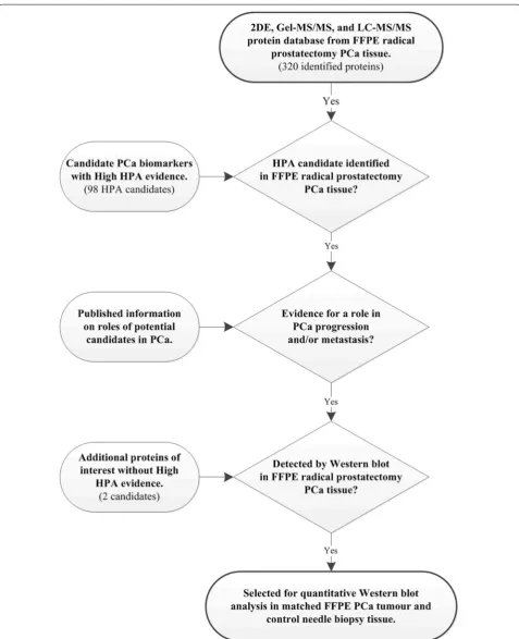
Initially, a protein database was established using 2DE, Gel-MS/MS, and LC–MS/MS of proteins extracted from tumor regions of archival FFPE prostate tissue. Although 2DE separations gave relatively few discrete protein spots (Additional file 1) MALDI MS of tryptic digests of pro-tein spots from these gels resulted in identification of 28 unique proteins including PSA (kallikrein-3). Western
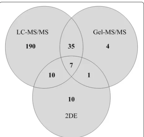
blotting with anti-PSA suggested multiple forms of this protein (Additional file 2), or of related kallikreins, but only the most abundant form was identified by MS. Gel-MS/MS identified 47 non-redundant proteins with at least one matched peptide, and using LC–MS/MS 242 non-redundant proteins were identified. Of the 242 pro-teins identified by LC–MS/MS, 190 were not detected by either of the other methods. Identity scores and % cov-erage for protein identification were often low, prob-ably due to covalent modification of proteins during tissue processing [4]. In total, 320 non-redundant pro-teins were identified (Fig. 1, Additional files 3, 4, 5). The greatest numbers of identifications were of high abun-dance cytoplasmic, cytoskeletal and organelle matrix proteins including nuclear histones, ribosomal proteins and organelle Rabs that are detected during proteomic analysis of many mammalian tissues including pros-tate. Given the greater number of proteins identified by LC–MS/MS, strategies including LC–MS/MS of bands excised from SDS PAGE gels that enhance detection of proteins including integral membrane proteins should be investigated in future studies. For comparison, there are relatively few published analyses of FFPE prostate tis-sue since the initial publication of Hood et al. who used
LC–MS/MS to examine tryptic digests of extracts from benign prostate hyperplasia and cancer regions of a FFPE prostate block [5]. Subsequent reports using frozen tissue include 2D-DIGE analysis of prostatectomy specimens from patients with or without relapse [6], and an iTRAQ comparison of tissue from localized and advanced pros-tate cancer patients [7].
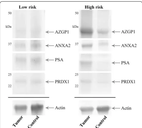
Selection of a set of potential prognostic biomarkers was carried out by interfacing the database of identi-fied proteins with a search of the Human Protein Atlas (http://www.proteinatlas.org/ version 12) for candidates with known immunological reactivity with PCa (Fig. 2). Human Protein Atlas proteins corresponding to keyword “Prostate”; protein class “Candidate cancer biomarkers”; HPA evidence “High”, and filtered for IHC staining in PCa, when interfaced with the MS datasets gave a set of 12 proteins common to both lists (Additional file 6). Five of these candidates were selected on the basis of known associations with PCa (see below) for initial examination by Western blotting of FFPE extracts of radical prosta-tectomy tissue but only four were detected at sufficient abundance for subsequent analysis. This included PSA and three other proteins that have been associated with PCa: annexin A2 (ANXA2) [8–11], zinc-alpha-2-glyco-protein (AZGP1, ZAG) [12–14] and peroxiredoxin-1 (PRDX1) [15, 16]. Positive reactions at the expected molecular masses were obtained for each of these pro-teins, and after optimising primary and secondary anti-body dilutions a multiplexing strategy was developed by blotting with a mixture of antibodies to all four pro-teins plus actin as a loading control (Fig. 3). The multi-plex strategy allowed detection of several proteins from a single blot and facilitated analysis of the small amount of tissue that was harvested from some FFPE blocks. Heat shock protein beta-1 was also detected by Western blot-ting but often with low intensity signals near the thresh-old for detection and was not included in the statistical analysis.
Tumor and control tissue regions excised from FFPE sections from 16 patients, independent of tissue used for the prior MS analyses, were examined using a mul-tiplexing method. The samples for multiplexed West-ern blotting were chosen to represent a range of disease outcomes (time to biochemical failure) with five cases >100 months, five cases ≤40 months, and six “intermedi-ate” 50–87 months. Measured protein abundances were normalized to actin amounts and the tumor/control abundance ratio for each protein was used for analysis of association with time to biochemical failure. The ratios of tumor to control tissue values were used to correct for possible inter-individual differences in protein abun-dance not related to PCa. In most cases the amount of tissue available from the 16 cases was only sufficient for
a single extraction and Western blot. Following detec-tion of the associadetec-tions of ANXA2 and PSA with disease outcome (see below), analytical reproducibility for the two proteins was examined by replicate analyses of pro-teins extracted from FFPE radical prostatectomy tissue. Four separate protein extractions were carried out from a radical prostatectomy block and the set of four extracts was each analyzed twice by Western blotting using 15 μg of protein on each gel. CVs for the normalized, relative abundance values (ANXA2 or PSA normalized against the actin loading control) of the four extracts ranged from 4.4 to 9.5 % for ANXA2 and 2.1–6.5 % for PSA, with less than 6.5 % variation for either protein between the duplicate Western blots.
For each protein of interest in the 16 cases the Kaplan– Meier estimator curves of the “survival” (biochemical failure) function were stratified using cut points deter-mined before the analysis and the failure curves com-pared using the log-rank Chi squared test. For two of the proteins there was a significant difference in the failure curves between the strata predefined by differences in protein abundance measurements. The Kaplan–Meier curves for ANXA2 (Fig. 4) demonstrate that for men with greater than a two-fold increase in ANXA2 abundance in tumor relative to control tissue this appears to predict those men who will experience early biochemical failure during the trial period (log-rank Chi squared p = 0. 025). All such men experienced biochemical failure within 66 months. In contrast, 60 % of the men who exhibited less than a two-fold increase, or decrease, in ANXA2 abundance at the time of diagnosis were biochemical failure free after 66 months, and more than 35 % of them remained biochemical failure free at the trial end-point. The Kaplan–Meier curves for PSA using a three-fold cut-off for protein abundance in tumor relative to con-trol tissue also demonstrated a significant difference in failure risk (log-rank Chi squared p = 0. 016) (Additional file 7). All men with greater than a three-fold increase in PSA abundance experienced biochemical failure within 87 months, whereas more than 55 % of those with less than a three-fold increase remained biochemical failure free at the trial end-point.
Page 5 of 10 Dunne et al. Clin Proteom (2015) 12:24
expression seen in the univariate analysis was not sup-ported in the multivariate modeling.
ANXA2 is a 36 kDa peripheral membrane protein belonging to the 12 member annexin family of calcium
binding proteins. ANXA2 is found on endothelial cells, early myeloid cells, and a variety of tumor cell types [17–20], and has been demonstrated to mediate cellular processes that are essential for cancer metastasis such as tumor cell migration [21, 22], invasion [23, 24] and adhesion [25]. Increased expression of ANXA2 has been reported in tumors of the breast, liver, and pancreas [23, 26, 27]. There is debate surrounding the changes observed in ANXA2 abundance with different types of PCa. The expression of ANXA2 is reduced in high-grade prostatic intraepithelial neoplasia and in low- to moderate-grade PCa [28], but becomes elevated in high-grade PCa [10, 29, 30]. Of interest also is the observation of Hood et al. [5] that ANXA2 apparently did not differ in abundance between cancer and benign hyperplasia regions of an FFPE block, although their analysis differs of course from our study of protein change that predicts disease out-come. Shiozawa et al. demonstrated recently that ANXA2 and its receptor are involved in PCa metastasis by regulat-ing tumor cell migration and adhesion to osteoblasts and endothelial cells, as well as tumor cell proliferation at met-astatic sites [30]. In contrast, Hailermarian et al. [31] did not find a statistically significant association of ANXA2 or three other immunohistochemical markers with risk of PCa. Our demonstration that ANXA2 abundance in tumor versus control tissue at the time of diagnosis of localized, advanced PCa was significantly associated with disease progression therefore complements the immu-nohistochemical analyses of sections from PCa and high grade prostatic intraepithelial neoplasia.
In summary, in this study we analyzed archival FFPE tissue from men with locally advanced PCa for whom there was ten-year outcome data for disease progression. In general, information about protein modification and Fig. 3 Representative Western blot images of matched tumor and
control regions from patients with low or high risk of early bio-chemical failure. Proteins were separated by 1D-PAGE, transferred to Hybond-LFP membrane and probed for ANXA2, AZGP1, PSA and PRDX1, plus actin as a loading control. Detection with AlexaFluor-labelled second antibodies was captured using a FLA-5100 scanner
Fig. 4 Biochemical failure free survival as a function of Annexin A2 risk stratification group. Kaplan–Meier estimator curves of time to bio-chemical failure were constructed using an a priori selected two-fold increase in ANXA2 abundance in tumor relative to control tissue as the cut-point in each of the 16 cases. Statistical analysis of the failure demonstrated a significant difference in time to biochemical failure between the two groups (log-rank Chi squared p = 0. 025), indicating that a >2-fold change in ANXA2 abundance is predictive for early biochemical failure. Vertical lines on the Annexin A2 <2 curve denote censored cases
Table 1 Cox proportional hazard modeling of target pro-tein abundance and time to biochemical failure
a Dependent variable: time to biochemical failure (months)
b Biochemical failure did not occur within the trial period
c Radiotherapy plus either 3 or 6 months androgen deprivation therapy (Additional file 8)
Number Percent
Cases available in analysis Eventa 11 68.8
Censoredb 5 31.2
Total 16 100.0
Variables in the
equation Regression coefficient Standard error p value
ANXA2 0.427 0.181 0.018
AZGP1 −0.038 0.035 0.276
PRDX1 −0.165 0.108 0.127
PSA 0.135 0.101 0.181
Page 7 of 10 Dunne et al. Clin Proteom (2015) 12:24
abundance obtained by Western blotting complements histological description of the cellular localization of proteins. Our strategy was to use proteomic techniques to compile a database of abundant proteins for selec-tion of candidates for subsequent multiplex Western blot analysis. Pauly et al. [32] recently reported multiplexed antibody microarray analysis of proteins extracted from FFPE tissue. To our knowledge we describe the first application of multiplexed Western blotting for analysis of FFPE extracts from prostate biopsies. Our results com-plement those of Geisler et al. who used 2DE of frozen prostate specimens to show differences among proteins that included ANXA4 and A5, although they did not identify ANXA2, between patients with or without bio-chemical relapse [6].
Looking forward, use of ELISA or focused mass spec-trometry [33, 34] for measurement of individual or pan-els of proteins from biopsy tissue may be used both for validation of prospective biomarkers and subsequently as diagnostic assays to enhance management of pros-tate cancer. There may be additional potential to develop urine or blood-based assays, although for proteins including ANXA2 that are also elevated in other cancers [35] measurements on prostate biopsies would be needed for specific diagnosis.
Conclusions
In conclusion, this pilot proteomics study suggests that tumor expression of ANXA2 in diagnostic samples of a PCa may be predictive for the metastatic potential of that cancer. This study also suggests that PSA may have pre-dictive potential. A larger study is in progress to examine and extend these findings using FFPE specimens from the Trans-Tasman TROG 03.04 trial [36] that is subsequent to the 96.01 trial used in the current study.
Methods
Study design and sample processing
FFPE tissue blocks containing prostate biopsies from Wellington men who entered the TROG 96.01 clinical trial were used in the current study. Proteomic studies on the biopsies were approved by the Wellington Eth-ics Committee. The biopsies were collected from 1996 to 2000 at the time of initial diagnosis before treatment and were immediately processed to FFPE blocks. The study design for TROG 96.01 has been described [2]. Eight hundred and eighteen men (ages 41–87), from Aus-tralia and New Zealand, who had locally advanced PCa without distant metastases, received either radiotherapy alone or radiotherapy plus androgen deprivation for 3 or 6 months. Benefits of androgen deprivation included bet-ter control of serum PSA (biochemical control) and PCa
specific survival. For the current proteomic study, tissue was included from 16 men who were treated with radia-tion plus androgen deprivaradia-tion [2]. For each man, the presenting tumor characteristics are provided in Addi-tional file 8. The clinical endpoints were whether or not biochemical failure occurred, and if it did, the time from randomization to the event.
Initially, sections (20 µm thick, approximately 25 mm2)
cut from a FFPE radical prostatectomy tissue block were used for investigation and optimization of protein extraction, 1DE, 2DE, MS and Western blotting. Pro-tein recovery after incubation of 20 µm thick sections (approximately 25 mm2) in Extraction Buffer (see below)
was 142 ± 58 µg protein/mg dry tissue (n = 2). Subse-quently, Western blot analysis was carried out on pro-teins extracted from tumor and control regions of FFPE needle biopsies for statistical analysis of protein asso-ciation with disease outcome. Tissue harvesting for pro-teomics was carried out by pathologists (BD and PF). FFPE sections (5 µm) from each block were stained with hematoxylin/eosin and examined to localize the site of the tumor. Tumor and control regions were excised from adjacent unstained sections under a dissecting micro-scope. FFPE sections (10 µm) were deparaffinised at room temperature using 1 mL xylene (3× 2 min), fol-lowed by rehydration in ethanol series (100 %, 90 %, 80 %, 70 %, 5 min each) and air dried.
Deparaffinised, air-dried tissue samples were incubated in 250 µL of Extraction Buffer consisting of 40 mM Tris– HCl (pH 8.2), 2 % SDS, and 3 % DTT (dithiothreitol) for 1 h at room temperature followed by 20 min at 100 °C [37]. Samples were immediately centrifuged at 14,000×g
Gel‑MS/MS
Protein extracts (30 µg) from FFPE tissue blocks were separated in triplicate by 1DE on 8 × 8 cm 4–12 % NuPAGE gels and fixed for 90 min at room temperature in 50 % ethanol:3 % orthophosphoric acid, then washed 3× with water. Each sample lane was sliced horizon-tally into 30–35 slices that were then transferred to the wells of a 96-well microtiter plate and processed in a GE Healthcare Ettan digester. Each gel piece was diced into approximately 1 mm3 cubes, destained using
50 % meth-anol:50 mM NH4HCO3 (3× 45 min), and air-dried for
2 h. Proteins were digested for 6 h using 10 µL trypsin solution (2.5 ng/µL trypsin in 20 mM NH4HCO3) per
well. Digest peptides were extracted using ACN (acetoni-trile):0.1 % TFA (trifluoroacetic acid) 1:1 (3× 45 min), air-dried, then resuspended in 2 µL 10 mg mL−1 CHCA
(alpha-cyano-4-hydroxycinnamic acid) in ACN:0.1 % TFA 1:1 and spotted onto an ABSCIEX 5800 MALDI TOF/TOF target plate.
LC–MS/MS
Total protein extracts (50 µg) were reconstituted over-night at 4 °C in 50 µL of 50 mM Tris–HCl (pH 8. 5), 8 M urea, 0.1 % SDS. Proteins were reduced in 5 mM DTT for 1 h, alkylated for 30 min in 10 mM iodoacetamide, and quenched in 15 mM DTT, all at room temperature. Samples were diluted 10-fold by the addition of 450 µL of 50 mM Tris–HCl (pH 8.5) and digested overnight at room temperature using 2.5 µg of trypsin per sam-ple. Digests were lyophilised, resuspended in 500 µL 0.1 % TFA and purified using Omix C18 reverse-phase 100 µL tips (Agilent Technologies, Santa Clara, CA). Peptides were eluted into 100 µL of ACN:0.1 % TFA 7:3, lyophilised, and resuspended in 30 µL 0.1 % TFA. Reverse-phase LC-MALDI was performed in triplicate using a TEMPO LC-MALDI spotter system (AB SCIEX, Framingham, MA) with a 150 × 0.1 mm Chromolith® CapRod® RP-18e monolithic column (Merck
Milli-pore, Billerica, MA). Five microlitres of each sample was injected into a 3 µL sample loop at 1 µL min−1. Peptide
separation was achieved using a mobile phase system comprised of 2 % ACN, 0.1 % TFA (Reagent A) and 98 % ACN-0.1 %TFA (Reagent B) with a 36 min linear gradient of 0–80 % Reagent B at 1 µL min−1. Eluted peptides were
mixed on-line with MALDI matrix (CHCA in ACN-0.1 % TFA 1:1 at 1 µL min−1) and spotted at 16 s intervals onto
an ABSCIEX 5800 target plate.
Mass spectrometry and database searches
Gel-MS/MS and LC–MS/MS spectra were collected using an ABSCIEX 5800 MALDI TOF/TOF mass spec-trometer in positive ion mode. TOF/TOF data files were searched against UniProtKB human sequences (88,473
sequences, final searches 17 January 2014) using Protein-Pilot v3.0 (AB SCIEX) with the Paragon algorithm [39]. Search parameters were maximum one missed trypsin cleavage, maximum 50 ppm and 0.2 Da mass tolerances for MS and MS/MS spectra respectively, cysteine carba-midomethylation as a fixed modification, and methionine oxidation as a variable modification. Paragon searches were conducted in “Thorough Mode” using a reversed sequence database to obtain 95 % peptide identification confidence. The mass spectrometry proteomics data have been deposited to the ProteomeXchange Consortium (http://www.proteomexchange.org) via the PRIDE part-ner repository [40] with the dataset identifier PXD000963 and doi 10.6019/PXD000963.
Western blotting
Page 9 of 10 Dunne et al. Clin Proteom (2015) 12:24
Statistical methods
SPSS software was used for statistical analysis (IBM SPSS Statistics 22.0—August 2013). The association of protein abundance measurements with time to biochemical fail-ure was assessed in a univariate way using the Kaplan– Meier estimator for the survival function with cut-points chosen a priori while multivariate associations with bio-chemical failure were assessed by application of Cox pro-portional hazard modeling.
Western blot signals were undetectable in 9.4 % of the 160 individual protein relative abundance measurements (four target proteins plus actin measured in tumor and control tissue for each the 16 cases). Missing values were imputed with a value that was approximately 50 % of the lowest detectable value for any protein in either tissue type.
Abbreviations
1DE: one-dimensional electrophoresis; 2DE: two-dimensional electrophoresis; ACN: acetonitrile; ANXA2: annexin A2; AZGP1: zinc-alpha-2-glycoprotein; CHCA: alpha-cyano-4-hydroxycinnamic acid; DTT: dithiothreitol; FFPE: formalin-fixed paraffin-embedded; LC: liquid chromatography; MS: mass spectrometry; PAGE: polyacrylamide gel electrophoresis; PCa: prostate cancer; PRDX1: peroxiredoxin-1; PSA: prostate-specific antigen; TFA: trifluoroacetic acid; TROG: Trans-Tasman Radiation Oncology Group.
Authors’ contributions
DSL, BD, JN and TWJ designed and initiated the study. DSL and JM carried out sample selection. Pathologists BD and PF examined sections and excised samples for analysis. JD carried out the proteomics and Western blot analysis. SS carried out additional Western blotting. PB provided the statistical analysis. JD, TWJ, DSL and PB wrote the manuscript. All authors read and approved the final manuscript.
Author details
1 Centre for Biodiscovery, School of Biological Sciences, Victoria University
of Wellington, PO Box 600, Wellington, New Zealand. 2 Prostate Cancer Trials
Unit, Department of Pathology and Molecular Medicine, University of Otago Wellington, Wellington, New Zealand. 3 Department of Surgery and
Anaesthe-sia, University of Otago Wellington, Wellington, New Zealand.
Acknowledgements
Funding support from the Prostate Cancer Foundation of New Zealand and the Cancer Society of New Zealand is gratefully acknowledged. We are
Additional files
Additional file 1: Two-dimensional electrophoresis of proteins extracted from FFPE tissue.
Additional file 2: Two-dimensional Western blot analysis of PSA.
Additional file 3: Summary of proteins identified by MALDI MS/MS of spots excised from 2DE gels.
Additional file 4: Summary of proteins identified in FFPE prostate tumor extracts using Gel-MS/MS.
Additional file 5: Summary of proteins identified in FFPE prostate tumor extracts using LC-MS/MS.
Additional file 6: Summary of the proteins identified in FFPE PCa tissue that were selected for Western blot (WB) analysis.
Additional file 7: Biochemical failure free survival as a function of PSA risk stratification group.
Additional file 8: Summary of patient samples analysed.
grateful to Lesley Hooson for technical assistance, to Danyl McLauchlan for bioinformatics support, and to the PRIDE team at EBI for support with the submission of the mass spectrometry proteomics data to the ProteomeX-change Consortium (http://www.proteomexchange.org) via the PRIDE partner repository.
Compliance with ethical guidelines
Competing interests
The authors declare that they have no competing interests.
Received: 22 April 2015 Accepted: 9 September 2015
References
1. Jemal A, Bray F, Center MM, Ferlay J, Ward E, Forman D. Global cancer statistics. CA-A Cancer J Clin. 2011;61:69–90.
2. Denham JW, Steigler A, Lamb DS, Joseph D, Turner S, Matthews J, et al. Short-term neoadjuvant androgen deprivation and radiotherapy for locally advanced prostate cancer: 10-year data from the TROG 96.01 randomised trial. Lancet Oncol. 2011;12:451–9.
3. Steigler A, Denham JW, Lamb DS, Spry NA, Joseph D, Matthews J, et al. Risk stratification after biochemical failure following curative treatment of locally advanced prostate cancer: data from the TROG 96.01 trial. Prostate Cancer. 2012;. doi:10.1155/2012/814724.
4. Maes E, Valkenborg D, Mertens I, Broeckx V, Baggerman G, Sagaert X, et al. Proteomic analysis of formalin-fixed paraffin-embedded colorec-tal cancer tissue using tandem mass tag protein labeling. Mol BioSyst. 2013;9:2686–95.
5. Hood BL, Darfler MM, Guiel TG, Furusato B, Lucas DA, Ringeisen BR, et al. Proteomic analysis of formalin-fixed prostate cancer tissue. Mol Cell Proteomics. 2005;4:1741–53.
6. Geisler C, Gaisa NT, Pfister D, Fuessel S, Kristiansen G, Braunschweig T, et al. Identification and validation of potential new biomarkers for pros-tate cancer diagnosis and prognosis using 2D-DIGE and MS. Biomed Res Int. 2015;2015:454256.
7. Khan AP, Poisson LM, Bhat VB, Fermin D, Zhao R, Kalyana-Sundaram S, et al. Quantitative proteomic profiling of prostate cancer reveals a role for miR-128 in prostate cancer. Mol Cell Proteomics. 2010;9:298–312. 8. Zhang XH, Liu SQ, Guo CM, Zong JW, Sun MZ. The association of annexin
A2 and cancers. Clin Transl Oncol. 2012;14:634–40.
9. Nadiminty N, Dutt S, Tepper C, Gao AC. Microarray analysis reveals poten-tial target genes of NF-kappa B2/p52 in LNCaP prostate cancer cells. Prostate. 2010;70:276–87.
10. Inokuchi J, Narula N, Yee DS, Skarecky DW, Lau A, Ornstein DK, et al. Annexin A2 positively contributes to the malignant phenotype and secretion of IL-6 in DU145 prostate cancer cells. Int J Cancer. 2009;124:68–74.
11. Das S, Shetty P, Valapala M, Dasgupta S, Gryczynski Z, Vishwanatha JK. Sig-nal transducer and activator of transcription 6 (STAT6) is a novel interactor of annexin A2 in prostate cancer cells. Biochemistry. 2010;49:2216–26. 12. Henshall SM, Horvath LG, Quinn DI, Eggleton SA, Grygiel JJ, Stricker PD,
et al. Zinc-alpha2-glycoprotein expression as a predictor of metastatic prostate cancer following radical prostatectomy. J Natl Cancer Inst. 2006;98:1420–4.
13. Hassan MI, Waheed A, Yadav S, Singh TP, Ahmad F. Zinc alpha 2-glycopro-tein: a multidisciplinary protein. Mol Cancer Res. 2008;6:892–906. 14. Descazeaud A, de la Taille A, Allory Y, Faucon H, Salomon L, Bismar T, et al.
Characterization of ZAG protein expression in prostate cancer using a semi-automated microscope system. Prostate. 2006;66:1037–43. 15. Chhipa RR, Lee KS, Onate S, Wu Y, Ip C. Prx1 enhances androgen receptor
function in prostate cancer cells by increasing receptor affinity to dihy-drotestosterone. Mol Cancer Res. 2009;7:1543–52.
17. Falcone DJ, Borth W, Khan KMF, Hajjar KA. Plasminogen-mediated matrix invasion and degradation by macrophages is dependent on surface expression of annexin II. Blood. 2001;97:777–84.
18. Hajjar KA, Guevara CA, Lev E, Dowling K, Chacko J. Interaction of the fibrinolytic receptor, annexin II, with the endothelial cell surface—essen-tial role of endonexin repeat. J Biol Chem. 1996;271:21652–9.
19. Mai JX, Waisman DM, Sloane BF. Cell surface complex of cathepsin B/ annexin II tetramer in malignant progression. Biochim Biophys Acta Protein Struct Mol Enzymol. 2000;1477:215–30.
20. Menell JS, Cesarman GM, Jacovina AT, McLaughlin MA, Lev EA, Hajjar KA. Annexin II and bleeding in acute promyelocytic leukemia. N Engl J Med. 1999;340:994–1004.
21. Zhang W, Zhao P, Xu XL, Cai L, Song ZS, Cao DY, et al. Annexin A2 pro-motes the migration and invasion of human hepatocellular carcinoma cells in vitro by regulating the shedding of CD147-harboring microvesi-cles from tumor cells. PLoS One. 2013;8:e67268. doi:10.1371/journal. pone.0067268.
22. An JH, Lee SY, Jeon JY, Cho KG, Kim SU, Lee MA. Identification of gliotropic factors that induce human stem cell migration to malignant tumor. J Proteome Res. 2009;8:2873–81.
23. Sharma MR, Koltowski L, Ownbey RT, Tuszynski GP, Sharma MC. Angiogenesis-associated protein annexin II in breast cancer: selective expression in invasive breast cancer and contribution to tumor invasion and progression. Exp Mol Pathol. 2006;81:146–56.
24. Diaz VM, Hurtado M, Thomson TM, Reventos J, Paciucci R. Specific interaction of tissue-type plasminogen activator (t-PA) with annexin II on the membrane of pancreatic cancer cells activates plasminogen and promotes invasion in vitro. Gut. 2004;53:993–1000.
25. Tressler RJ, Updyke TV, Yeatman T, Nicolson GL. Extracellular annexin-II is associated with divalent cation-dependent tumor-cell endothelial-cell adhesion of metastatic RAW117 large-endothelial-cell lymphoma-endothelial-cells. J Cell Biochem. 1993;53:265–76.
26. Vishwanatha JK, Chiang YP, Kumble KD, Hollingsworth MA, Pour PM. Enhanced expression of annexin-II in human pancreatic-carcinoma cells and primary pancreatic cancers. Carcinogenesis. 1993;14:2575–9. 27. Mohammad HS, Kurokohchi K, Yoneyama H, Tokuda M, Morishita A, Jian
G, et al. Annexin A2 expression and phosphorylation are up-regulated in hepatocellular carcinoma. Int J Oncol. 2008;33:1157–63.
28. Chetcuti A, Margan SH, Russell P, Mann S, Millar DS, Clark SJ, et al. Loss of Annexin II heavy and light chains in prostate cancer and its precursors. Cancer Res. 2001;61:6331–4.
29. Banerjee AG, Liu J, Yuan Y, Gopalakrishnan VK, Johansson SL, Dinda AK, et al. Expression of biomarkers modulating prostate cancer angiogenesis: differential expression of annexin II in prostate carcinomas from India and USA. Mol Cancer. 2003;2:34–47.
30. Shiozawa Y, Havens AM, Jung YH, Ziegler AM, Pedersen EA, Wang JC, et al. Annexin II/Annexin II receptor axis regulates adhesion, migration, hom-ing, and growth of prostate cancer. J Cell Biochem. 2008;105:370–80. 31. Hailemariam S, Vosbeck J, Cathomas G, Zlobec I, Mattarelli G, Eichen-berger T, et al. Can molecular markers stratify the diagnostic value of high-grade prostatic intraepithelial neoplasia? Hum Pathol. 2011;42:702–9.
32. Pauly F, Dexlin-Mellby L, Ek S, Ohlin M, Olsson N, Jirstrom K, et al. Protein expression profiling of formalin-fixed paraffin-embedded tissue using recombinant antibody microarrays. J Proteome Res. 2013;12:5943–53. 33. Hembrough T, Thyparambil S, Liao W-L, Dafler MM, Abdo J, Bengali
KM, et al. Application of selected reaction monitoring for multiplex quantification of clinically validated biomarkers in formalin-fixed, paraffin-embedded tumor tissue. J Mol Diagn. 2013;15:454–65.
34. Sprung RW, Martinez MA, Carpenter KL, Ham A-JL, Washington MK, Arteaga CL, et al. Precision of multiple reaction monitoring mass spec-trometry analysis of formalin-fixed, paraffin-embedded tissue. J Proteome Res. 2012;11:3498–505.
35. Sun Y, Gao G, Cai J, Wang Y, Qu X, He L, et al. Annexin A2 is a discrimina-tive serological candidate in early hepatocellular carcinoma. Carcinogen-esis. 2013;34:595–604.
36. Denham JW, Joseph D, Lamb DS, Spry NA, Duchesne G, Matthews J, et al. Short-term androgen suppression and radiotherapy versus intermediate-term androgen suppression and radiotherapy, with or without zoledronic acid, in men with locally advanced prostate cancer (TROG 03.04 RADAR): an open-label, randomised, phase 3 factorial trial. Lancet Oncol. 2014;15:1076–89.
37. Addis MF, Tanca A, Pagnozzi D, Crobu S, Fanciulli G, Cossu-Rocca P, et al. Generation of high-quality protein extracts from formalin-fixed, paraffin-embedded tissues. Proteomics. 2009;9:3815–23.
38. Rawson P, Stockum C, Peng LF, Manivannan B, Lehnert K, Ward HE, et al. Metabolic proteomics of the liver and mammary gland during lactation. J Proteomics. 2012;75:4429–35.
39. Shilov IV, Seymour SL, Patel AA, Loboda A, Tang WH, Keating SP, et al. The paragon algorithm, a next generation search engine that uses sequence temperature values and feature probabilities to identify peptides from tandem mass spectra. Mol Cell Proteomics. 2007;6:1638–55. 40. Vizcaino JA, Cote RG, Csordas A, Dianes JA, Fabregat A, Foster JM, et al.
The Proteomics Identifications (PRIDE) database and associated tools: status in 2013. Nucleic Acids Res. 2013;41:D1063–9.
41. Schneider CA, Rasband WS, Eliceiri KW. NIH Image to ImageJ: 25 years of image analysis. Nat Methods. 2012;9:671–5.
Submit your next manuscript to BioMed Central and take full advantage of:
• Convenient online submission • Thorough peer review
• No space constraints or color figure charges • Immediate publication on acceptance
• Inclusion in PubMed, CAS, Scopus and Google Scholar • Research which is freely available for redistribution