RESEARCH
The cancer-associated CTCFL/BORIS
protein targets multiple classes of genomic
repeats, with a distinct binding and functional
preference for humanoid-specific SVA
transposable elements
Elena M. Pugacheva
2, Evgeny Teplyakov
1, Qiongfang Wu
1, Jingjing Li
1, Cheng Chen
1, Chengcheng Meng
1,
Jian Liu
1, Susan Robinson
2, Dmitry Loukinov
2, Abdelhalim Boukaba
1, Andrew Paul Hutchins
3,
Victor Lobanenkov
2and Alexander Strunnikov
1*Abstract
Background: A common aberration in cancer is the activation of germline-specific proteins. The DNA-binding pro-teins among them could generate novel chromatin states, not found in normal cells. The germline-specific transcrip-tion factor BORIS/CTCFL, a paralog of chromatin architecture protein CTCF, is often erroneously activated in cancers and rewires the epigenome for the germline-like transcription program. Another common feature of malignancies is the changed expression and epigenetic states of genomic repeats, which could alter the transcription of neighboring genes and cause somatic mutations upon transposition. The role of BORIS in transposable elements and other repeats has never been assessed.
Results: The investigation of BORIS and CTCF binding to DNA repeats in the K562 cancer cells dependent on BORIS for self-renewal by ChIP-chip and ChIP-seq revealed three classes of occupancy by these proteins: elements cohabited by BORIS and CTCF, CTCF-only bound, or BORIS-only bound. The CTCF-only enrichment is characteristic for evolu-tionary old and inactive repeat classes, while BORIS and CTCF co-binding predominately occurs at uncharacterized tandem repeats. These repeats form staggered cluster binding sites, which are a prerequisite for CTCF and BORIS co-binding. At the same time, BORIS preferentially occupies a specific subset of the evolutionary young, transcribed, and mobile genomic repeat family, SVA. Unlike CTCF, BORIS prominently binds to the VNTR region of the SVA repeats in vivo. This suggests a role of BORIS in SVA expression regulation. RNA-seq analysis indicates that BORIS largely serves as a repressor of SVA expression, alongside DNA and histone methylation, with the exception of promoter capture by SVA.
Conclusions: Thus, BORIS directly binds to, and regulates SVA repeats, which are essentially movable CpG islands, via clusters of BORIS binding sites. This finding uncovers a new function of the global germline-specific transcriptional regulator BORIS in regulating and repressing the newest class of transposable elements that are actively transposed in human genome when activated. This function of BORIS in cancer cells is likely a reflection of its roles in the germline.
© 2016 The Author(s). This article is distributed under the terms of the Creative Commons Attribution 4.0 International License (http://creativecommons.org/licenses/by/4.0/), which permits unrestricted use, distribution, and reproduction in any medium, provided you give appropriate credit to the original author(s) and the source, provide a link to the Creative Commons license, and indicate if changes were made. The Creative Commons Public Domain Dedication waiver (http://creativecommons.org/ publicdomain/zero/1.0/) applies to the data made available in this article, unless otherwise stated.
Open Access
*Correspondence: [email protected]
1 Molecular Epigenetics Laboratory, Guangzhou Institutes of Biomedicine and Health, Guangzhou 510530, Guangdong, China
Background
Transposable elements (TEs) play active roles in nor-mal genome evolution in humans [1] and in primates in general [2], as well as in sporadic genome rearrange-ment [3–5] including deleterious events associated with pathology [6–12]. Multiple polymorphisms and intron evolution in normal human populations are largely facili-tated by TE insertions [13, 14]. A substantial and distinct role of satellite repeats was also recently demonstrated for double-strand breaks (DSBs) incidence upon replica-tion stress [15]. Active families of TEs (L1, Alu, and SVA) account for a large number of germline mutations [16]. In cancer, insertions of mobile element and the recom-bination between them have been identified as causes of many cancers [12, 17, 18], with some repeats shown to become aberrantly expressed [17, 19] to acquire a potential to change the regulation of neighboring genes [17, 20, 21] and to destabilize chromosomes [7, 22]. The effect of repeated DNA in the origins and progression of cancer and tumor cell physiology could be two-pronged: the induced change of expression in neighboring or tar-geted genes [22–24] and the structural destabilization of the epigenetic landscape of chromosomes [2, 25]. These two effects are interrelated, as epigenetic changes in the repeats open chromosomal domains for both aberrant changes in gene expression and elevated somatic recom-bination. Some elements were also shown to act as bona fide enhancers [26].
The presence of a strong epigenetic component in such repeats and TE-mediated genome regulation and insta-bility is well established [20, 27–30]. In cancer cells, there is likely a higher epigenetic impact of TEs, compared to the norm [12], as promoters of expressed mobile ele-ments become hypomethylated and their transcription elevated [22, 31, 32].
The array of epigenetic changes leading to repeat deregulation in cancer cannot be understood without molecular analysis of repeats’ chromatin. This brings to light the role of CTCF and its paralog CTCFL/BORIS in these processes. In addition to serving as a bona fide transcription factor, CTCF reads the epigenetic marks [33–36] and plays a key role in the formation of topologi-cally associated domains (TADs) in chromatin [37–39], in remodeling chromatin structure [40], and in the for-mation of chromatin boundaries [29, 41]. CTCF was also shown to have multiple binding sites embedded in TEs [42, 43]. CTCF target sites (CTSs) are also important for telomere repeat stability [44, 45]. Furthermore, the fact that CTCF control of gene expression and recombina-tion requires physical contacts between different CTSs via looping [46–49] indicates that CTCF sites in repeats are not inert in the chromatin architecture, as indeed was demonstrated at some instances [50–53].
Taking into account the important role of CTCF in regulating TE expression and epigenetic maintenance, it is possible that the aberrant activation of its ger-mline paralog CTCFL/BORIS in cancer has an impact on repeat physiology and genome stability. BORIS is a cancer testis (CT) gene [54], and its ectopic expression could be lethal or inhibitory for somatic cells because BORIS, being a germline transcription factor, activates gene expression of germline-specific genes on its own or in cooperation with CTCF [55]. Nevertheless, some cancer cells undergo adaptation/addiction to BORIS activation and incorporate the BORIS protein into their physiology [55, 56]. BORIS also interferes with a variety of other CTCF-specific functions in somatic cells, such as in the organization of chromatin loops that are alterna-tive to the chromatin configuration of normal cells [55]. The ultimate molecular and physiological role of BORIS in cancer is still poorly understood, however, beyond the association with stemness [56], phenocopying of germline-specific gene expression pattern, and the corre-sponding 3D chromatin organization [55]. In particular, it is not clear how some cancer cells became dependent on BORIS for their proliferation, making BORIS a poten-tial anticancer target [57, 58].
While many genomic repeats are heavily methylated and BORIS has a probable role in DNA demethylation [57, 59–61], the role of BORIS in repeat biology has not been studied. Incidentally, even the most comprehensive genome-wide studies on CTCF tended to ignore the pos-sible simultaneous presence of BORIS in cells studied, be it cancer or embryonic stem cells [48, 50, 62–64]. In this present study, we attempted to assess the specific pattern of BORIS recognition of genomic repeats in cancer cells and to link it to TE expression. As a result, we uncovered a surprising association of BORIS with one of the evo-lutionary youngest families of actively transcribed and mobile repeats in human genome, the SVA family of TEs. Follow-up analysis of the modulation of BORIS expres-sion revealed that it predominately acts as one of the mechanisms repressing the expression of these elements.
Results
BORIS expression in K562 forms a specific pattern of repeat binding
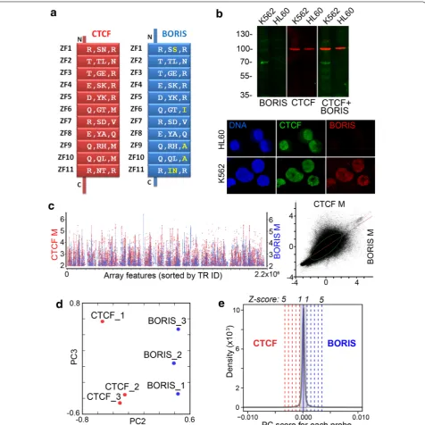
(NGS) reads to massively repeated DNA, while micro-array analysis has well documented limitations of its own. Here, we employed a similar two-step approach in reverse; the repeats’ enrichment by DNA-binding pro-teins was first assessed by ChIP-chip and then validated by ChIP-seq. We used mainly the established cancer cell line K562 as a model for the coexistence of CTCF and BORIS stably expressed at a relatively the same level, as assayed by RT-PCR [55], to assess genome repeat occu-pancy by these two proteins. K562 retains a set of prop-erties characteristic for cancer stem cells, e.g., the ability to initiate tumors in graft models, and the propensity to differentiate in response to exogenous stimuli [66]. As CTCF and BORIS have essentially the same composition of the DNA-binding domain, including the number of ZF and their spacing, as well as residues involved in DNA contacts (Fig. 1a), they show the virtually identical DNA-binding specificity in vitro, albeit not in native chromatin [55]. Therefore, it was important to use a cell line where two proteins are expressed in equivalent amounts, such as K562. Unlike most established cancer cell lines or pri-mary non-germline tumor cells, where the expression of BORIS is low, with only a minor subset of cells character-ized by high BORIS expression [56], K562 expresses high level of BORIS largely localized to the nuclei (Fig. 1b). BORIS was also confirmed to be incorporated into tran-scription regulation in K562 and to be required for its self-renewal [55].
For the initial analysis, by ChIP-chip, anti-CTCF and anti-BORIS immunoprecipitations were conducted and microarray hybridization was performed as described in Methods. The plot of normalized ChIP-chip fluores-cence intensities showed indications of distinct binding patterns for BORIS and CTCF on highly enriched tiles (Fig. 1c). Significance analysis of microarray (SAM) indi-cated that over 40,000 tiles were enriched differentially by CTCF and BORIS, but provided little clue about the occupancy of the rest of the repeats. The principal com-ponent analysis (PCA) of arrays hybridized to CTCF and BORIS ChIP samples confirmed the presence of differ-entially bound genomic repeats (Fig. 1d). The PCA also revealed the three expected scenarios of occupancy: binding by BORIS only, by CTCF only, and BORIS and CTCF co-binding being by far the largest group (Fig. 1e). As CTCF and BORIS have essentially the same DNA-binding specificities in vitro, the differences in occupancy observed in vivo must be largely driven by the epigenetic factors.
Prior to proceeding further with analyses of repeat binding sequences, we conducted a validation of ChIP-chip data using an alternative high-throughput procedure, ChIP-seq, as conventional qPCR valida-tion methods are not applicable or scalable to the TRs
genome-wide. We set out to validate the three identi-fied subsets: first, repeats preferentially enriched by CTCF (Fig. 1e), second, repeats preferentially enriched by BORIS (Fig. 1e), and, third, repeats equally enriched by both CTCF and BORIS (Fig. 1e, a subset of the mid-dle group). Based on detailed PCA analysis, an additional cutoff across the three groups was applied to make uni-form criteria for selecting the representative subsets for validation. For co-bound repeats we chose the 4× enrich-ment for both proteins in all three ChIP-chip replicates, while for the Z5 groups we used 4× enrichment for one protein, with no enrichment for the other, also in all three replicates. Drawing the threshold at such a relatively high level also significantly reduced repeat redundancy in the TR dataset. For the ChIP-seq validation, we considered a ChIP-chip-positive repeat validated, if any tile from that repeat was reproducibly enriched at least twofold in ChIP-seq datasets with 95 % DNA match. Thus, all the
repeats discussed below are repeats identified by ChIP-chip and validated by ChIP-seq.
Co‑binding of BORIS and CTCF is characteristic for the simple tandem repeats
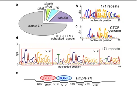
The simultaneous binding of BORIS and CTCF genome-wide in cancer cell lines was shown to reset, at least par-tially, the functions of CTSs in transcriptional regulation in accordance with germline-like program [55]. Thus, from the standpoint of cancer biology, it was important to characterize repeats bound by both CTCF and BORIS (CTCF and BORIS repeats, Additional file 1: Table S2), especially as they outnumbered other classes (Fig. 1e). The 171 distinct repeats in the CTCF and BORIS class were mostly represented by uncharacterized simple repeats, which can also be classified as VNTRs, and a small fraction of TEs, with the telomeric satellite TAR1
unit does not enclose a bona fide cluster CTS, the tan-dem arrangement of this class sets a potentially multiple/ staggered binding mode for CTCF and BORIS at these elements potentially generating a cluster site, if the tan-dem structure is long enough (Fig. 2e). Therefore, we can hypothesize that co-binding of CTCF and BORIS to the same site, as in this group of repeats, is facilitated when two binding regions are juxtaposed in cis, as hap-pens in the rest of the genome [55]. The fact that multiple uncharacterized simple repeats were found in this class indicates that these elements should have a regulatory function in the epigenome mediated by dual binding by CTCF and BORIS.
Analysis of CTCF and BORIS co-binding at repeated DNA would have been incomplete without assessing the least characterized region of human epigenome— the chromatin of nucleolar organizer (NOR, or rDNA repeats). The bona fide human genomic rDNA has a very complex structure with multiple intervening sequences [68], and the NOR sequence from any human chromo-some still remains to be determined. Therefore, human
rDNA was not represented at TRF database and was not present on our microarrays. While we did not vali-date rDNA binding by CTCF and BORIS in ChIP-chip, it is known that the repeat unit contains a strong hotspot for CTCF binding facilitating CTCF’s interaction with PolI transcription machinery [69]. We used a “consensus” human rDNA repeat, as in [65], to align ChIP-seq reads and assess the potential differences between CTCF and BORIS binding (Additional file 2: Figure S2B). Compar-ing BORIS and CTCF bindCompar-ing showed that CTCF has a single binding site upstream of rDNA PolI promoter, consistent with published data in mice [70]. At the same time, BORIS appeared to have some enrichment at addi-tional sites (Addiaddi-tional file 2: Figure S2A). These loca-tions, however, corresponded to low-complexity regions (Additional file 3: Table S1), which were also present else-where in the genome. Unlike the established CTCF bind-ing site, the two selected BORIS sequences that appeared to be enriched in ChIP-seq were not confirmed to bind BORIS by EMSA in vitro (Additional file 2: Figure S2C). Thus, one may assume that such sites likely represent an
artifact of short reads’ alignment to tandemly repeated DNA, and the additional such sites were not tested. The presence of BORIS at the main Pol I regulatory site in rDNA, however, indicates that BORIS might be involved in ribosome biogenesis in cancer cells by virtue of co-reg-ulating the rDNA transcription with CTCF.
CTCF‑only enrichment is found in older repeat classes
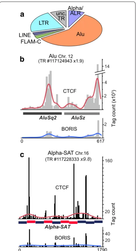
The CTCF-only binding sites have a still unknown func-tion in the genome, possibly unrelated to transcrip-tion [55]. PCA results in Fig. 1e enabled us to separate the CTCF-bound repeats that were refractory to BORIS intrusion (Fig. 3a). Thirty-eight individual CTCF-only repeats in this group were validated by ChIP-seq (Addi-tional file 2: Table S2). This set includes major known types of repeats with long evolutionary history, while evolutionary young and simple TRs were largely absent. This agrees well with the studies, indicating that some CTCF-only binding sites in repeats are conserved in evolution [67]. Two examples of ChIP-seq analysis for repeats in this class, a TR of two Alu elements (Fig. 3b) and a run of divergent centromeric alpha-satellites (Fig. 3c), showed a robust enrichment by CTCF as com-pared to BORIS. As the enrichment of alpha-satellites by CTCF did not appear to be very strong, it is possible that a substantial fraction of alphoid elements in the K562 genome are not occupied by CTCF. Combined with the fact that CTCF binding does not appear to be correlat-ing with CENP-B box presence (Fig. 3c), this may even indicate that only non-centromeric alpha-satellites are bound by CTCF. The absence of strong BORIS binding to this group of repeats agrees well with the underrepresen-tation of clustered CTS consensuses in this repeat group (not shown).
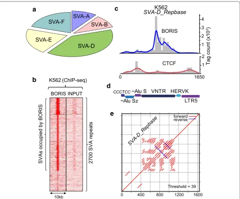
A movable and evolutionary youngest class of TEs is specifically enriched in BORIS binding
The BORIS-only repeats, where BORIS binds without the equivalent presence of CTCF, are the most revealing with respect to BORIS biology in cancer cells, as they are directly involved in the transcriptional regulation of the non-repeated part of the genome [55]. Remarkably, in this group, the only 10 TRF classes that were validated fell within a single repeat type: the SVA family (Fig. 4a; Additional file 1: Table S2). The SVA repeats are a hom-inid-specific family, which is still currently mobile in the human genome owing to L1 activity [71, 72]. Overall, ChIP-seq analysis indicates that as much as 70 % of SVA elements could be occupied by BORIS in K562 (Fig. 4b). When this preference for SVA repeats was dissected for individual genomic repeat sequences, it became appar-ent that the enrichmappar-ent by BORIS peaked in the cappar-entral part of the element composed of the GC-rich VNTRs
(Fig. 4c–e). VNTRs in SVA are GC-rich sequences with unknown molecular function. The patterns of CTCF and BORIS occupancy at SVA elements were distinct (Fig. 4c), unlike in other elements analyzed in Fig. 3. This might indicate the exceptional specialization of the
a
b
Alpha/ ALR
Alu FLAM-C
LINE LTR
unc. TR
c
Tag count (x1
0
3)
Alu Chr. 12 (TR #117124943 x1.9)
2 2 14
7 1 6 0
CTCF
BORIS
Alpha-SAT Chr.16 (TR #117228333 x9.8)
CTCF
BORIS Alpha-SAT
1790 0
20 40 20 160 AluSq2 AluSz
Tag count
4
VNTRs for BORIS binding in cancer cells. In order to exclude the possibility that SVA enrichment by BORIS is a specific property of K562, myeloid cells, or the female epigenome in general, we conducted ChIP-seq analy-sis of an unrelated cancer cell line with aberrantly acti-vated BORIS, Delta-47 cells [55]. Although the difference between BORIS and CTCF enrichment was not as dra-matic as in K562, the preference of BORIS was evident (Additional file 4: Figure S1A), notwithstanding the lower level of BORIS in Delta-47 [55]. Considering that the SVA’s VNTRs are dynamic in number and composition
themselves [73], the finding of a global regulator BORIS bound to a mobile and extremely variable repeat class could be indicative of an additional germline-specific function of BORIS.
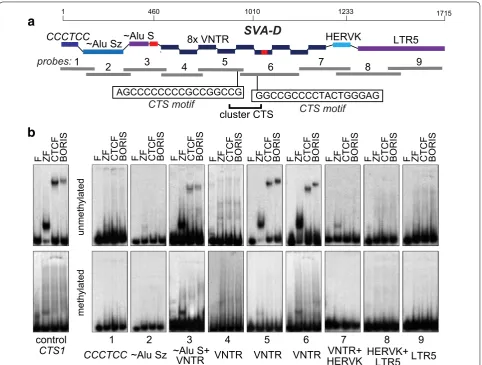
In order to map the locations of BORIS binding sites in SVA elements with higher precision, we designed nine probes corresponding together to a full-size SVA-D ele-ment (Fig. 5a) and analyzed them by EMSA with BORIS and CTCF proteins produced by in vitro translation. EMSA assay showed that the weak binding found in the AluS part can be attributed to a short unique sequence
a
c
d
SVA-A
SVA-B
SVA-D
SVA-E
SVA-F
0 400 800 1200 1600 forward reverse
Threshold = 39
K562
SVA-D_Repbase
VNTR ~Alu Sz
CCCTCC~Alu S HERVK
LTR5
SVA-D_Repbase
e
1 2 3 4
1 2
Tag count (x1
0
3 )
0 5 6 1 0
CTCF BORIS
b
2700 SVA repeat
s
BORIS INPUT
K562 (ChIP-seq)
10kb
SVAs occupied by BORI
S
there (Fig. 5b). The central core of VNTR region, rep-resented by two probes (5 and 6) in an EMSA, showed reproducible binding to both BORIS and CTCF proteins (Fig. 5b). Based on the EMSA data and CTCF motif anal-ysis (Fig. 5b), these two VNTR sites juxtaposed to each other together form a cluster CTS, which is required for BORIS-only binding [55]. The 83-bp unique sequence embedded in the probe 6 in Fig. 5 was by itself unable to bind either protein (not shown). Not surprisingly, no discernible difference was detected between CTCF and BORIS in binding in vitro (Fig. 5b). This indicates that the BORIS’ preference for SVA binding observed in chroma-tin (ChIP data) is likely determined by epigenetic factors. As CTCF is known to have both DNA methylation-sensi-tive and methylation-insensimethylation-sensi-tive binding sites, we verified
whether BORIS is able to bind VNTRs when CpGs are methylated. EMSA analysis with methylated probes (Additional file 4: Figure S1B) showed that both CTCF and BORIS binding were abolished by full CpG methyla-tion (Fig. 5b). This likely indicates that the preference of these sites for BORIS binding in chromatin, even if par-tially controlled by DNA methylation, must be fine-tuned with respect to specific CpGs methylation.
What could be BORIS activity at SVA elements? Our previous results on the genome-wide consequences of modulation of BORIS expression indicated that BORIS could serve as an activator as well as repressor [55]. The distinct preference of the aberrantly expressed BORIS for SVA elements may potentially indicate that BORIS has some regulatory activity at these elements in germline
F ZF CTCF BORI
S
1 CCCTCC
2 ~Alu Sz
3 ~Alu S+
VNTR 4 VNTR
7 VNTR+ HERVK
8 HERVK+
LTR5 9 LTR5 5
VNTR 6 VNTR
F ZF CTCF BORI
S
F ZF CTCF BORI
S
F ZF CTCF BORI
S
F ZF CTCF BORI
S
F ZF CTCF BORI
S
F ZF CTCF BORI
S
F ZF CTCF BORI
S
F ZF CTCF BORI
S
F ZF CTCF BORI
S
8x VNTR ~Alu Sz
CCCTCC
1
~Alu S HERVK LTR5
1010
1 460 1233 1715
6 5
4 3
2 7 8 9
SVA-D
probes:
control CTS1
a
b
unmethylated
methylated
AGCCCCCCCCGCCGGCCG GGCCGCCCCTACTGGGAG
CTS motif CTS motif
cluster CTS
and/or in cancer cells. As there is little doubt that SVAs mobilization is detrimental to genome stability, because they are under a strong repression in primates [73–76], a possible BORIS involvement in the regulation of SVA transcription must be biologically important. Indeed, the transcription is required for SVA transposition, and it could also have a regulatory role in the expression of neighboring genes.
BORIS acts as a transcriptional co‑repressor of a significant proportion of SVAs in K562 cells
While the transcription unit of SVAs is not well charac-terized [76, 77], the Alu-derived sequences are the chief drivers of transposition in SVA [78]. Thus, SVAs contain sequences potentially transcribed by both RNA Pol III and Pol II, either of which can drive retrotransposition [79]. At the same time, based on structural considera-tions, it is unlikely that SVA elements are actually tran-scribed by Pol III [77]. We tested whether there was a difference in the occupancy of RNA Pol III factors at SVA elements between the publicly available ChIP-seq datasets for BORIS-positive K562 and BORIS-negative NHEK. Incidentally, we found no notable enrichment at any SVA elements for POLR3G, BDP1, BRF1, BRF2, or RPC155 (data not shown).
Next, we focused on the RNA Pol II transcription of SVAs and first took advantage of CAGE datasets available for K562 (BORIS positive) and NHEK (BORIS negative). The CAGE reads were aligned to the genome, and the extended areas corresponding to SVA elements were ana-lyzed separately. However, the levels of SVA transcription were low, and SVA transcription in BORIS-positive K562 cells was mostly well correlated with the BORIS-nega-tive NHEK cells (Pearson correlation 0.98). At the same time, RNA-seq data available for human testis suggest that some SVA elements could be highly expressed; how-ever, the two full-length (FL) SVA elements with highest expression in human testis showed no ChIP-seq enrich-ment for BORIS at the VNTRs (Fig. 6a). The extension of analysis in Fig. 6a to 59 additional SVA elements with various degrees of BORIS occupancy showed only mar-ginal levels of expression without any correlation with BORIS presence at the VNTRs (not shown). Thus, it is highly unlikely that BORIS bound to VNTRs serves as a transcription activator of SVA transcription in K562 cells.
At this point, one may hypothesize that the affin-ity of BORIS to VNTRs of SVA elements demonstrated in K562 is a reflection of its role in germline pertaining to these elements and that this role is likely a repressive one. Indeed, we recently showed that despite BORIS previously perceived as an activator, BORIS upregula-tion was linked to the repression of some genes and,
vice versa, BORIS downregulation has resulted in some gene being activated [55]. Therefore, we investigated the K562 cells with downregulated BORIS. As SVA elements might be rapidly repressed by some other mechanism in the absence of BORIS, we could not rely on BORIS KO data [55], as the points of comparison there were sepa-rated by a long period of time. Instead, we experimented with the downregulation of BORIS expression in K562 cells for a short period of time using inducible shRNA. This approach enabled us to assess immediate down-stream effects of BORIS downregulation. We constructed K562 cell lines with two alternative inducible anti-BORIS shRNA constructs stably integrated into the genome and conducted RNA-seq experiments after BORIS KD for 48 h. Neither the degree of BORIS depletion nor the time span of the experiment was sufficient to induce the dif-ferentiation, as was described for BORIS KO [55]. While genome-wide expression of genes responding to BORIS KD was almost evenly divided between up- and down-regulation of transcription (data not shown), SVA ele-ments longer than 1 kb were notably activated (Fig. 6d). In order to address whether any SVA were actually down-regulated upon BORIS KD, we isolated the subclass of SVA elements that were already expressed in K562 and compared their expression to BORIS KD cells. As shown in Additional file 5: Figure S3A, the 70 SVA elements that were expressed did not significantly change their expres-sion upon the downregulation of BORIS.
- +
BORIS
Tubulin
c
- + - + - +
K562 vectorsite1 site2sh-BORIS1 1 0.8 0.9 1 0.2 0.8 0.2
0 0.2 0.4 0.6 0.81
Dox+
K562vector
KD BORIS
Relative mRNA
KD site
2
KD site
1
a
b
chr.10
chr.15
1 kb
SVA-B
SVA-D
BORIS Dox:
BORIS
77,509.4 77,511.3
4 . 6 8 5 , 5 4 6
. 4 8 5 , 5 4
SVA count
KD BORIS
5-Aza
DZNep
d
2223 SVA mRNA fold change (log2)
e
KD BORIS
DZNep
KD BORIS
DZNep
761 SVA (inactive in control) mRNA fold change (log2)
SVA count
f
KD BORIS
mRNA fold change (log
2
)
DZNep mRNA fold change (log2)
14.1
20.7 6.3
15.6
9.0 34.4
SVA-F
12.7 14.2
6.4
10.8
38.4
SVA-F
SVA-A
SVA-A
SVA-B
SVA-B
SVA-D
SVA-D
SVA-E
SVA-E
SVA-C
SVA-C
17.4 2223 SVA analyzed471 SVA coactivated by BORIS KD and DZNep
g
-3 -2 -1 0 1 2 3 4 5
-3 -2 -1 0 1 2 3 4 5
r=0.77
200 800
-5 -1 0 1 6 -5 -1 0 1 6 -5 -1 0 1 6
200 800
200 800
100
-4 0 1 7
100
-4 0 1 7
100
-4 0 1 7
0 0 2 0
A distinct type of BORIS function at the SVA‑F1 TEs
The prevalent repressive role of BORIS on SVAs does not exclude the possibility that under certain conditions it could actually serve as an SVA activator. One such case could be the MAST2/SVA-F exon trap [85–87]. The capturing of MAST2 sequence by SVA-F resulted in the formation of a novel family (SVA-F1), represented by 81 members in the hg19 human genome assembly [85, 88] The 5′ flanking region of SVA-F1 family is the result of a fusion between the first exon of MAST2, a gene expressed in testes, with the SVA-F repeat. Thus, it is conceivable that in testis BORIS acts as an activator of SVA-F1. This is possible as the binding of BORIS to SVA-A through SVA-F is within the VNTR region, but for SVA-F1 BORIS preferentially binds within the 5′ flanking region of the SVAs, upstream of the hexamer repeat region (Fig. 7a– c). It is worth noting that the first exon of MAST2 is not just occupied by BORIS in K562 cells but is also aber-rantly expressed in cancer cells together with BORIS expression (Additional file 6: Figure S4A). Thus, BORIS binding outside of SVA elements may serve as an exter-nal promoter for SVA-F1 expression. The numbers of nucleotides captured from the MAST2 exon by SVA-F1 vary from 36 to 382, with potentially four BORIS binding sites incorporated into 382 bp-promoter sequence (Addi-tional file 6: Figure S4B). That may create a possibility for multiple TSSs starting from any of four BORIS binding sites. It may also explain the presence of MAST2 SVA-F1 sequences of varying length. Indeed, the common feature of nearly all SVA-F1 transduced sequences is the pres-ence of at least one BORIS binding site. In agreement with multiple BORIS binding sites in the transduced sequence the BORIS occupancy significantly correlates
with the length of transduced sequence (Additional file 6: Figure S4B). While SVA-F1 sequences are strongly expressed in testis, they remain methylated in other instances of substantial hypomethylation of the genome [89]. Their expression is also quite low in BORIS-pos-itive cell lines (Fig. 7d). Neither did the KO of BORIS in K562 cells change the overall expression of SVA-F1 (Fig. 7e). Nevertheless, the ectopic BORIS expression in BORIS-negative cells appears to have a slight activating effect on SVA-F1 (Fig. 7f). We also analyzed the puta-tive promoter-trapping events similar to the MAST2
case throughout human genome and identified several putative locations of such occurrences. For example, we found that NDUFV2, FDX1, PHKA1, WDR33, RHOT1,
ZNF488, ZNF487, PHLPP2, TOM1L2, ARL4A, and
MPPE1 promoters were trapped by SVA repeats and used for SVA expression in K562 cells (Additional file 7: Fig-ure S5; Additional file 8: Table S3). One of the common features of all these promoters is the presence of BORIS binding sites inside the trapped sequences, occupied by BORIS in K562 cells and transcribed in BORIS-positive cells (Additional file 6: Figure S4; Additional file 7: Figure S5). Based on such data, one would be compelled to con-clude that the capture of BORIS binding sites by SVAs is beneficial for their transcription. The trapping of BORIS binding sites within the promoter region of SVA repeats may also be indicative of an existing pathway for non-random SVA integration.
In conclusion, it appears that BORIS acts as a co-repressor of SVA transcription in K562 cells, alongside DNA methylation and heterochromatinization. It is therefore likely that BORIS plays a similar role in the ger-mline, with the exception of promoter-trapping events.
SVA-A
SVA-B
SVA-C
SVA-D
SVA-E
SVA-F
SVA-F1
5’ flanking 1kb ~2kb 3’ flanking 1kb
~4kb
BORIS ChIP (average tag density)
Input
FAM19A2
SVA-A BORIS CTCF
Input
10 10 10
10 10 10
SVA-B
SVA-C 15
10 10
10 10 10 ANKRD4
AKAP10
SVA-D
ZNF28
SVA-E 10
10 10
SVA-F
SVA-F1
10 10
10
30 10 10
MAST2
a
VNTRAlu-like hex repeats
SINE-R
chr2:63,947,640-63,951,640
chr5:75,465,909-75,469,909 VNTR
VNTR
VNTR
VNTR
VNTR
VNTR
BORIS
CTCF Input
BORIS
CTCF Input
BORIS
CTCF Input
BORIS
CTCF Input
BORIS
CTCF Input
BORIS
CTCF Input
K562 ChIP examples
JHDM1D
b
c
BORIS
0 5 10 15 20 25
-Teste s
K562 Delta
47
SVA-F1
SVA-F1
0 1
-2 - SVA-F1
K562 EVK562 KD
MCF7 EVMCF7 cl 7
MCF7 cl 4
+BORIS
d
e
f
Expression
relative to Delta47
Expression
relative to MCF7
0 1 2
Relative expression
These findings indicate a potential biological role of BORIS as a regulator of active TEs in human genome.
Discussion
The “explosive” chromosome instability is confirmed to be one of the defining features of cancer genome [90, 91]. This notion has sparked multiple attempts to find either a unify-ing mechanism or a set of concurrent mechanisms for this process [92, 93]. The early onset of chromosome instability in cancer and pre-cancer cells strongly indicates the epige-netic roots of the destabilization. In this context, the roles of chromatin states of genomic repeats in cancer are of sig-nificant interest because they directly bridge the epigenetic landscape with a potential to destabilize genome via trans-position and/or recombination. TEs that can pose a danger to genome integrity tend to be silenced for recombination and retrotransposition by epigenetic mechanisms [17, 73,
94]. Here, we found evidence of BORIS involvement in the co-regulation of TEs. The established role of BORIS as a transcriptional regulator in cancer [55, 95] and as activator of testis-specific genes [70, 96, 97] might also be applicable to the states of genomic repeats in cancer cells. Neverthe-less, the role of BORIS with respect to genomic repeats was previously totally unknown, despite the significant recent progress in understanding the transposition as the primary venue of genome evolution pertaining to the dis-tribution of CTCF binding sites [67].
In this study, we established that BORIS, upon its acti-vation at a relatively high level in cancer cells, has a sub-stantial capacity to occupy the same sites in the repeated elements as CTCF (Fig. 1e). We can presume, with a high level of certainty, that it is a manifestation of the BORIS’ co-functions with CTCF in the normal germline [55, 70]. While co-binding is generally expected due to the DNA-binding properties of the two proteins in vitro, the recent discovery of cluster sites being a prerequisite for CTCF and BORIS co-binding or binding of BORIS alone [55] suggests that a significant fraction of such repeats have cluster site configuration. Indeed, the assessment of DNA consensus characteristic for BORIS and CTCF co-bound repeat sites (Fig. 2c) showed no significant devia-tion from the basic unit of CTCF consensus derived from the genome-wide binding studies (Fig. 2b), but revealed the presence of a staggered arrangement (Fig. 2d), which potentially enables such TR locations to become super-cluster sites with ample co-binding capacity. The char-acterization of repeats that are co-occupied by CTCF and BORIS showed that the bulk of co-binding seems to be associated with the low-copy simple TRs (Fig. 2a). These elements have a relatively narrow length distribu-tion, most are longer that 50 nt, indicating that they are under selection, possibly by the requirement to bind CTCF or BORIS. While expansion of short TRs is known
to cause disease in a number of studied cases [98, 99], their genome-wide biological role is obscure. Thus, it is likely that BORIS and CTCF co-binding there uncovered a putative regulatory role for these elements in germline and/or cancer transcription.
The few repeat types that show a significant bias toward CTCF-only binding are rather enigmatic, as the function of CTCF-only sites genome wide is not well character-ized [55]. The most notable case here is the centromeric repeats, where recombination is highly undesirable [100], but the transcription was nevertheless found to be of paramount importance for normal kinetochore forma-tion [101]. While CTCF’s binding at alpha-satellites and its involvement in centromeric transcription were not studied, the interaction between CTCF and some centro-meric proteins has been invoked at ectopic sites [102].
The most distinctive result generated by this study is the high preference of SVA repeats for BORIS binding, as com-pared to binding by CTCF in K562 (Fig. 4). Unfortunately, in the absence of ChIP data for BORIS from human testis one cannot be absolutely sure that it is also the situation in normal testis. The functions for SVA that are described so far are attributed to the disruption/features of insertion sites rather than to the transcription originating within the insertion [103, 104]; yet the finding of BORIS binding hints at the regulatory role of SVA VNTRs themselves. The presence of several BORIS binding sites within the VNTR repeats (Figs. 4c, f, 5), which are actually required for SVA transposition [78], indicates that the BORIS protein and SVA elements may have even undergone co-evolution, as has been recently suggested for other ZF proteins [73]. Thus, one may expect the SVA elements to play a nota-ble regulatory role in germline development and genome evolution in primates. In that regard, the recent studies on gibbon genome [2, 105] provided some invaluable insight into the new level of plasticity that SVA-like elements LAVA infused into primate genomes. At present, one can-not conclude whether SVA TEs merely represent a genetic load or actually have a physiological role in germline. Despite human SVAs being associated with at least some chromosomal breaks [106], we could probably exclude the direct contribution of SVA elements into the meiotic recombination, as DSB maps of human meiosis [107] did not correspond to SVA locations (not shown).
that BORIS participates in the repression of SVA ele-ments that are located in the heterochromatin-like regions of epigenome. This BORIS-mediated tier of SVA repression could have an exceptional significance in male germline, where the rounds of DNA demethylation [108] could potentially open SVA retrotransposons for a tran-sient activation leading to germline mutations, as it has been found in pluripotent cells [109].
The addition of BORIS to cancer cells’ chromatin con-stitutes a potent epimutation, as it could introduce a sub-stantial change into CTCF’s functions [36]. Some of these changes were recently documented, particularly with respect of recapitulating the germline pattern of gene regulation [55]. With respect to the genomic repeats, the associated rewiring of epigenetic regulatory network, which is normally embodied by CTCF alone in somatic cells, may greatly alter the functional role of inserted repeats themselves, e.g., their expression and transposi-tion, as well as their propensity to regulate neighboring genes and chromatin domains.
Conclusions
As a result of this study, by employing ChIP-chip and ChIP-seq approaches, we characterized CTCF and BORIS binding patterns of genomic repeat binding upon aberrant BORIS expression in the K562 cancer cell line, which is dependent on BORIS for proliferation. This study showed that, while CTCF-only enrichment is found in most known repeat classes, BORIS and CTCF bind together predominately to the uncharacterized simple TRs, which likely form compound cluster binding sites. We discov-ered that the SVA elements, a presently active family of TEs in human genome with a strong mutagenic poten-tial and a role in transcription regulation, are specifically enriched in BORIS, with binding concentrated at the VNTR region. Furthermore, RNA-seq analysis of BORIS KD in K562 showed that BORIS acts to repress multiple SVA, alongside the transcriptionally repressive histone modification and DNA methylation. These finding uncov-ered a novel function of BORIS in controlling the levels of TE transcription in cancer cells and likely in the germline.
Methods
Cell culture, transfection, and lentiviral infection
K562, Delta-47, and HL60 cell lines were grown in IMDM (Hyclone) supplemented with 10 or 20 % Tet-approved-FBS. HEK293T/17 cell line was grown in DMEM (Hyclone) supplemented with 10 % FBS. Trans-fection was done according to manufacturer’s instruc-tions using X-tremeGENE 9 DNA Transfection Reagent (Roche). To package lentivirus, HEK293T/17 cells were cotransfected with the vector Tet-pLKO-Neo (Addgene) or anti-BORIS shRNA derivatives and two packaged
plasmids psPAX2 and Pmd2.G. Lentivirus stocks were collected 72 h post-transfection and used to infect K562 at 40–50 % confluence using 500 µl lentivirus stock and 8 µg/ml polybrene (Sigma). The media were then changed 12 h after infection to include 600 µg/ml G418, and the cells were selected for G418 resistance for at least 4 weeks. The resistant clones were selected in 96-well plates and analyzed by RT-qPCR and immunoblotting. The stable clones were induced by 200 ng/ml doxycycline to activate the Tet-On promoter.
The tiling repeat microarray
The design for this custom array [65] was conducted at Roche/Nimblegen using tiling approach. As a source for the design, we used a catalogue of human TRs generated by TR finder [110, 111]. The version of TRF algorithm used for the design of the array generated 947,696 dis-tinct repeat instances based on the human genome. The tentative estimate of redundancy conducted by apply-ing the most strapply-ingent versions of TRF suggests that the repeat dataset had about 40 % sequence redundancy. The repeats were broken into 50-base tiles using the following rules: Tiles were picked based on the predicted hybridi-zation normalihybridi-zation; when the repeat was shorter than 50 nucleotides, it was extended in tandem fashion. Our tiling approach has generated some additional redun-dancy within tiles themselves because long homogeneous repeats produced a number of identical tiles. The redun-dancies within the array did not interfere with microar-ray data analysis, as the primary hybridization signal was recorded for each tile independently of any other. The final array design contained 2,166,672 features, including two control sets: 29,161 random sequence tiles and 181 tiles from the rDNA locus of Saccharomyces cerevisiae.
ChIP‑chip and ChIP‑seq
For the ChIP-chip and ChIP-seq, CTCF and anti-BORIS ChIP were conducted from at least 50 million cells growing asynchronously. ChIP-seq preparation and analysis were done essentially as described in [55]. The specificity of ChIP reactions was validated by qPCR for known targets: the TSP50 and CST promoters for BORIS, and the MYC promoter sites for CTCF as in [96, 97].
immunoprecipitate was washed, cross-links reversed, pro-tein component was digested with propro-teinase K, and DNA was extracted using phenol/chloroform/isoamyl alcohol. DNA concentration was measured by Qubit (Life Tech-nologies) and/or Nanodrop (Thermo Scientific) fluorim-eters. For ChIP-chip, the immunoprecipitated DNA was amplified using the Phi29 strand-displacement procedure (GE Bioscience) following the concatemerization of precip-itated DNA fragments via ligation to double-strand adap-tors containing BamHI overhangs and internal SapI sites. Both amplified and non-amplified samples showed essen-tially the same relative enrichment for known sites of CTCF and BORIS binding. Following the amplification, adapters were removed by SapI digestion and agarose gel purifica-tion. Input DNA was used as a hybridization reference for the hybridization of amplified ChIP DNA to a set of cus-tom TR arrays (Roche-Nimblegen). Raw intensities for each channel were centered against the mean of control features set, including random oligonucleotides and yeast rDNA. Then, Lowess smoothing was applied to two-channel data to generate corrected M values that were used in subse-quent analyses. The Lowess normalization, SAM, and PCA calculations were done using publicly available R scripts. For downstream analysis of ChIP-seq data, the Illumina reads (50 bp) were aligned to human repeat subgenome generated by TRF [111] using BLAT [112] (allowing 95 % identity) and/or Bowtie [113] (with parameters -v 2 --best --strata --tryhard). seqMINER [114] was used to analyze and plot CAGE expression data from published data-sets. Motif Elicitation (MEME) software [115] was used to derive consensus sequences from genomic repeats with parameters (-mod oops -revcomp -w 20) to identify motifs on both DNA strands.
Analysis of public high‑throughput genomic data
ENCODE/RIKEN data (GSE34448) for K562 and NHEK cell lines were used in this study. The DSB maps of human meiosis were derived from [107].
Immunoblotting
Protein extracts were prepared by lysing cells SDS-PAGE sample buffer after washing with PBS supplemented with 1× protease inhibitor cocktail (Roche Applied Science). Protein samples were separated by SDS-PAGE, trans-ferred to a PVDF membrane, and incubated with the appropriate primary antibodies, followed by detection using LiCor secondary antibodies fused to fluorochromes. Photoluminescent images were captured by scanning and processed for quantification using LiCor workstation.
Immunofluorescent cell staining
K562 and HL60 cells were spun down in Cytospin cen-trifuge (Thermo Scientific) onto poly-Lysine-coated
coverslips and fixed with 4 % paraformaldehyde for 10 min, followed by cold methanol for 10 min. Cells were permeabilized with 0.1 % Triton X-100/PBS for 10 min and then blocked with BSA for 30 min, after which they were incubated with primary antibodies. After washes, the anti-rabbit or anti-mouse secondary antibodies con-jugated to either Alexa Fluor 647 or Alexa Fluor 488 were applied. Cells were mounted for microscopy in mount-ing media containmount-ing DAPI and images captured usmount-ing either confocal (Zeiss) or wide-field (Olympus) inverted microscopes.
Electrophoretic mobility shift assay (EMSA)
To map CTCF and BORIS binding sites in SVA repeats, the SVA subfamily D repeat (chr11: 107,782,495– 107,784,189, GRCh37/hg19) was covered with nine overlapping DNA probes either amplified by PCR or syn-thesized as oligonucleotides (Additional file 3: Table S1). PCR amplified products were cloned into the pCR2.1 TOPO vector (Invitrogen), and the sequence was con-firmed by DNA sequencing. DNA fragments were labeled with [γ-32P] ATP at the 5′ ends by T4 polynu-cleotide kinase per Invitrogen protocol. Labeled DNA fragments were gel purified, and equal amount of each fragment was used for EMSAs. FL human CTCF, 11ZF domain of CTCF, and FL human BORIS were synthesized from pCITE expression vectors (EMD Millipore), using the reticulocyte lysate-coupled in vitro transcription-translation system (TNT, Promega). Binding reactions for EMSA were for 1 h at 23 °C with 4 µl of in vitro syn-thesized DNA-binding proteins in binding buffer [25 mM HEPES pH7.6, 100 mM KCl, 2 mM MgCl2, 10 % glycerol, 0.5 µg poly(dIdC) × poly(dIdC)]. DNA–protein com-plexes were resolved on 5 % non-denaturing polyacryla-mide gels in 0.5× Tris-borate-EDTA buffer. Gal3ST1
promoter fragment was used in EMSA as a positive control for both CTCF and BORIS binding [97]. To test methylation sensitivity of protein binding, all labeled probes used in EMSA were methylated using SssI methyl-transferase (New England BioLabs) by the following pro-tocol: 200 ng of each oligonucleotide was combined with 2.7 μl of NEBuffer 2, 3 μl (12 U) of SssI methylase and 1 μl of S-adenosylmethionine (32 mM). After 3 h of incu-bation at 37 °C, 0.5 μl of NEBuffer 2, 3 µl (12 U) of SssI methylase, and 1 μl of S-adenosylmethionine (32 mM) were added, and the reaction incubated for an additional 3 h at 37 °C. The completion of methylation was assessed by digesting them with the methylation-sensitive enzyme AciI (Additional file 2: Figure S2B).
RT‑PCR and quantitative PCR
with genomic DNA Eraser (perfect real time) (TaKaRa) according to the manufacturer’s protocol. Quantitative PCR (qPCR) was performed using SYBR Premix Ex Taq™ (TaKaRa) and the Mx30005P QPCR System (Agilent).
RNA‑seq analysis
For the RNA-seq experiments, inducible BORIS knock down (KD) and control cell lines were created by infect-ing K562 cells with 3 different Tet-on lentivirus constructs: empty vector pLKO-Tet-ON-neo [116], and two alterna-tive anti-BORIS shRNA constructs. Several stable clones of each infected cell line were selected using 600 µg/ml G418. BORIS KD vectors were constructed to express the follow-ing shRNA templates: GGAAATACCACGATGCAAATT (Site 1) and GGTGTGAAATGCTCCTCAACA (Site 2). For lentivirus vectors construction, the annealed oligonu-cleotides were inserted into the pLKO-Tet-On-neo vec-tor between AgeI and EcoRI restriction sites. After 72-h induction by doxycycline, BORIS mRNA was reproduc-ibly showing 2.5-fold to threefold reduction, while BORIS protein levels were robustly decreased over fivefold (Fig. 6c, d). For RNA analysis, these K562-inducible stable shRNA cells were plated in 10-cm plates at 40–50 % confluence in DMEM media and left to grow in the presence of doxycy-cline (200 ng/ml) for 96 h. For the 5-aza-deoxycytidine and DZNep experiments, cells were identically pretreated with doxycycline, harvested, and re-plated at 50–60 % conflu-ence to grow 48 h in the presconflu-ence of either 500 nM 5-aza-2′-deoxycytidine, 1 µM DZNep or DMSO. The degree of genomic DNA demethylation was assessed using DNA IP with anti-5-methylcytosine mAb MABE146, clone 33D3 (EMD Millipore), and qPCR against known targets. The effectiveness of DZNep treatment was assessed by immu-noblotting against the EZH2 protein with D2C9 rabbit mAb (Cell Signaling Technology). The cells were then col-lected, frozen, and outsourced for Illumina sequencing to RiboBio (Guangzhou). The amount of RNA submitted for each individual run was on average 85 µg (Nanodrop). The quality of RNA was assessed by the Agilent 2200 TapeSta-tion. About 20 million reads were obtained for each individ-ual experiment. Four biological replicates were produced and analyzed for each set of experimental conditions. The results of all RNA-seq experiments were analyzed for con-sistency and reproducibility using Cufflinks 2.0.0 [117] fol-lowing reads alignment to the human reference genome (hg38) using TopHat2, with the default parameter set-ting. Upon that validation, for SVA alignment to RNA-seq data, a sub-genome file of 2223 SVA elements was assem-bled from elements mapped in hg38 that were longer than 1 kb, i.e., to ensure that VNTRs were included. The SVA elements were aligned to RNA-seq reads with Bowtie (-v0), and read counts per each element were normalized according to total read numbers in each experiment. Then,
fold-enrichment ratios relative to the averaged normalized reads in the empty vector experiments were calculated.
Additional files
Additional file 1: Table S2. Classes of repeats co-occupied or differen-tially occupied by CTCF and BORIS.
Additional file 2: Figure S2. BORIS binds at the same regulatory site as CTCF in rDNA. (A) The distribution of Chip-seq tags across the consensus rDNA repeat is shown for the input, CTCF ChIP-seq and BORIS ChIP-seq. The sites chosen for EMSA are indicated with brackets. (B) The correspond-ing structure of “canonical” rDNA repeat. The long arrow corresponds to the Pol I transcript; the short arrow—noncoding RNA; NTS—non-transcribed spacers. (C) EMSA of the chosen rDNA sites confirming that the known CTCF site in PolI promoter is co-occupied by CTCF in BORIS, while sampling of BORIS-only putative sites shows that there is no BORIS binding to these sites in vitro. (D) The assessment of CpG methylation for probe #3 used in (C) using the diagnostic digestion with Aci I endonucle-ase, before and after methylation.
Additional file 3: Table S1. EMSA oligonucleotides.
Additional file 4: Figure S1. Extended analyses of SVA-D. (A) BORIS binding peaks at SVA VNTRs in Delta-47 cells. The ChIP-seq tag density distribution for the full-length SVA-D element from Repbase indicates that BORIS retains preference for VNTR region event in this cell line completely unrelated to K562 and with a substantially lower BORIS expression level. The normalized counts were binned along the DNA sequence (histo-gram bars) with the smoothing line added. (B) The assessment if CpG methylation of oligonucleotides used in EMSA. DNA fragments were digested by the methylation-sensitive endonuclease Aci I before and after methylation.
Additional file 5: Figure S3. The expression of SVA elements that are transcribed in K562 cells is not affected by BORIS dosage. RNA-seq dif-ferential ratio distribution for 75 SVA elements, which were apparently transcriptionally active in the untreated K562 cells (i.e., over 10 normalized counts in empty vector control). Only elements longer than 1 Kb were included in analysis. Shown are the graphs for BORIS KD K562 cells, K562 treated with DZNep and the combination of both treatments.
Additional file 6: Figure S4. SVA elements capture BORIS binding sites from unique gene promoters. (A) BORIS ChIP-seq and deep CAGE (Cap Analysis of Gene Expression)-seq (ENCODE data) coverage tracks for the MAST2 gene (upper track) and for the SVA-F1 element (lower track) in K562 cells. BORIS occupancy at the MAST2 first exon sequence coincided with the multiple transcription start sites (TSS) for MAST2 and SVA-F1 family expression in K562 cells. The black arrows show the direction of transcrip-tion based on CAGEs enrichment on plus strand. The red double-headed arrows show the MAST2 sequence captured by SVA-F1 family from the
MAST2 gene. (B) ChIP-seq enrichment of BORIS occupancy depends on
the number of BORIS binding sites in the transduced sequences. The top panel is the schematic representation of SVA-F1 elements with different numbers of BORIS binding sites depending on the length of 5’-transduced sequence. The bottom panel is the plot showing the average tag density of BORIS ChIP-Seq across the transduced sequences of different length.
Additional file 7: Figure S5. Examples of BORIS binding at promoters trapped by SVA elements. The gene tracks represent cases of BORIS bind-ing sites within genes’ promoters trapped by the indicated SVA element. The red double-headed arrows show the sequences trapped by SVAs and occupied by BORIS in K562 cells. BORIS ChIP-seq coverage and the CAGE tracks are shown for K562 cells. Expression from either minus or plus strands is shown by blue and red CAGE tracks, respectively. The particular examples are: BORIS binding site as the part of FDX1 promoter trapped by SVA-D, RHOT1 trapped by SVA-A, NDUFV2—by SVA-D, WRD33—SVA-F,
PHKA1 and MMPE1—by SVA-D.
Abbreviations
CAGE: cap analysis of gene expression; ChIP: chromatin immunoprecipita-tion; ChIP-chip: microarray analysis of ChIP; ChIP-seq: NGS analysis of ChIP; CT: cancer testis (genes); CTS: CTCF target sites; DSB: double-strand breaks; DZNep: 3-deazaneplanocin A; EMSA: electrophoretic mobility shift assay; KD: knock down; KO: knock out; NGS: next-generation sequencing; NOR: nucleolar organizer; PCA: principal component analysis; rDNA: ribosomal RNA genes; RT-qPCR: reverse transcription and quantitative polymerase chain reaction; SVA: SINE, VNTR, and Alu (transposable element); SVD: singular value decomposition; TAD: topologically associated domains; TE: transposable elements; TR: tandem repeat; TRF: tandem repeat finder; TSSs: transcription start sites; VNTR: variable number tandem repeat; ZF: zinc finger; 5-AzadCyD: 5-Aza-2′-deoxycytidine.
Authors’ contributions
AS, EMP, DL and VL conceived and designed the experiments; EMP, QFW, JJL, CC, CCM, JL, and AB performed experiments; AS, EMP, ET, JL, and APH conducted data analysis; SR and DL contributed reagents and tools; and AS, EMP, APH, DL and VL wrote the paper. All authors read and approved the final manuscript.
Author details
1 Molecular Epigenetics Laboratory, Guangzhou Institutes of Biomedicine and Health, Guangzhou 510530, Guangdong, China. 2 Laboratory of Immu-nogenetics, NIH, NIAID, Rockville, MD 20852, USA. 3 Department of Biology, Southern University of Science and Technology of China, Shenzhen 518055, Guangdong, China.
Acknowledgements
Authors would like to acknowledge the Drug Discovery Center of the Guang-zhou Institutes of Biomedicine and Health for logistical support. It was funded by the Guangzhou sciences and technology Grant 201508020131.
Competing interests
The authors declare that they have no competing interests.
Availability of supporting data
NGS data were deposited to the Gene Expression Omnibus (GEO) reposi-tory with the accession number GSE70764. The TRF microarray design and the ChIP-chip datasets were deposited at the GEO with accession number GSE84326.
Funding
This work was supported by the PRC government’s “1000 Talents Program” grant to AS, the Guangdong provincial government’s “Guangdong High Talent” award to AS, and the Intramural Program of the National Institute of Allergy and Infectious Diseases for VL.
Received: 31 May 2016 Accepted: 18 August 2016
References
1. Alkan C, Ventura M, Archidiacono N, Rocchi M, Sahinalp SC, Eichler EE. Organization and evolution of primate centromeric DNA from whole-genome shotgun sequence data. PLoS Comput Biol. 2007;3:1807–18. 2. Carbone L, Harris RA, Gnerre S, Veeramah KR, Lorente-Galdos B,
Hud-dleston J, Meyer TJ, Herrero J, Roos C, Aken B, et al. Gibbon genome and the fast karyotype evolution of small apes. Nature. 2014;513:195–201. 3. Schumann GG, Gogvadze EV, Osanai-Futahashi M, Kuroki A, Munk C,
Fujiwara H, Ivics Z, Buzdin AA. Unique functions of repetitive transcrip-tomes. Int Rev Cell Mol Biol. 2010;285:115–88.
4. Huang CR, Burns KH, Boeke JD. Active transposition in genomes. Annu Rev Genet. 2012;46:651–75.
5. Hutchins AP, Pei D. Transposable elements at the center of the cross-roads between embryogenesis, embryonic stem cells, reprogramming, and long non-coding RNAs. Sci Bull. 2015;60:1722–33.
6. Sen SK, Han K, Wang J, Lee J, Wang H, Callinan PA, Dyer M, Cord-aux R, Liang P, Batzer MA. Human genomic deletions mediated
by recombination between Alu elements. Am J Hum Genet. 2006;79:41–53.
7. Han K, Lee J, Meyer TJ, Remedios P, Goodwin L, Batzer MA. L1 recombi-nation-associated deletions generate human genomic variation. Proc Natl Acad Sci USA. 2008;105:19366–71.
8. Cordaux R. The human genome in the LINE of fire. Proc Natl Acad Sci USA. 2008;105:19033–4.
9. Schneider AM, Duffield AS, Symer DE, Burns KH. Roles of retrotrans-posons in benign and malignant hematologic disease. Cellscience. 2009;6:121–45.
10. Gray LT, Fong KK, Pavelitz T, Weiner AM. Tethering of the conserved piggyBac transposase fusion protein CSB-PGBD3 to chromosomal AP-1 proteins regulates expression of nearby genes in humans. PLoS Genet. 2012;8:e1002972.
11. Gasior SL, Wakeman TP, Xu B, Deininger PL. The human LINE-1 retrotransposon creates DNA double-strand breaks. J Mol Biol. 2006;357:1383–93.
12. Rodic N, Steranka JP, Makohon-Moore A, Moyer A, Shen P, Sharma R, Kohutek ZA, Huang CR, Ahn D, Mita P, et al. Retrotransposon insertions in the clonal evolution of pancreatic ductal adenocarcinoma. Nat Med. 2015;21:1060–4.
13. Stewart C, Kural D, Stromberg MP, Walker JA, Konkel MK, Stutz AM, Urban AE, Grubert F, Lam HY, Lee WP, et al. A comprehensive map of mobile element insertion polymorphisms in humans. PLoS Genet. 2011;7:e1002236.
14. Wang D, Su Y, Wang X, Lei H, Yu J. Transposon-derived and satellite-derived repetitive sequences play distinct functional roles in Mamma-lian intron size expansion. Evol Bioinform. 2012;8:301–19.
15. Crosetto N, Mitra A, Silva MJ, Bienko M, Dojer N, Wang Q, Karaca E, Chiarle R, Skrzypczak M, Ginalski K, et al. Nucleotide-resolution DNA double-strand break mapping by next-generation sequencing. Nat Methods. 2013;10:361–5.
16. Baillie JK, Barnett MW, Upton KR, Gerhardt DJ, Richmond TA, De Sapio F, Brennan PM, Rizzu P, Smith S, Fell M, et al. Somatic retrotrans-position alters the genetic landscape of the human brain. Nature. 2011;479:534–7.
17. Ting DT, Lipson D, Paul S, Brannigan BW, Akhavanfard S, Coffman EJ, Contino G, Deshpande V, Iafrate AJ, Letovsky S, et al. Aberrant overex-pression of satellite repeats in pancreatic and other epithelial cancers. Science. 2011;331:593–6.
18. Chenais B. Transposable elements and human cancer: a causal relation-ship? Biochim Biophys Acta. 2013;1835:28–35.
19. Goodier JL. Retrotransposition in tumors and brains. Mob DNA. 2014;5:11.
20. Estecio MR, Gallegos J, Dekmezian M, Lu Y, Liang S, Issa JP. SINE retrotransposons cause epigenetic reprogramming of adjacent gene promoters. Mol Cancer Res. 2012;10:1332–42.
21. Babatz TD, Burns KH. Functional impact of the human mobilome. Curr Opin Genet Dev. 2013;23:264–70.
22. Lee E, Iskow R, Yang L, Gokcumen O, Haseley P, Luquette LJ 3rd, Lohr JG, Harris CC, Ding L, Wilson RK, et al. Landscape of somatic retrotransposi-tion in human cancers. Science. 2012;337:967–71.
23. Soriano P, Gridley T, Jaenisch R. Retroviruses and insertional mutagen-esis in mice: proviral integration at the Mov 34 locus leads to early embryonic death. Genes Dev. 1987;1:366–75.
24. Kim DS, Kim TH, Huh JW, Kim IC, Kim SW, Park HS, Kim HS. LINE FUSION GENES: a database of LINE expression in human genes. BMC Genomics. 2006;7:139.
25. Hancks DC, Kazazian HH Jr. Active human retrotransposons: variation and disease. Curr Opin Genet Dev. 2012;22:191–203.
26. Nakanishi A, Kobayashi N, Suzuki-Hirano A, Nishihara H, Sasaki T, Hirakawa M, Sumiyama K, Shimogori T, Okada N. A SINE-derived ele-ment constitutes a unique modular enhancer for mammalian dience-phalic Fgf8. PLoS One. 2012;7:e43785.
27. Jaenisch R, Schnieke A, Harbers K. Treatment of mice with 5-azacytidine efficiently activates silent retroviral genomes in different tissues. Proc Natl Acad Sci USA. 1985;82:1451–5.
29. Rebollo R, Miceli-Royer K, Zhang Y, Farivar S, Gagnier L, Mager DL. Epi-genetic interplay between mouse endogenous retroviruses and host genes. Genome Biol. 2012;13:R89.
30. Casa V, Gabellini D. A repetitive elements perspective in Polycomb epigenetics. Front Genet. 2012;3:199.
31. Belancio VP, Roy-Engel AM, Deininger PL. All y’ all need to know ‘bout retroelements in cancer. Semin Cancer Biol. 2010;20:200–10.
32. Rebollo R, Horard B, Hubert B, Vieira C. Jumping genes and epigenetics: towards new species. Gene. 2010;454:1–7.
33. Maurano MT, Wang H, John S, Shafer A, Canfield T, Lee K, Stamatoyan-nopoulos JA. Role of DNA Methylation in modulating transcription factor occupancy. Cell Rep. 2015;12:1184–95.
34. Landan G, Cohen NM, Mukamel Z, Bar A, Molchadsky A, Brosh R, Horn-Saban S, Zalcenstein DA, Goldfinger N, Zundelevich A, et al. Epigenetic polymorphism and the stochastic formation of differentially methylated regions in normal and cancerous tissues. Nat Genet. 2012;44:1207–14. 35. Wang H, Maurano MT, Qu H, Varley KE, Gertz J, Pauli F, Lee K, Canfield T,
Weaver M, Sandstrom R, et al. Widespread plasticity in CTCF occupancy linked to DNA methylation. Genome Res. 2012;22:1680–8.
36. Ong CT, Corces VG. CTCF: an architectural protein bridging genome topology and function. Nat Rev Genet. 2014;15:234–46.
37. Gomez-Marin C, Tena JJ, Acemel RD, Lopez-Mayorga M, Naranjo S, de la Calle-Mustienes E, Maeso I, Beccari L, Aneas I, Vielmas E, et al. Evolution-ary comparison reveals that diverging CTCF sites are signatures of ancestral topological associating domains borders. Proc Natl Acad Sci USA. 2015;112:7542–7.
38. Lupianez DG, Kraft K, Heinrich V, Krawitz P, Brancati F, Klopocki E, Horn D, Kayserili H, Opitz JM, Laxova R, et al. Disruptions of topological chroma-tin domains cause pathogenic rewiring of gene–enhancer interactions. Cell. 2015;161:1012–25.
39. Ji X, Dadon DB, Powell BE, Fan ZP, Borges-Rivera D, Shachar S, Weintraub AS, Hnisz D, Pegoraro G, Lee TI, et al. 3D chromosome regulatory land-scape of human pluripotent cells. Cell Stem Cell. 2016;18:262–75. 40. Weth O, Paprotka C, Gunther K, Schulte A, Baierl M, Leers J, Galjart N,
Renkawitz R. CTCF induces histone variant incorporation, erases the H3K27me3 histone mark and opens chromatin. Nucleic Acids Res. 2014;42:11941–51.
41. Liu M, Maurano MT, Wang H, Qi H, Song CZ, Navas PA, Emery DW, Stamatoyannopoulos JA, Stamatoyannopoulos G. Genomic discovery of potent chromatin insulators for human gene therapy. Nat Biotech-nol. 2015;33:198–203.
42. Bourque G, Leong B, Vega VB, Chen X, Lee YL, Srinivasan KG, Chew JL, Ruan Y, Wei CL, Ng HH, Liu ET. Evolution of the mammalian transcrip-tion factor binding repertoire via transposable elements. Genome Res. 2008;18:1752–62.
43. Schwalie PC, Ward MC, Cain CE, Faure AJ, Gilad Y, Odom DT, Flicek P. Co-binding by YY1 identifies the transcriptionally active, highly con-served set of CTCF-bound regions in primate genomes. Genome Biol. 2013;14:R148.
44. Deng Z, Wang Z, Stong N, Plasschaert R, Moczan A, Chen HS, Hu S, Wikramasinghe P, Davuluri RV, Bartolomei MS, et al. A role for CTCF and cohesin in subtelomere chromatin organization, TERRA transcription, and telomere end protection. EMBO J. 2012;31:4165–78.
45. Stong N, Deng Z, Gupta R, Hu S, Paul S, Weiner AK, Eichler EE, Graves T, Fronick CC, Courtney L, et al. Subtelomeric CTCF and cohesin binding site organization using improved subtelomere assemblies and a novel annotation pipeline. Genome Res. 2014;24:1039–50.
46. Holwerda S, de Laat W. Chromatin loops, gene positioning, and gene expression. Front Genet. 2012;3:217.
47. Shih HY, Verma-Gaur J, Torkamani A, Feeney AJ, Galjart N, Krangel MS. Tcra gene recombination is supported by a Tcra enhancer- and CTCF-dependent chromatin hub. Proc Natl Acad Sci USA. 2012;109:E3493–502.
48. Sanyal A, Lajoie BR, Jain G, Dekker J. The long-range interaction land-scape of gene promoters. Nature. 2012;489:109–13.
49. Phillips-Cremins JE, Sauria ME, Sanyal A, Gerasimova TI, Lajoie BR, Bell JS, Ong CT, Hookway TA, Guo C, Sun Y, et al. Architectural protein subclasses shape 3D organization of genomes during lineage commit-ment. Cell. 2013;153:1281–95.
50. Horakova AH, Calabrese JM, McLaughlin CR, Tremblay DC, Magnuson T, Chadwick BP. The mouse DXZ4 homolog retains Ctcf binding and
proximity to Pls3 despite substantial organizational differences com-pared to the primate macrosatellite. Genome Biol. 2012;13:R70. 51. Ottaviani A, Schluth-Bolard C, Gilson E, Magdinier F. D4Z4 as a
proto-type of CTCF and lamins-dependent insulator in human cells. Nucleus. 2010;1:30–6.
52. Horakova AH, Moseley SC, McLaughlin CR, Tremblay DC, Chadwick BP. The macrosatellite DXZ4 mediates CTCF-dependent long-range intrachromosomal interactions on the human inactive X chromosome. Hum Mol Genet. 2012;21:4367–77.
53. Arnold R, Maueler W, Bassili G, Lutz M, Burke L, Epplen TJ, Renkawitz R. The insulator protein CTCF represses transcription on binding to the (gt)(22)(ga)(15) microsatellite in intron 2 of the HLA-DRB1(*)0401 gene. Gene. 2000;253:209–14.
54. Wang C, Gu Y, Zhang K, Xie K, Zhu M, Dai N, Jiang Y, Guo X, Liu M, Dai J, et al. Systematic identification of genes with a cancer-testis expression pattern in 19 cancer types. Nat Commun. 2016;7:10499.
55. Pugacheva EM, Rivero-Hinojosa S, Espinoza CA, Mendez-Catala CF, Kang S, Suzuki T, Kosaka-Suzuki N, Robinson S, Nagarajan V, Ye Z, et al. Comparative analyses of CTCF and BORIS occupancies uncover two distinct classes of CTCF binding genomic regions. Genome Biol. 2015;16:161.
56. Alberti L, Losi L, Leyvraz S, Benhattar J. Different effects of BORIS/CTCFL on stemness gene expression, sphere formation and cell survival in epithelial cancer stem cells. PLoS One. 2015;10:e0132977. 57. Vatolin S, Abdullaev Z, Pack SD, Flanagan PT, Custer M, Loukinov DI,
Pugacheva E, Hong JA, Morse H III, Schrump DS, et al. Conditional expression of the CTCF-paralogous transcriptional factor BORIS in nor-mal cells results in demethylation and derepression of MAGE-A1 and reactivation of other cancer-testis genes. Cancer Res. 2005;65:7751–62. 58. Dougherty CJ, Ichim TE, Liu L, Reznik G, Min WP, Ghochikyan A,
Agadjanyan MG, Reznik BN. Selective apoptosis of breast cancer cells by siRNA targeting of BORIS. Biochem Biophys Res Commun. 2008;370:109–12.
59. Bhan S, Negi SS, Shao C, Glazer CA, Chuang A, Gaykalova DA, Sun W, Sidransky D, Ha PK, Califano JA. BORIS binding to the promoters of cancer testis antigens, MAGEA2, MAGEA3, and MAGEA4, is associated with their transcriptional activation in lung cancer. Clin Cancer Res. 2011;17:4267–76.
60. Messerschmidt DM, Knowles BB, Solter D. DNA methylation dynamics during epigenetic reprogramming in the germline and preimplantation embryos. Genes Dev. 2014;28:812–28.
61. Ehrlich M, Lacey M. DNA hypomethylation and hemimethylation in cancer. Adv Exp Med Biol. 2013;754:31–56.
62. Neph S, Stergachis AB, Reynolds A, Sandstrom R, Borenstein E, Stama-toyannopoulos JA. Circuitry and dynamics of human transcription factor regulatory networks. Cell. 2012;150:1274–86.
63. Teif VB, Vainshtein Y, Caudron-Herger M, Mallm JP, Marth C, Hofer T, Rippe K. Genome-wide nucleosome positioning during embryonic stem cell development. Nat Struct Mol Biol. 2012;19:1185–92. 64. Handoko L, Xu H, Li G, Ngan CY, Chew E, Schnapp M, Lee CW, Ye C, Ping
JL, Mulawadi F, et al. CTCF-mediated functional chromatin interactome in pluripotent cells. Nat Genet. 2011;43:630–8.
65. Samoshkin A, Dulev S, Loukinov D, Rosenfeld JA, Strunnikov AV. Condensin dysfunction in human cells induces nonrandom chromo-somal breaks in anaphase, with distinct patterns for both unique and repeated genomic regions. Chromosoma. 2012;121:191–9. 66. Tsiftsoglou AS, Pappas IS, Vizirianakis IS. Mechanisms involved
in the induced differentiation of leukemia cells. Pharmacol Ther. 2003;100:257–90.
67. Schmidt D, Schwalie PC, Wilson MD, Ballester B, Goncalves A, Kutter C, Brown GD, Marshall A, Flicek P, Odom DT. Waves of retrotransposon expansion remodel genome organization and CTCF binding in multiple mammalian lineages. Cell. 2012;148:335–48.
68. Caburet S, Conti C, Schurra C, Lebofsky R, Edelstein SJ, Bensimon A. Human ribosomal RNA gene arrays display a broad range of palindro-mic structures. Genome Res. 2005;15:1079–85.
70. Sleutels F, Soochit W, Bartkuhn M, Heath H, Dienstbach S, Bergmaier P, Franke V, Rosa-Garrido M, van de Nobelen S, Caesar L, et al. The male germ cell gene regulator CTCFL is functionally different from CTCF and binds CTCF-like consensus sites in a nucleosome composition-depend-ent manner. Epigenet Chromatin. 2012;5:8.
71. Raiz J, Damert A, Chira S, Held U, Klawitter S, Hamdorf M, Lower J, Stratling WH, Lower R, Schumann GG. The non-autonomous retrotrans-poson SVA is trans-mobilized by the human LINE-1 protein machinery. Nucleic Acids Res. 2012;40:1666–83.
72. Hancks DC, Goodier JL, Mandal PK, Cheung LE, Kazazian HH Jr. Retro-transposition of marked SVA elements by human L1s in cultured cells. Hum Mol Genet. 2011;20:3386–400.
73. Jacobs FM, Greenberg D, Nguyen N, Haeussler M, Ewing AD, Katzman S, Paten B, Salama SR, Haussler D. An evolutionary arms race between KRAB zinc-finger genes ZNF91/93 and SVA/L1 retrotransposons. Nature. 2014;516:242–5.
74. Zhao K, Du J, Han X, Goodier JL, Li P, Zhou X, Wei W, Evans SL, Li L, Zhang W, et al. Modulation of LINE-1 and Alu/SVA retrotransposi-tion by Aicardi–Goutieres syndrome-related SAMHD1. Cell Rep. 2013;4:1108–15.
75. Rowe HM, Friedli M, Offner S, Verp S, Mesnard D, Marquis J, Aktas T, Trono D. De novo DNA methylation of endogenous retrovi-ruses is shaped by KRAB-ZFPs/KAP1 and ESET. Development. 2013;140:519–29.
76. Quinn JP, Bubb VJ. SVA retrotransposons as modulators of gene expres-sion. Mob Genet Elem. 2014;4:e32102.
77. Hancks DC, Kazazian HH Jr. SVA retrotransposons: evolution and genetic instability. Semin Cancer Biol. 2010;20:234–45.
78. Hancks DC, Mandal PK, Cheung LE, Kazazian HH Jr. The minimal active human SVA retrotransposon requires only the 5′-hexamer and Alu-like domains. Mol Cell Biol. 2012;32:4718–26.
79. Kroutter EN, Belancio VP, Wagstaff BJ, Roy-Engel AM. The RNA polymer-ase dictates ORF1 requirement and timing of LINE and SINE retrotrans-position. PLoS Genet. 2009;5:e1000458.
80. Jones PA, Taylor SM. Cellular differentiation, cytidine analogs and DNA methylation. Cell. 1980;20:85–93.
81. Jones PA. Effects of 5-azacytidine and its 2′-deoxyderivative on cell dif-ferentiation and DNA methylation. Pharmacol Ther. 1985;28:17–27. 82. Stresemann C, Lyko F. Modes