http://www.arjournals.org/index.php/ijpm/index
Original Research Article
ISSN: 0975-0185
Nobiletin Increased Cytotoxic Activity Of Doxorubicin On Mcf-7 Cells
But Not On T47d Cells
Edy Meiyanto*, Adam Hermawan, Sendy Junedi, Aditya Fitriasari, Ratna Asmah Susidarti
*Corresponding author:
Edy Meiyanto Faculty of Pharmacy Universitas Gadjah Mada Sekip Utara II, Yogyakarta 55281, Indonesia
Tel./Fax.:+62-274-543120 Email : meiyan_e@ugm.ac.id
http://ccrc.farmasi.ugm.ac.id
`
Abstract
Nobiletin, a citrus flavonoid, shows strong cytotoxic effect in several cancer cell lines. The aim of this research was to investigate cytotoxic activities of nobiletin alone and in combination with doxorubicin. Cell viability assay of nobiletin, doxorubicin, and combination treatments were carried out by using MTT assay. Apoptosis assay was done using double staining method using Ethidium Bromide-Acridine Orange. Cell cycle distribution was determined by flowcytometry FACS-Calibur and data was analyzed by using ModFit LT 3.0 program. Nobiletin (5, 10 and 15 µM) increased cytotoxic effect of doxorubicin compared with doxorubicin alone in MCF-7 cells but not in T47D cells. The strongest cytotoxic activity was showed by the combination of 200 nM doxorubicin and 15 µM nobiletin in MCF-7 cells. Single treatment of doxorubicin 200 nM, nobiletin 15 µM and their combination induced G2/M accumulation in MCF-7 cells. Combination of 15 µM and nobiletin 200 nM doxorubicin showed strong synergism on apoptosis induction of MCF-7 cells. Single treatment of nobiletin 15 µM and doxorubicin 7.5 nM induced T47D cell accumulation in G1 phase and G2/M phase respectively, while their combination induced accumulation of T47D cells in G2/M phase. Nobiletin is potentially to be developed as co-chemotherapeutic agent for breast cancer, while molecular mechanism need to be explored.
Key words: Nobiletin, doxorubicin, breast cancer, cell cycle, apoptosis.
Introduction
The incidency of breast cancer is increasing in several decades and become major as death caused cancer in women [1]. Several methods for breast cancer therapy such as surgery, chemotherapy, hormonal therapy and radiotherapy had been established. Doxorubicin is the common chemotherapeutic agent used in breast cancer therapy, but its effectivity is limited because of drug resistance problem and its toxicity in normal cells. To improve the antitumor efficacy of chemotherapy, a combination with chemopreventive agent (co-
chemotherapy) would be interesting to evaluate [2]
Nobiletin (5,6,7,8,3',4'-hexamethoxy flavone)has been proved to inhibit some kinds of tumor cells proliferation. Nobiletin blocked carcinogenesis on mice induced DMBA skin [3]. Nobiletin inhibited cell proliferation, and also induced apoptosis on HepG2 cells [4]. Nobiletin showed cytotoxic effect, induced apoptosis and modulated cell cycle on TMK-1, MKN-45, MKN-74 and KATO-III cells [5]. Nobiletin induced G1 arrest on MDA-MB-435, MCF-7 and HT-29 [6], and the newest study mentioned that
130 nobiletin has chemopreventive effects against colon carcinogenesis [7]. However, knowledge of the effect of nobiletin in combination with doxorubicin on breast cancer cells is unknown. Therefore, the present study was carried out to address the issue if nobiletin has potency as co-chemotherapeutic agent of doxorubicin.
Those researches showed the potency of nobiletin as chemopreventive agent and became a basic for the development of nobiletin as co-chemotherapeutic agent to increase the cytotoxic activity and reduce the side effects of doxorubicin. Therefore, the purpose of this research is to examine the effect of nobiletin alone and combination with doxorubicin on cytotoxicity, cell cycle and apoptosis induction of MCF-7 and T47D breast cancer cells.
Materials and Methods
MaterialsNobiletin was obtained from Sigma Aldrich Chemie GmBH, Steinheim, Germany (Cat No. N1538) while doxorubicin was obtained from Ebewe. A DMSO (Sigma Aldrich Chemie GmBH, Steinheim, Germany) solution of nobiletin was used for in vitro experiment by diluting appropriate consentration.
Doxorubicin Ebewe ((vial 10mg/5ml) obtained from P.T. Ferron Par Pharmaceutical (Cikarang, Indonesia)) was diluted directly in culture medium. The final DMSO concentration was set at less then 0.1 %.
Cell Lines
MCF-7 and T47D cells were cultured in Dulbecco’s Modified Eagle’s Medium (DMEM) containing Fetal Bovine Serum (FBS) 10% (v/v) (FBS qualified, Gibco, InvitrogenTM USA) and penicillin-streptomycin 1 % (v/v) (Gibco, Invitrogen Corporation, Grand Island, NY, 14072, USA). Those cell lines were kindly provided by Prof. Tatsuo Takeya (Nara Institute of Science and Technology, Nara, Japan).
Cytotoxic Assay (MTT Method)
The method was modified from Mosmann (1983). Cells (5x10³ cells/well) were transfered to 96-well plate (Iwaki) and incubated for 24 hours (70-80% confluent). Cultures were maintained in a humidified incubator at 37 oC in an atmosphere of 5% CO2 and 95% air. Cells were treated by
nobiletin, doxorubicin, and their combination, and incubated for 24 hours. At the end of the incubation, 0,5 mg/ml MTT [3-(4,5-dimethylthiazol-2-yl)-2,5-diphenyl tetrazolium bromide] (Sigma, Sigma-Aldrich Corp, St. Louis, MO, USA) were added to each wells and the cells were incubated for 4 hours in 37°C. Viable cells reacts with MTT to form purple formazan crystal. After 4 hours, stopper sodium dodesil sulphate 10% in 0,1 N HCl solution were added to dissolve formazan crystal. Cells were incubated over night and protected from light. Cells were shaken for 10 minutes before read by ELISA reader at λ 595 nm. The absorbance of each well converted to percentage of viable cells :
Figure 1. Chemical structure of nobiletin.
Cell cycle Analysis
Cells (5x10⁵ cells/well) were transferred into 6-well plate (Iwaki) and incubated until the cells return to normal condition. Cells were treated by nobiletin, doxorubicin, and their combination, and incubated for 24 hours. Cultures were maintained in a humidified incubator at 37 oC in an atmosphere of 5% CO2 and 95% air. At the
end of the incubation, the media containing free cells suspension were taken and transferred into 1.5 ml eppendorfs, then it were centrifugated (2000 rpm, 3 minutes) and the supernatant were removed. The cells in 6-well plate were added by PBS, and the PBS was transferred into previous eppendorfs. The eppendorfs were centrifugated and the supernatant were removed again. This steps were repeated before the cells harvested by trypsin-EDTA 0.25% (Gibco, Invitrogen, Canada).
Harvested cells were transferred into the eppendorfs and centrifugated (2000 rpm, 30 seconds). The remaining cells in the 6-well plate were rinsed with PBS and transferred into the eppendorfs. The eppendorfs were centrifugated and the supernatant were removed. Pellet cells in eppendorfs washed by cold PBS and added by propidium iodide (PI) reagents. The eppendorfs were wrapped in aluminum foil and incubated in 37ºC for 10 minutes. After 10 minutes, cell suspension was homogenated and transferred into the flowcyto-tube to be analyzed by FACS flowcytometer.
Statistical Analysis
Statictical analysis was performed using Student’s t-test and results considered significant at the p<0.05 level.
Result and Discussion
Cytotoxic AssayIn this study we explored the effect of nobiletin alone and combination with doxorubicin on cytotoxicity, cell cycle and apoptosis induction of MCF-7 and T47D breast cancer cells. Nobiletin have been proved to have cytotoxic and antiproliferative effect against several cancer cell lines such as HepG2, TMK-1, 45, MKN-74 and KATO-III cells. Our study showed that single treatment of nobiletin did not show cytotoxic effect on MCF-7 and T47D cells because nobiletin is slightly soluble in culture medium. The maximum soluble concentration of nobiletin is 15 µM. Our previous study showed that doxorubicin showed strong cytotoxic effect on MCF-7 and T47D breast cancer cell lines with IC50 value 400 and 15 nM, respectively
(Hermawan et al., 2010; Junedi et al., 2010). This is due to the characteristic of MCF-7 which is resistant to doxorubicin by overexpressing anti-apoptotic protein Bcl-2 and P-glycoprotein efflux pump [8]. While T47D cells resistance to apoptosis because of mutation of p53 gene. To increase the sensitivity of MCF-7 and T47D cells towards doxorubicin, we combined doxorubicin with nobiletin.
Combination of doxorubicin with nobiletin 5, 10, and 15 μM showed higher inhibition of proliferation than single treatment of doxorubicin in MCF-7 breast cancer cell lines (Figure 2), but not increase doxorubicin cytotoxicity on T47D cells (Figure 3). This result suggested that this combination give synergistic effect on MCF-7 cells. This synergistic effect is occured due to apoptotic induction and cell cycle modulation.
Figure 2. The effect of nobiletin and doxorubicin to the viability of MCF-7 breast cancer cells.
The combination effect of nobiletin and doxorubicin to the proliferation of MCF-7 (A), control (B), doxorubicin 200 nM (C), nobiletin 15 µM (D), combination doxorubicin 200 nM-nobiletin 15 µM (E). The assay performed by incubating 5x10³ cells/well with nobiletin (5-15 µM) and doxorubicin (200 nM) on MCF-7 for 24 hours. After 24 hours, cells were added by MTT reagent to calculate the absorbance which represent viable cells. Cell viability profile was shown from average ± standard of error (SE) of 3 experiment. Death cell was pointed by red arrow. Cell morphology was examined by using inverted microscope with magnification 400 x. Nobiletin increased cytotoxicity of doxorubicin 200 nM.
Figure 3. The effect of nobiletin and doxorubicin to the viability of T47D breast cancer cells.
The combination effect of nobiletin and doxorubicin to the proliferation of T47D (A), control (B), doxorubicin 7.5 nM (C), nobiletin 15 µM (D), combination doxorubicin 7.5 nM-nobiletin 15 µM (E). The assay performed by incubating 5x10³ cells/well with nobiletin (5-15 µM) and doxorubicin (7.5 nM) on T47D for 24 hours. After 24 hours, cells were added by MTT reagent to calculate the absorbance which represent viable cells. Cell viability profile was shown from average ± standard of error (SE) of 3 experiment. Death cell was pointed by red arrow. Cell morphology was examined by using inverted microscope with magnification 400 x. Nobiletin did not increase cytotoxicity of doxorubicin 7.5 nM.
Apoptosis Assay
We next investigated wheter the nobiletin increased apoptosis induction of doxorubicin in MCF-7 and T47D cells. The results from cytotoxic assay of doxorubicin and nobiletin were parallel with apoptotic assay using double staining method. All cells in control cell showed green fluorescence means there was no death cell. Orange fluorescence cells represent apoptotic cells that loss cell membrane permeability and form apoptotic bodies. The nuclear of several cells were fragmented and formed apoptotic bodies. On MCF-7 (Figure 4) cells single treatment of doxorubicin 200 nM induced apoptosis, but there were several viable cells, while single treatment of nobiletin 15 µM did not
show apoptosis induction. Combination of doxorubicin 200 nM and nobiletin 15 µM increased the incidence of apoptosis, compared with single treatment of doxorubicin 200 nM on MCF-7 cells.
On T47D cells (Figure 5) cells single treatment of doxorubicin 7.5 nM induced apoptosis in almost of cells, while single treatment of nobiletin 15 µM did not show apoptosis induction. Combination of doxorubicin 7.5 nM and nobiletin 15 µM did not increase the incidence of apoptosis, compared with single treatment of doxorubicin on T47D cells. Probably the apoptosis incidence in combinational treatment is dominantly because of doxorubicin 7.5 nM.
Figure 4. Apoptotic effect of doxorubicin, nobiletin and their combination on MCF-7 cells.
MCF-7 cell were treated by doxorubicin, nobiletin and their combination for 15 hours and stained by etidium bromide-acridine orange. (A) Cell control, (B) Doxorubicin 200 nM, (C) Nobiletin 15 μM, (D) Combination of nobiletin 15 μM and doxorubicin 200 nM. Viable cells give green fluorescence, apoptotic cells give orange fluorescence (poinetd by red arrow). Examination of apoptosis was done by using fluorescent microscope with magnification 400 x. Nobiletin increased apoptosis induction of doxorubicin on MCF-7 cells.
Figure 5. Apoptotic effect of doxorubicin, nobiletin and their combination on T47D cells.
T47D cell were treated by doxorubicin, nobiletin and their combination for 15 hours and stained by etidium bromide-acridine orange. (A) Cell control, (B) Doxorubicin 7.5 nM, (C) Nobiletin 15 μM, (D) Combination of nobiletin 15 μM and doxorubicin 7.5 nM. Viable cells give green fluorescence, apoptotic cells give orange fluorescence (poinetd by red arrow). Examination of apoptosis was done by using fluorescent microscope with magnification 400 x. Nobiletin did not increase apoptosis induction of doxorubicin on T47D cells.
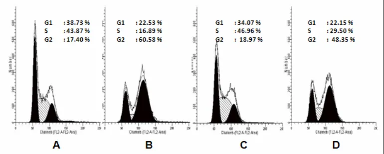
Figure 7. T47D cell cycle analysis after treatment of doxorubicin, nobiletin and their combination.
T47D cell were treated by doxorubicin, nobiletin and their combination for 24 hours and stained by PI reagent before analyzed by flowcytometer. (A) Cell control, (B) doxorubicin 7.5 nM, (C) nobiletin 15 μM, (D) Combination of doxorubicin 7.5 nM-nobiletin 15 μM.
Cell Cycle Analysis
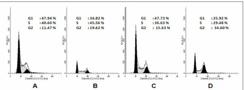
In the next series of experiments, we explored the possibility of cell cycle perturbation by nobiletin, doxorubicin and their combination in MCF-7 and T47D cells. Synergistic effect of combination between nobiletin and doxorubicin could be occured via MCF-7 cell cycle modulation (Table 1 and Figure 6). Cell cycle analysis of MCF-7 breast cancer cell lines showed that single treatment of Doxorubicin 200 nM induced G2/M
arrest. Single treatment of nobiletin 15 µM induced G1 arrest, and combination of doxorubicin 200 nM-nobiletin 15 µM induced G2/M arrest on MCF-7 cells. T47D cell cycle profile (Table 2 and Figure 7) after treatment of doxorubicin 7.5 nM induced G2/M arrest. Single treatment of nobiletin 15 µM induced S arrest, and combination of doxorubicin 7.5 nM-nobiletin 15 µM induced G2/M arrest on T47D cells.
Table 1. MCF-7 Cell distribution after treatment of doxorubicin, nobiletin and their combination for 24 hours.
Treatment Concentration G1 (%) S (%) G2/M (%)
control - 47,94 40,60 11,47
doxorubicin 200 nM 34,82 45,56 19,62
nobiletin 15 μM 47,73 36,63 15,63
doxorubicin and nobiletin 200 nM -15 μM 35,92 29,48 34,60
Table 2. T47D Cell distribution after treatment of doxorubicin, nobiletin and their combination for 24 hours
Treatment Concentration G1 (%) S (%) G2/M (%)
control - 38,73 43,87 17,40
doxorubicin 7.5 nM 22,53 16,89 60,58
nobiletin 15 μM 34,07 46,96 18,97
doxorubicin and nobiletin 7.5 nM-15 μM 22,15 29,50 48,35
135
Figure 6. MCF-7 cell cycle analysis after treatment of doxorubicin, nobiletin and their combination.
MCF-7 cell were treated by doxorubicin, nobiletin and their combination for 24 hours and stained by PI reagent before analyzed by flowcytometer. (A) Cell control, (B) doxorubicin 200 nM, (C) nobiletin 15 μM, (D) Combination of doxorubicin 200 nM-nobiletin 15 μM.
MCF-7 cells showed lower sensitivity to doxorubicin than T47D cells [9]. Over expression of Bcl-2 and P-glyco Protein (PgP) in MCF-7 cells caused cells resistance to apoptosis triggered by doxorubicin. Over expression of Bcl-2 caused cells evading the apoptosis mechanism [10] while PgP transport the drug outside the cells so that concentration of chemotherapeutic agent inside the cells will be low and failed to kill the cancer cells. T47D is one one cancer cell model with mutant p53, and doxorubicin treatment in increasing dose in this cells induced drug resistance phemonema via upregulation of Akt and PgP [11,12]. Drug resistant in cancer cells could be occured by cell cycle arrest that will repair the DNA damage caused by chemotherapeutic agent, so combination of chemotherapeutics with an agent which modulate cancer cell cycle is interesting to be studied.
Single treatment of nobiletin did not show cytotoxic effect, but the combination with doxorubicin increased cytotoxicity of doxorubicin in MCF-7 cells. Single treatment of nobiletin did not induced apoptosis, but the combination with doxorubicin increased apoptosis induction of doxorubicin in MCF-7 cells. The combination also increased G2/M arrest of MCF-7 cells. The mechanism of increasing of doxorubicin cytotoxicity by
nobiletin probably because of up-regulation of p53 and its related pathway. Doxorubicin induced DNA damage and trigger the apoptosis pathway or DNA repair depend on the degree of DNA damage [13]. In the cells with wild type p53 e.g. MCF-7 cells, upregulation of p53 could increase apoptosis induction or G2/M arrest. [14] mentioned nobiletin induced apoptosis via p53 up-regulation and those phenomena was correlated with G2/M arrest in A549 lung cancer cells. Our reults is similar to the previous study, but the mechanism need to be explored more details in MCF-7 cells.
This result showed the potency of nobiletin to be delevoped as co-chemotherapeutic agent for doxorubicin by inducing apoptosis and cell cycle arrest. The use of doxorubicin together with nobiletin is expected to increase the activity and reduce the side effects of doxorubicin. However, the molecular mechanism of apoptotic induction and cell cycle arrest by this combination need to be explored further.
Conclusion
This research showed that combination of nobiletin and doxorubicin synergically increase the effect of doxorubicin through apoptotic
induction and cell cycle modulation. Based on
this result, nobiletin is potential to be delevoped as co-chemotherapeutic agent for doxorubicin in breast cancer therapy.
Author’s Contribution
EM have made conception and design of this study, acquisition of data, data collection, analysis and interpretation and statistical data, drafted, and corresponding author the manuscript. All author have already read and approved the final revision of this manuscript.
Acknowledgement
This study was funded by Post Graduate Research Grant from DP2M DIKTI (Directorate
of Higher Education) Ministry of Education, Indonesia 2009 and 2010.
References
1. Jemal A, Siegel R, Xu J and Ward E. Cancer Statistics, 2010, CA Cancer J Clin, doi: 2010;10:3322/caac.20073.
2. Fimognari C, Nusse MN, Lenzi M, Sciuscio D, Cantelli-Forti G and Hrelia P. Sulforaphane Increases The Efficacy of Doxorubicin in Mouse Fibroblasts Characterized by p53 Mutations, Mutation Research, 2006;601:92–101.
3. Murakami A, Nakamura Y, Torikai K, Tanaka T, Koshiba T, Koshimizu KKuwahara S, Takahashi Y, Ogawa K, Yano M, Tokuda H, Nishino H, Mimaki Y, Sashida Y,
Kitanaka S and Ohigashi H. Inhibitory Effect of Citrus Nobiletin on Phorbol Ester-Induced Skin Inflammation, Oxidative Stress and Tumor Promotion in Mice, Cancer Res., 2000;60:5059–5066
4. Ohnishi H, Asamoto M, Tujimura K, Hokaiwado N, Takahashi S, Ogawa K, Kuribayashi M, Ogiso T, Okuyama H and Shirai T. Inhibition of Cell Proliferation by Nobiletin, A Dietary Phytochemical, Associated With Apoptosis and Characteristic Gene Expression, But Lack Of Effect on Early Rat Hepatocarcinogenesis In Vivo, Cancer Sci., 2004;95(12):936-942.
5. Yoshimizu N, Otani Y, Saikawa Y, Kubota T, Yoshida M, Furukawa T, Kumai K, Kameyama K, Fujii M, Yano M, Sato T, Ito A and Kitajima M. Anti-tumour Effects of Nobiletin, A Citrus Flavonoid, on Gastric Cancer Include: Antiproliferative Effects, Induction of Apoptosis and Cell Cycle Deregulation, Aliment Pharmacol Ther, 2004;1:95-101.
6. Morley KL, Ferguson PJ and Koropatnick J. Tangeretin and Nobiletin Induce G1 Cell Cycle Arrest But Not Apoptosis In Human Breast and Colon Cancer Cells, Cancer Lett, 2007;251(1):168-78.
7. Miyamoto S, Yasui Y, Tanaka T, Ohigashi H and Murakami A. Suppressive Effects of Nobiletin on Hyperleptinemia and Colitis-Related Colon Carcinogenesis In Male ICR Mice, Carcinogenesis, 2008;29(5):1057-1063. 8. Davis JM, Navolanic PM,
Weinstein-Oppenheimer CR, Steelman LS, Wei H, Konopleva M, Blagosklonny MV and McCubrey JA. Raf-1 and Bcl-2 Induce Distinct and Common Pathways That Contribute to Breast Cancer Drug Resistance. Clin. Canc. Res. 2003;9:1161-1170.
9. Zampieri L, Bianchi P, Ruff P and Arbuthnot P. Differential Modulation by Estradiol of P-glycoprotein Drug Resistance Protein Expression in Cultured MCF7 and T47D Breast Cancer Cells, Anticancer Res., 2002;22:2253-2259.
12
137 10. Hanahan D and Weinberg RA. The Hallmarks
of Cancer. Cell, 2000;100:57-70.
11. Emami A, Zeinali S, Motahari Z and Azizi E. 2005. Resistance to Adriamycin Alters the MDR1/P-gp, Topoisomerase II-alpha Gene and Protein Expression Levels in T47D Human Breast Cancer Cells, Conference Module of 2005 AAPS Annual Meeting and
Exposition, downloaded from: www.aapsj.orgabstractsAM_2005AAPS2005
-001197.pdf, on accessed in September 2009.
. Li X, Lu Y, Liang K, Liu B and Fan Z. Differential Responses to Doxorubicin-induced Phosphorylation and Activation of Akt in Human Breast Cancer Cells, Breast Cancer Research, 2005;7:589-597.
13. Minotti G, Menna P, Salvatorelli E, Cairo G and Gianni L. Anthracyclins: Molecular Advances and Pharmacologic Developments in Antitumor Activity and Cardiotoxicity. Pharmacol Rev., 2004;56:185-228.