Introduction
Thrombosis of cerebral veins and of intracranial sinuses is a rare condition that manifests itself through a variety of symptoms with the possibility of dramatic reversals and with a grave prognosis. It occurs in middle-aged and younger individuals, mostly in women. Cerebral venous thrombosis (CVT) is an infrequent cause of sudden-onset headache and is also a recognized cause of subarachnoid haemor-rhage (8). Various risk factors can be identified in 70–80 % of patients, such as local infections (middle ear or facial skin infections), thrombophilic states (factor V Leiden gene mutation; deficit of antithrombin III, protein C, and pro-tein S; factor II G20210A mutation; hyperhomocyspro-teinemia with or without elevated factor VIII levels; and antiphos-pholipid syndrome), systemic inflammatory diseases (for example Behcet’s disease), medication treatment (contra-ception), pregnancy, and the puerperium (3,10,18). There has been a recent steep upward trend in the diagnosis of central venous thrombosis (CVT). This trend is linked to the development of sophisticated neuroradiology methods, which are now widely accessible, especially magnetic reso-nance imaging (MRI) angiography. Furthermore, this trend is linked to a better awareness concerning this method and an increased interest in its use. Finally, this phenomenon is associated with the fact that seriously ill patients are amas-sed in specialized centers. Although pregnancy and the puerperium are significant risk factors in their own right, pregnancy ended with a cesarean delivery represents a further significant increase in this risk (the incidence is 7–14 %, mainly in the puerperium) (16).
Patients
The objective of our report is to discuss two cases of young women who, after a cesarean delivery, were admitted to the department of neurology over the course of 3 months and who were diagnosed as having CVT.
Case 1
A 29-year-old secundipara underwent CS due to breech presentation and premature rupture of the membranes at 37 gestational week (GW). Treatment with enoxaparine 0.4 ml s.c. was started in the moment of CS and continued for 6 days. Severe headaches gradually developed 10 days after a cesarean delivery. Three days later, she developed a sen-sation of dullness in the left half of the face with gradual development of left-sided hemihypesthesia and light hemi-paresis. The following day, she was admitted to a neurology department. Brain MRI and venous magnetic resonance an-giography (MRA) demonstrated thrombosis of ventral parts of the superior sagittal sinus, small gyriform subacute venous infarction in a high frontodorsally located area on the right side of the central region as well as small sub-arachnoidal hemorrhage in the same location but on the left side (Fig. 1 and Fig. 2). Stomatological and ENT exa-minations, CT of paranasal sinuses and laboratory tests excluded local cause of CVT. The patient was started on a full dose of low-molecular-weight heparin (enoxaparin 2x 0.6 ml subcutaneously; 68 kg – up to the weight of 75 kg the dose is 1.2 ml per day). The same day, the patient had a generalized epileptic seizure with a subsequent postictal
CASE REPORT
CEREBRAL VENOUS THROMBOSIS AFTER A CESAREAN DELIVERY
Edvard Ehler1, Aleš Kopal1, Milan Mrklovský2, Milan Košťál3
Pardubice Regional Hospital, Pardubice, Czech Republic: Department of Neurology1 Department of Obstetrics and
Gynecology3; Radiology Center – MultiScan, Pardubice, Czech Republic2
Summary:Cerebral venous thrombosis (CVT) is a serious condition affecting mostly women. This report concerns two cases of women who developed CVT within 14 days of cesarean delivery. Magnetic resonance angiography of the brain (venous phase) is the best modality to diagnose the condition, and parenteral application of low-molecular-weight heparin is the most beneficial treatment. The first patient was found to have an elevated factor VIII level. In the second patient, homozygosity of the C677T mutation in the 5,10-methylenetetrahydrofolate reductase gene was found. The puerperal pe-riod and Cesarean Section (CS) are risk factors for thrombotic complications, including CVT. It is necessary to search for risk factors in a patient’s history and within the group of at-risk patients to prolong preventive administration of low mo-lecular weight heparin (LMWH). CVT (including puerperium related) is not a detrimental to future pregnancies.
Fig. 1: A fluid attenuated inversion recovery (FLAIR) ima-ge in the axial plane demonstrated hyperintense blood in a sulcus on the left side of the central region and small hy-perintense venous infarction on the right side of the central region.
Fig. 3: During a follow-up examination, sagittal MIP image of venous 2D TOF magnetic resonance angiography de-monstrated recanalization of the thrombosed superior sa-gittal sinus.
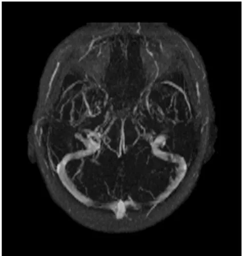
Fig. 2: Sagittal maximal-intensity projections (MIP) image of venous 2D time-of-flight (TOF) magnetic resonance an-giography demonstrated thrombosis of the superior sagittal sinus.
right-sided hemiparesis, which quickly resolved. Electroen-cephalography (EEG) revealed an abnormality, including bilateral slow activity recorded in frontodorsal and central electrodes with a slight predominance on the right side and without the presence of specific graphoelements. Because the patient was breastfeeding, she was started on lamotri-gine, up to a dose of 100 mg per day. Over the next 4 days, the headaches as well as the subtle brain symptomatology resolved, and the patient did not have any other epileptic seizure. Family history revealed that on three occasions, the patient‘s father had experienced a myocardial infarction and that once he had developed a pulmonary embolism. Prior to this hospitalization, the patient had not experienced any serious illnesses; this was her second delivery after a normal pregnancy. The patient was discharged on the 15th
day, 28 days after CS, on dicumarol treatment (INR 2.–2.5) Six weeks after the patient’s symptoms started, follow-up MRI and MRA of the brain were performed, which showed normal findings (Fig. 3). Then the therapy with di-cumarol was concluded.
The patient was found to have an elevated level of fac-tor VIII (380 %, normal: up to 200 %). Other laborafac-tory re-sults were within normal limits (haemocoagulation tests: TEKO, APCR, PC, PS, LAp, LAk, LA, factor VII, VIII, protein C, S, ANCA, RF, thyroid function: fT4, TSH, ge-netic testing)
Case 2
Approximately 14 days after a preterm cesarean delivery (at 29 GW, due to fetal hypotrophy and severe preeclampsia,
enoxaparine 0.4 ml s.c. was administered in the moment of SC and then for 4 postoperative days), the 31-year-old woman gradually developed a strong headache in bilateral parieto-occipital regions. The patient was being treated for arterial hypertension, which had developed during her pregnancy. Brain computed tomography (CT) raised suspicion of thrombosis of the sinus rectus (a dense strip was presented along the falx). The investigation was complemented by MRI and venous MRA of the brain, which demonstrated thrombosis of the internal cerebral veins and the great cere-bral vein of Galen. Furthermore, these examinations revealed partial thrombosis of the sinus rectus and the transverse si-nus (Fig. 4). Work-up for local inflammatory cause of CVT yielded normal (laboratory tests, X-ray, stomatological and otorhilaryngological examinations). The patient was started on LMWH (enoxaparine 2x 0.6 ml subcutaneously; weight 72 kg). Follow-up MRA of the brain, performed 8 days later, confirmed complete recanalization of deep veins and a decrease in the size of the partial thrombosis of the sinus rectus. Moreover, the patency of both transverse sinuses was almost completely restored (Fig. 5). The patient’s head-aches disappeared within 10 days and at the same time with-out other brain symptoms. The patient was discharged on the 14thday (the 28thday after CS). She was treated for next
2 months with dicumarol (INR 2–2.5).
The patient’s history was unremarkable. She had not been seriously ill before she became pregnant. It was confirmed that the patient had the homozygous C677T mutation in the 5,10-methylenetetrahydrofolate reductase (MTHFR) gene. Other laboratory results were within normal limits (haemocoagulation, genetic).
Discussion
Cerebral venous thrombosis is a serious condition. The incidence of CVT is estimated to be 4–8 cases per 100,000 people/year, with a mortality rate of between 10–20 %. Purulent processes, including penetrating head injury, be-long to the known causes of CVT; however, due to the existence of modern effective antibiotics, these causes are becoming less frequent and, nowadays, they are present in fewer than 10 % of the cases. Congenital thrombophilic states represent 10–15 % of the cases, and between 20–30 % of CVT cases have an unclear cause. However, other risk factors are known, which are of varying significance and whose effects may be additive (1,3,5,11). The discussed pa-tients were confirmed to have an elevated level of factor VIII and the homozygous form of the C677T mutation in the MTHFR gene. Thus far, the C677T mutation in the MTHFR gene has not been considered as an independent risk factor causing CVT even though this mutation is fre-quently accompanied by hyperhomocysteinemia, a signifi-cant and strong risk factor for the development of CVT (3). Homocystein level of our patient was within normal limits. However, in combination with other risk factors, the proba-bility of CVT is increased. During pregnancy and the
perium, the incidence of cerebrovascular accident (CVA) and CVT is increased, and these conditions occur only slightly less frequently than CVA of different etiology (brain ischemia and hemorrhage). Depending on the sources of the literature selected, geographic locations as well as the length of the period studied, the incidence rate of CVA (of other than venous origin) fluctuates between 13.1–35/ 100.000 deliveries and that of CVT fluctuates between 11.6–14; at the same time, the prevalence rate of all CVA’s ranges from 8.9–67.1 (9,12,13,15). A number of risk factors have been identified; furthermore, a coefficient (an odds ra-tio) was determined, which is used to multiply the basic risk of developing CVT in pregnancy and the puerperium (4,6). Cesarean delivery was shown to be an independent risk factor that had a significant coefficient (3.1). Other factors associated with the development of CVT include mainly oral contraceptives (the odds ratio is as high as 13), and in combination with a prothrombotic defect, the odds ratio is as high as 30. Furthermore, spinal analgesia with a subarachnoidally administered anesthetic represents a sig-nificant risk factor (10). An important factor is hyperten-sion that develops during pregnancy, as was the case in our patient (12).
In comparing a group of women with CVT that occurred during pregnancy and the puerperium with another group of women with CVT that occurred in a period other than pregnancy and the puerperium, we find that women be-longing to the first group were younger (aged 26 versus 36), the onset was acute in more cases (82 % versus 54 %) but the deficit stabilized more quickly (70 % versus 45 %) and the outcome was better (80 % versus 58 %) (7).
In young women who develop brain symptomatology after giving birth, evaluation of their clinical condition should raise suspicion of CVT or the possibility of CVT should at least be considered in the differential diagnosis. MRI with venous phase MRA would logically be the first neurodia-gnostic test. However, in many cases, brain CT is performed first, either in the context of suspicion for cerebrovascular accident or because of lesser accessibility to MRI (1,5). At the same time, suspicion of sinus thrombosis (a hyperden-se streak in the sinus) or an actual confirmation of CVT (the “delta sign” in superior sagittal sinus thrombosis) are present only in approximately 20 %. However, a finding of multiple intracerebral hematomas that do not follow arte-rial territories raise suspicion of CVT. In our first patient, we immediately performed MRI/venous MRA. In the se-cond patient, CT of the brain was performed on admission. Because finding raises suspicion of sinus thrombosis, MRI and venous MRA followed to complement the investigation. The first step in CVT treatment consists of initiating full anticoagulant treatment by administering heparin or, quite frequently nowadays, by administering LMWH. Not even a rather extensive hemorrhage in venous cerebral infarcts is a contraindication to parenteral administration of heparin. The duration of heparin administration varies–it ranges from several days (17) to three weeks (1,11). After this
pe-riod, transition to an orally administered anticoagulant is recommended, with treatment duration of 3–6 months (11, 17). In addition to evaluating common contraindications to anticoagulant treatment, patients after a cesarean section require an assessment of local findings (the surgical wound, the uterus, and hematomas) by a gynecologist. If extensive brain edema occurs, antiedematous therapy is considered. In cases of clinical deterioration after heparin therapy and in absence of venous and sinus recanalization, local throm-bolysis using a microcatheter is indicated, possibly with mechanical fragmentation of thrombi (2,14). Furthermore, in cases of cerebellar infarction or deep vein thrombosis, ventricular drainage must be considered. Decompression craniotomy is indicated in cases of unmanageable intracra-nial hypertension. In our patients after a cesarean section, LMWH treatment was used, which was safer than other methods, and its duration was extended to 6 weeks. The pa-tients were switched to dicumarol after this period.
Conclusion
Birth and the puerperium are instances of increased risk, during which the incidence of thrombotic events in-creases six-fold (600 %). Birth by Cesarean Section is an-other serious risk factor, even when coupled with the administration of a standard antithrombotic preventive mea-sure such as LMWH intra- and post-operatively. During such times, it is important to consider the possibility of CVT in patients who have displayed cerebral indicators. In preventing CVT, it is imperative to influence those risk fac-tors that can be affected, to uncover hidden risk facfac-tors, and to increase the duration of preventive administration of LMWH in cases of predisposed patients.
We believe that CVT (including puerperium-related CVT) is not a contraindication for future pregnancies.
Conflict of interest statement We declare that we have no conflict of interest.
References
1. Asad A, Barney S. Cerebral venous thrombosis: a review. Neurologist 2006; 12:32–38.
2. Basama FMS, Granger K. Case report: post partum class 1 HELLP syndrome. Arch Gynecol Obstet 2007;275:187–9.
3. Bousser MG, Ferro JM. Cerebral venous thrombosis: an update Lancet Neurol 2007;6:162–70.
4. Cantu C, Barinagarremeteria F. Cerebral venous thrombosis associated with pregnancy and puerperium: review of 67 cases. Stroke 1993;24:1880–4. 5. Chrobok V, Pellant A, Ehler E, et al. Sinus sigmoides thrombosis – contemporary
view of diagnostics and therapy. Cesk Slov Neurol N 2007;70/103:424–8. 6. Cole B, Criddle LM. A case of postpartum cerebral venous thrombosis.
J Neurosci Nurs 2006;38:350–3.
7. De Bruijn SFTM, Budde M, Teunisse S, De Haan RJ, Stam J. Long-term outcome of cognition and functional health after cerebral venous sinus thrombosis. Neurology 2000;54:1687–9.
8. Jaiser SR, Raman A, Maddison P. Cerebral venous thrombosis as a rare cause of thunderclap headache and non-aneurysmal subarachnoid haemorrhage. J Neurol 2008;255:448–9.
cere-bral vein thrombosis after subarachnoid analgesia for labour. Can J Anaesth 2006;53:1015–19.
11. Kimber J. Cerebral venous sinus thrombosis Q J Med 2002;95:137–42. 12. Lanska DJ, Kryscio RJ. Risk factors for peripartum and postpartum stroke and
intracranial venous thrombosis. Stroke 2000;31:1274–82.
13. Lanska DJ, Kryscio RJ. Stroke and intracranial venous thrombosis during preg-nancy and puerperium. Neurology 1998;51:1622–8.
14. Lee W, Cchen CC, Nguyen TH. Cerebral venous congestion as indication for th-rombolytic treatment. Cardiovasc Intervent Radiol 2007;30:675–87.
15. Liang CC, Chang SD, Lai SL, Hsieh CC, Chuen HY, Lee TH. Stroke compli-cating pregnancy and the puerperium. Eur J Neurol 2006;13:1256–60. 16. Masuhr F, Mehrein S, Einhäupl K. Cerebral venous and sinus thrombosis.
J Neurol 2004;251:11–13.
17. Niclot P, Bousser MG. Cerebral venous thrombosis. Current Treatment Options in Neurology 2000;2:343–52.
18. Procházka M, Procházka V, Lubušký M, Procházková J, Hrbáč T. Cerebral ve-nous thrombosis in the users of hormonal contraceptives. Cesk Slov Neurol N 2007;70/103(6):678–84.
Corresponding author:
Edvard Ehler, M.D., Department of Neurology, Pardubice Regional Hospital, Kyjevská 44, 530 03 Pardubice, Czech Republic; e-mail: eda.ehler@tiscali.cz