Efficacy of Sodium Carboxymethyl in Mandibular
Extraction Sockets
1Doddarayapete N Umashankar, 2Partha S Saha, 3Narasimhamurthy Srinath, 4R Mahesh Kumar 5Chaitra Patil, 6Priyanka D Jesabel
ORIGINAL ARTICLE
1Reader, 2Consultant, 3Head, 4Professor, 5Senior Lecturer 6Postgraduate Student
1,3-6
Department of Oral and Maxillofacial Surgery, Krishnadevaraya College of Dental Sciences & Hospital, Bengaluru, Karnataka India
2Department of Oral and Maxillofacial Surgery, Hayat Hospital Guwahati, Assam, India
Corresponding Author: Doddarayapete N Umashankar, Reader Department of Oral and Maxillofacial Surgery, Krishnadevaraya College of Dental Sciences & Hospital, Bengaluru, Karnataka India, e-mail: drumshankar1978@gmail.com
ABSTRACT
Aim: The present study was carried out to assess the wound healing, bone formation, and preservation after placing sodium carboxymethyl starch in mandibular extraction sockets.
Materials and methods: A prospective study with a sample size of 60 where 30 patients received sodium carboxymethyl on one of the mandibular extraction sockets as the test group and con-tralateral side was used as the control group. Student’s paired t-test and McNemar test were used for statistical tabulation.
Results: On assessing bone density, the test group showed more mean values of bone density 2.33 HU, p-value of 0.14 in 3rd month, and 2.37 HU, p-value of 0.04 in 6th month postopera-tive respecpostopera-tively.
Conclusion: Placement of sodium carboxymethyl starch significantly increases the bone density during regeneration of extraction sockets which might promise us good quality of bone formation. However, further research in the material and a longer follow-up period are desirable for a definitive conclusion. Keywords: Bone density and preservation of bone, Bone healing, Sodium carboxymethyl, Wound healing.
How to cite this article: Umashankar DN, Saha PS, Srinath N, Kumar RM, Patil C, Jesabel PD. Efficacy of Sodium Carboxy -methyl in Mandibular Extraction Sockets. Int J Prev Clin Dent Res 2018;5(1):48-54.
Source of support: Nil
Conflict of interest: None
INTRODUCTION
The alveolar bone is a complex and constantly changing tissue which is capable of self-repairing and adaptation to new loads. It consists of an outer layer of cortical bone, an inner cancellous bone, and alveolar bone proper. Together with the root, cementum, and the periodontal membrane, the alveolar bone constitutes the dental attachment appa-ratus.1 Due to the presence of these dental elements, there
will be pushing and pulling a stimulus, which allows the maintenance of bone shape and density (Wolff’s law).2
Pain, infection, bone loss, or fracture of the tooth are the most common reasons for the extraction of teeth.3
Once the tooth is extracted, the alveolar bone that holds the tooth in place (the socket) is often damaged or under-goes a three-dimensional bone resorption. And the resorp-tion is lifelong, irreversible, chronic, and cumulative.4
Bone loss after tooth extraction shows marked osseous changes of the residual alveolar ridge which includes severe bone alterations both in height and in width.
These remodeling jeopardizes the prosthetic reha-bilitation for three main reasons: firstly, the absence of adequate bone levels makes implant placement difficult; Secondly, esthetic problems in the fabrication of implant-supported restoration could be caused by serious bone reabsorption and delayed healing of bone.5 However, it
is possible to minimize such problems by simply carrying out socket preservation procedures in extraction sockets using bone graft materials. But the scarcity of adequate donor tissues, donor site morbidity, the risk of disease transmission, and other allergic reaction has triggered to search for new modalities of grafting to reduce bony resorption and to rehabilitate the missing tooth using implant by faster bone formation.
The socket preservation is an indispensable procedure; the aim is to prevent bone loss following tooth extraction. Preservation is the maintenance of the socket, which is essentially the height and width of the gap that is left after the tooth is removed. It is done by placing a graft material or scaffold immediately into the socket of an extracted tooth to preserve bone height, width, and density.
Various materials are used in modern dental and maxillofacial surgery for bone tissue substitution and reconstruction, which includes autogenic, allogenic (freeze-dried bone allograft, demineralized freeze-dried bone allograft), xenogenic (Bio-oss-osteohealth, Shirley, NY), and synthetic (hydroxyapatite, tricalcium phos-phate, calcium sulfate). Autogenous bone is still regarded as the gold standard due to its osteoinductive and osteo-conductive properties.6
Efficacy of Sodium Carboxymethyl in Mandibular Extraction Sockets
IJPCDR
biodegradability, regenerative characteristics, and also they have osteoinduction, osteoconduction, osteogenesis, and osteointegration than those of synthetic materials.7
One such material is purified sodium carboxymethyl starch, which is a natural-based material, a polysaccharide, used for biomedical purpose as a hemostatic agent due to its properties which allow it not only to rapidly clot the blood but also faster bone regeneration by stabilizing and delivering the growth factors, and is also responsible for the growth of collagen and mesenchymal cells in the extraction socket.8
AIMS AND OBJECTIVES
To evaluate the efficacy of sodium carboxymethyl starch (HaemoCer®), a hemostatic material in mandibular
sockets healing; clinical assessment of wound healing and radiograph assessment of bone formation and bone pres-ervation are done following the placement of HaemoCer.
MATERIALS AND METHODS Study Setting
The study was done on patients who visited the Depart-ment of Oral and Maxillofacial Surgery, Krishnadeva-raya College of Dental Science and Hospital, Bengaluru, Karnataka, India.
Study Design
In this study, the sample size was 60 where 30 patients between age groups of 18 and 40 years both female and male requiring bilateral removal of mandibular teeth were taken. All the 30 patients received sodium carboxymethyl on one of the mandibular extraction sockets as test the group and other extraction socket was used as the control group where sodium carboxymethyl was not used.
All patients were informed about the study and consent was taken for the same. Routine blood investigations were carried out. Preoperative orthopantomogram was taken. All patients underwent extraction of the teeth atraumatically under local anesthesia with adrenaline. The extraction site was randomly allotted as a test group and a control group. Antibiotics and analgesics were administered postopera-tively. Patients were examined clinically for wound healing by assessing pain, swelling, and secondary infection on 3rd, 5th, and 7th postoperative days. And radiographic examination was done for bone density and preservation of bone on 3rd and 6th month postoperatively.
Material used
Sodium carboxymethyl starch (HaemoCer) is a polysac-charide hemostatic agent. It is chemically characterized as the sodium salt of the carboxymethyl ether of potato starch
(as purified Na carboxymethyl starch). The material is able to absorb water up to 18 times its own weight (Fig. 1).
MODE OF ACTION
It dehydrates quickly and achieves a high concentration of platelets, erythrocytes, and the factors of the coagu-lation. After this initial phase, sodium carboxymethyl starch forms a gel-like adhesive mass which serves as a preliminary mechanical barrier against further bleeding. During the subsequent healing phase, the starch particles of carboxymethyl are chemically dissolved completely, absorbed, and metabolized. This sodium carboxymethyl starch is nothing but polysaccharide which on fibroblastic stage of wound healing phase acts on stabilizing and cementing collagen fibers together. This then delivers the growth factors and is also responsible for the growth of mesenchymal cells which ultimately produce osteoblasts.
SURGICAL PROCEDURE
Standard scrubbing, painting, and draping procedures were done.
Preoperative orthopantomograph and intraoral extraction site shown in Figure 2 respectively.
• Administration of local anesthesia (2% lignocaine with 1:2,00,000 epinephrine) by classical inferior alveolar nerve block with lingual nerve block and long buccal nerve was given. Modified wards incision was done. • Reflection of full thickness mucoperiosteal flap was
done.
• Bone removal using bur technique with constant copious saline irrigation.
• Tooth delivery in toto or tooth division depending upon individual case.
• Debridement of socket with saline and povidone iodine irrigation.
• Same procedure was done on contralateral side (Fig. 3). • For teeth other than third molar, regular extraction
procedure was carried out.
• The test group sockets were filled with sodium car-boxymethyl starch and control group did not receive anything (Fig. 4).
• Hemostasis was achieved on both extraction socket. • Closure was done using 3.0 silk, simple interrupted
sutures (Fig. 5).
• Postoperative instructions were given.
• Immediate postoperative radiograph and 6 month postoperative radiograph shown in Figs 6 and 7 respectively.
PARAMETERS
• Clinical evaluation: Assessment of wound healing – Postoperative pain
– Postoperative swelling – Infection
Figs 2A and B: (A) Preoperative radiograph; (B) intraoral photograph
Fig. 3: Bilateral atraumatic extraction done
Figs 4A to C: (A) Dispensing sodium carboxymethyl starch into 46 socket (test group); (B) control group no powder placed; and (C) test group
• Radiographic evaluation: – Assessment of bone density – Assessment of bone preservation
Statistical Analysis
The following methods of statistical analyses have been used in this study. The results were averaged for each parameter.
Descriptive Statistics
Gender distribution analysis was done on all the patients and the results are expressed in percentage (Table 1).
Influence Test
The mean difference between test and control groups in order to determine the visual analog scale (VAS) score of pain, bone density, and bone preservation was done using Student’s t-test. The mean difference of swelling between test and control groups was done by McNemar’s test. The level of significance of p-value was fixed at 0.05.
RESULTS
The patients were clinically evaluated for wound healing under the following parameters: pain, swelling, and secondary infection. Secondly, bone density and
A B
Efficacy of Sodium Carboxymethyl in Mandibular Extraction Sockets
IJPCDR
bone preservation were evaluated through radiographic analysis.
• The patient was evaluated for pain every 4 hours after the procedure. The result showed that pain is equal in test and control groups in the 4th hour. Then, postoperatively in the 8th, 12th, 24th, and 36th hour, the pain is more in the test group than in the control group (Table 2 and Graph 1).
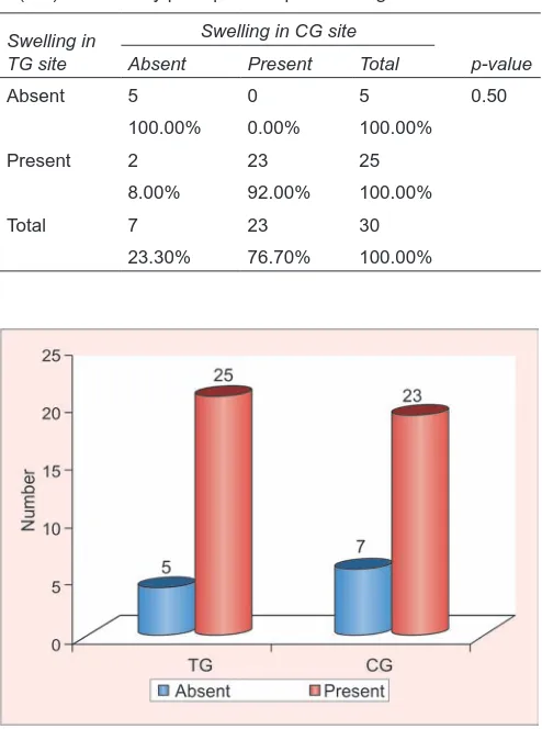
• Swelling was seen in both test and control groups postoperatively on the 3rd day (Table 3 and Graph 2). But postoperatively, on the 5th day, the swelling was more in the test group than in the control group (Table 4). On the 7th day, the swelling was reduced in both test and control groups (Table 5).
• No secondary infection was present in any of the groups postoperatively, i.e., 3rd, 5th, or 7th day (Graph 3). • In radiographic analysis, bone density was evaluated
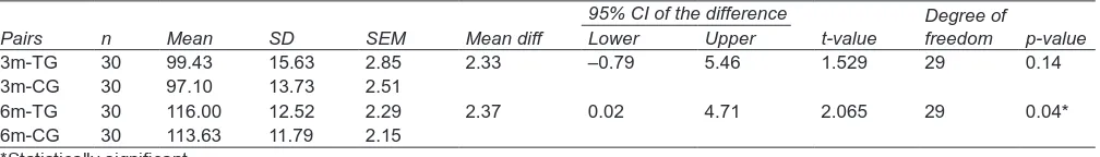
using gray value histogram in the 3rd month post-operatively; it showed equal amount of bone density in both test and control groups. However, in the 6th month postoperatively, the test group showed an increase in bone density compared with the control group (Table 6 and Graph 4).
Table 2: Comparison of mean VAS scores of pain between test (TG) and control groups (CG) using Student’s paired t-test
Pairs n Mean SD SEM Mean diff
95% CI of the difference
t-value Degree of freedom p-value Lower Upper
4h-TG 30 7.53 1.87 0.34 0.80 0.34 1.26 3.525 29 0.001*
4h-CG 30 6.73 1.93 0.35
8h-TG 30 7.20 1.86 0.34 1.33 0.93 1.74 6.679 29 <0.001*
8h-CG 30 5.87 1.96 0.36
12h-TG 30 6.00 1.97 0.36 1.13 0.76 1.51 6.158 29 <0.001*
12h-CG 30 4.87 1.80 0.33
24h-TG 30 5.47 2.29 0.42 1.00 0.25 1.75 2.715 29 0.01*
24h-CG 30 4.47 2.15 0.39
36h-TG 30 4.00 1.97 0.36 1.13 0.46 1.80 3.458 29 0.002*
36h-CG 30 2.87 1.55 0.28
48h-TG 30 3.27 2.00 0.37 1.33 0.80 1.86 5.135 29 <0.001*
48h-CG 30 1.93 1.70 0.31
3d-TG 30 1.13 1.55 0.28 0.60 0.20 1.00 3.071 29 0.005*
3d-CG 30 0.53 1.17 0.21
5d-TG 30 0.67 1.42 0.26 0.53 0.14 0.92 2.804 29 0.009*
5d-CG 30 0.13 0.51 0.09
SD: Standard deviation; SEM: Standard error of mean; CI: Confidence interval; t-value cannot be computed for day 7 because the standard error (SE) of the difference is 0; *Statistically significant
Figs 5A and B: Closure done with 3-0 silk suture
Fig. 6: Immediate postoperative radiograph Fig. 7: Six months postoperative radiograph
A B
Table 1: Gender distribution of samples
Sex n Percent
Males 14 46.7
Females 16 53.3
• Radiographic image taken postoperatively in the 3rd month to assess the bone preservation using Digimizer software shows a slight decrease of bone height in the test group when compared with control. And postoperatively in the 6th month, the bone height was slightly increased (Table 7 and Graph 5).
DISCUSSION
The study was conducted to evaluate the efficacy of sodium carboxymethyl starch as a hemostatic agent in extraction socket. The results obtained testify to the fact that this material has significantly superior bone forming ability, lesser postoperative complications, is a good hemostatic agent, and helps in socket preservation compared with the synthetic materials.
Natural polymers have a wide range of applications in the medical field, e.g., drug delivery systems because they break down to form physiological metabolites.9
Polysaccharides are nothing but carbohydrates which are ubiquitous biopolymers built up from monosaccharides, and 99% are located in plants.10
Polysaccharides, in particular, have some excellent properties which make them the polymer group with
Swelling in
TG site Absent Present Total p-value
Absent 5 0 5 0.50
100.00% 0.00% 100.00%
Present 2 23 25
8.00% 92.00% 100.00%
Total 7 23 30
23.30% 76.70% 100.00%
Graph 1: Comparison of mean VAS scores of pain between test and control groups using Student’s paired t-test
Graph 2: Comparison of swelling in test and control groups at the 3-day postoperative period using McNemar’s test Table 4: Comparison of swelling in test (TG) and control groups
(CG) at the 5-day postoperative period using McNemar’s test Swelling in
TG Absent Swelling in CG sitePresent Total p-value
Absent 11 0 11 <0.001*
100.00% 0.00% 100.00%
Present 18 1 19
94.70% 5.30% 100.00%
Total 29 1 30
96.70% 3.30% 100.00%
*Statistically significant
Table 5: Comparison of swelling in test (TG) and control groups (CG) at the 7-day postoperative period using McNemar’s test Swelling in
TG site Absent Swelling in CG sitePresent Total p-value
Absent 29 0 29
100.00% 0.00% 100.00%
Present 0 1 1 1.00
0.00% 100.00% 100.00%
Total 29 1 30
96.70% 3.30% 100.00%
Efficacy of Sodium Carboxymethyl in Mandibular Extraction Sockets
IJPCDR
the longest and widest medical applications, which include experience of nontoxicity (monomer residues are not hazardous to health), water solubility or high water absorption capacity by simple chemical modification, chemically stable at varying pH, and a broad variety of chemical structures.11,12 This versatility makes these
materials able to overcome some disadvantages like low mechanical, temperature, and chemical stability, and proneness to microbial and enzymatic degradation, which, in some cases, can be used as an advantage.
There is abundant use of polysaccharides and their derivatives in the medicinal and pharmaceutical field. Alginate, amylase, glycogen, chitin, cellulose, and starch are the most common derivatives of polysaccharides.10 Starch
is the most important storage saccharide in plant cell.10
This sodium carboxymethyl starch is basically a polysaccharide hemostatic agent and chemically charac-terized as the sodium salt of the carboxymethyl ether of potato starch.13 Sodium carboxymethyl starch is used as
hemostatic agent which allows it not only to rapidly clot the blood but also faster bone regeneration by stabilizing and delivering the growth factors, and is also responsible for the growth of collagen and mesenchymal cells in the extraction socket.
It has wide applications in cardiovascular surgery for bleeding control of sternotomy edges in 37/40 patients.14
Then, in ear, nose, and throat surgery, some authors applied as a hemostatic agent in several neck dissec-tions, parotidectomy, tonsillectomy, and endoscopic sinus surgery.15-17
Some authors have reported good results in laparo-scopic prostatectomy, and persistent bleeding from neuro-vascular bundles was also stopped.18 Military aspects and
in battlefield the usability and results of this material are much better when compared with other agents.8
Regard-ing the figures for the bone formation rate at 7 weeks, the authors found that polysaccharides seem to promote bone healing compared with control region.
Graph 4: Comparison of mean bone density scores between test and control groups using Student’s paired t-test
Graph 5: Comparison of mean bone preservation scores between test and control groups using Student’s paired t-test Table 6: Comparison of mean bone density scores between test (TG) and control groups (CG) using Student’s paired t-test
Pairs n Mean SD SEM Mean diff 95% CI of the differenceLower Upper t-value Degree of freedom p-value
3m-TG 30 99.43 15.63 2.85 2.33 –0.79 5.46 1.529 29 0.14
3m-CG 30 97.10 13.73 2.51
6m-TG 30 116.00 12.52 2.29 2.37 0.02 4.71 2.065 29 0.04*
6m-CG 30 113.63 11.79 2.15
*Statistically significant
Table 7: Comparison of mean bone preservation scores between test (TG) and control groups (CG) using Student’s paired t-test
Pairs n Mean SD SEM Mean diff
95% CI of the difference
t-value Degree of freedom p-value Lower Upper
3m-TG 30 0.85 0.69 0.13 –0.07 –0.18 0.03 –1.401 29 0.17
3m-CG 30 0.92 0.74 0.13
6m-TG 30 3.00 10.61 1.94 1.97 –1.97 5.91 1.024 29 0.31
6m-CG 30 1.03 0.64 0.12
CONCLUSION
Sodium carboxymethyl starch has a capacity to increase the density of the bone during regeneration of extrac-tion sockets which might promise us good quality of bone formation. Hence, sodium carboxymethyl can be recommended for extraction sockets which are planned for implant prosthesis. Further to this result, its ability to maintain the alveolar bone height is not positive; maybe due to its powder form, it is not able maintain the alveolar height. Mixing sodium carboxymethyl with other xenograft materials might prove beneficial in maintaining alveolar bone height.16 However, further
research in the material and a longer follow-up period may recommend this material in extraction sockets on a regular basis.
REFERENCES
1. Tal H, Artzi Z, Kolerman R, Beitlitum I, Goshen G. Augmenta-tion and preservaAugmenta-tion of the alveolar process and alveolar ridge of bone, bone regeneration, Tal H, editor, ISBN: 978-953-51-0487-2, InTech; 2012. Available from: http://www.intechopen. com/books/bone-regeneration/augmentation-andpreserva-tion-of-the-alveolar-process-and-alveolar-ridge-of-bone. 2. Reddy MS, Geurs NC, Wang IC, Liu PR, Hsu YT, Jeffcoat RL,
Jeffcoat MK. Mandibular growth following implant restora-tion: does Wolff’s law apply to residual ridge resorption? Int J Periodontics Restorative Dent 2002 Aug;22(4):315-321. 3. Dimova C. Socket preservation procedure after tooth
extrac-tion. Key Eng Mater 2014;587:325-330.
4. Stefani CM, Achado MA, Sallum EA, Sallum AW, Toledo S, Nociti FH Jr. Platelet-derived growth factor/insulin-like growth factor-combination and bone regeneration around implants placed into extraction sockets: ahistometric study in dogs. Implant Dent 2000;9(2)126-131.
6. Allegrini S Jr, Koening B Jr, Allegrini MR, Yoshimoto M, Gedrange T, Fanghaenel J, Lipski M. Alveolar ridge sockets preservation with bone grafting—review. Ann Acad Med Stetin 2008;54(1)70-81.
7. Oryan A, Alidadi S, Moshiri A, Maffulli N. Bone regenerative medicine: classic options, novel strategies, and future direc-tions. J Orthop Surg Res 2014 Mar 17;9(1):18.
8. Robinson K, Bloomsburg RN. Controlling bleeding in the field: hemostatic powders and dressings debut in the prehospital setting. J Emerg Nurs 2004 Apr;30(2):160-161.
9. Klein S. Polysaccharides in oral drug delivery. 20 Dec 2009. doi:10.1021/bk-2009-1017.ch001. Available from: http://pubs. acs.orgPublication.
10. Supramolecular Biopolymers II—Polysaccharides. Available from: http://www.mpikg.mpg.de/886855/Polysaccharide.pdf. 11. Hon D-S. Cellulose and its derivatives: structures, reactions,
and medical uses. In: Dumitriu S, editor. Polysaccharides in medicinal applications. New York (NY): Marcel Dekker; 1996. pp. 87-105.
12. Miyamoto T, Takahashi S, Ito H, Inagaki H, Noishiki Y. Tissue biocompatibility of cellulose and its derivatives. J Biomed Mater Res 1989;23:125-133.
13. Mauro MA, Murphy K, Thomson K, Venbrux A, Zollikofer CL. Image-guided intervention. Elsevier Health Sciences; 2008. 14. Schmitz C, Sodian R. Use of plant based polysaccharide
haemostat for the treatment of sterna bleeding after median sternotomy. J Cardiothorac Surg 2015 Apr 24;10
15. Sindwani R. Use of novel hemostatic powder MPH for endo-scopic sinus surgery: initial impressions. Otolaryngol Head Neck Surg 2009 Feb;140(2):262-263.
16. Antisdel JL, West-Denning JL, Sindwani R. Effect of micropo-rous polysaccharide hemospheres (MPH) on bleeding after endoscopic sinus surgery: randomized controlled study. Otolaryngol Head Neck Surg 2009 Sep;141(3):353-357. 17. Phillips P. Arista powder efficacy in 100 nasal surgery patients.
Otolaryngol Head Neck Surg 2009 Sep;141(3).