Summer 2011, Volume 2, Number 4
Basic and Clinical
1. Introduction
he nucleus cuneiformis (NCF) is located just ventrolateral to the periaqueductal gray (PAG) (Gioia & Bianchi, 1987) and it is involved in supraspinal processing of
Capsazepine, a Transient Receptor Potential Vanilloid
Type 1 (TRPV1) Antagonist, Attenuates Antinociceptive
Effect of CB1 Receptor agonist, WIN55,212-2, in the Rat
Nucleus Cuneiformis
Asghar Parvishan 1,2, Zahra Taslimi 1, Mohammad Ebrahimzadeh 1, Abbas Haghparast 1,*
1. Neuroscience Research Center, Shahid Beheshti University of Medical Sciences, P.O. Box 19615-1178, Tehran, Iran 2. Physiology-Pharmacology Research Center, Rafsanjan University of Medical Sciences, Rafsanjan, Iran.
pain (Haghparast & Ahmad-Molaei, 2009; Haghparast, Gheitasi, & Lashgari, 2007; Haghparast, Ordikhani-Seyedlar, & Ziaei, 2008; Haghparast, Soltani-Hekmat, Khani, & Komaki, 2007; Rezvanipour, Milan, & Hagh-parast, 2006). Several lines of evidence have highlighted the importance of cannabinoids in nociceptive
process-* Corresponding Author:
Abbas Haghparast, Msc, PhD
Associate Professor of Neurophysiology, Single Unit Recording Laboratory, Neuroscience Research Center, Shahid Beheshti University of Medical Sciences, Evin St., Shahid Chamran Exp.way. P.O.Box: 19615-1178, Tehran, IRAN
Introduction: Nucleus cuneiformis (NCF), as part of descending pain inhibitory system, cooperates with periaqueductal gray (PAG) and rostral ventromedial medulla (RVM) in supraspinal modulation of pain. Cannabinoids have analgesic effects in the PAG, RVM and NCF. The transient receptor potential vanilloid type 1(TRPV1) can be activated by anandamide and WIN55,212-2 as a cannabinoid receptor agonist. The aim of the current study is to investigate the possible interplay between the cannabinoid and vanilloid systems for modulation of pain at the NCF.
Methods: In this study, a cannabinoid receptor agonist, WIN55,212-2 ( 15 μg/0.3 μl DMSO), and selective TRPV1 receptor antagonist, capsazepine (10, 25, 50 and100 nmol/0.3 μl DMSO), were microinjected bilaterally into the NCF, and tail-flick and formalin tests were used to assess the animal’s pain-related behaviors at 5-min intervals for a 60-min period.
Results: Our findings demonstrated that analgesic effect of WIN55,212-2 were dose-dependently attenuated by capsazepine in both tests. In the tail-flick test, capsazepine at both doses of 50 (P<0.01) and 100 (P<0.001) nmol could significantly prevent the antinociceptive effect of WIN55,212-2 while capsazepine, in formalin test, could decreased its antinociceptive effect at the dose of 50 nmol (P<0.05) as well. On the other hand, solely administration of the highest dose of capsazepine in both tests did not alter the pain-related behaviors.
Discussion: It suggests a possible role for TRPV1 receptors in NCF-mediated cannabinoid-induced antinociception.
A B S T R A C T
Article info:Received: 10 April 2011 First Revision: 22 April 2011 Accepted: 12 May 2011
Key Words:
Nucleus Cuneiformis, TRPV1 Receptor, Cannabinoids, Tail-Flick Test, Formalin Test, Rat
Cannabinoids have been shown to produce analgesia in neuroanatomical regions subserving transmission and modulation of pain signals, including the PAG, rostral ventrolateral medulla (RVM), basolateral nucleus of amygdala (BLA) (Hasanein, Parviz, Keshavarz, & Ja-vanmardi, 2007; Martin et al., 1999; Martin, Patrick, Coffin, Tsou, & Walker, 1995; Martin, Tsou, & Walk-er, 1998) and the NCF (Ebrahimzadeh & Haghparast, 2011). Our recent study showed that antinociceptive ef-fects of WIN55,212-2 in the NCF are mediated, at least partly, by CB1 receptors (Ebrahimzadeh & Haghparast, 2011) and it is suggested the non-cannabinoid recep-tors such as transient receptor potential vanilloid type 1 (TRPV1) receptors may be involved in this phenom-enon at the level of NCF.
TRPV1 receptor is activated by heat and capsaicin (Caterina et al., 1997). Other activators of TRPV1 re-ceptors are cannabinoids (Jeske et al., 2006). Electro-physiological and behavioral studies have provided evidence for participation of TRPV1 receptors in the transmission and modulation of nociceptive input (Kelly & Chapman, 2002a,b; Ohkubo, Shibata, & Taka-hashi, 1993). Mice lacking TRPV1 receptors failed to develop heat hyperalgesia after inflammation (Davis et al., 2000) and in response to microinjection of capsa-icin (TRPV1 receptor agonist) into the dorsolateral PAG produces hypoalgesia. The observed effects were absent in capsazepine-pretreated animals, indicating that the TRPV1 receptors may contribute to this antinociception (Palazzo et al., 2002). It has been shown that cannabi-noid agonists including WIN55,212-2, anandamide and cannabidiol, activate both CB1 and TRPV1 receptors (Bisogno et al., 2001; Jeske et al., 2006; Pertwee, 2006). The presence of TRPV1 receptors has been recognized in the prefrontal cortex, hippocampus, amygdala, hy-pothalamus, PAG, locus coeruleus, and cerebellum (Mezey et al., 2000; Roberts, Davis, & Benham, 2004; Sanchez, Krause, & Cortright, 2001; Toth et al., 2005).
Co-expression of TRPV1 and CB1 receptors in several brain regions (Cristino et al., 2006) raises this possibili-ty that cannabinoids could simultaneously act upon both receptors. TRPV1 receptors have a key role in the anti-nociceptive effect of anandamide at the level of spinal cord (Horvath, Kekesi, Nagy, & Benedek, 2008). In the ventrolateral PAG, cannabinoids modulate pain signals not only through CB1 receptors, but also via TRPV1 receptors (Maione et al., 2006). This study was designed
of bilateral microinjection of (R)-(+) –[2,3-dihydro-5-methyl]-3-(4-morpholinylmethyl) pyrrolo [1,2,3-de]-1,4-benzoxazin 6-yl]-1-naphthalenyl-methanone me-sylate (WIN55,212-2), with capsazepine (TRPV1 receptor antagonist), on behavioral manifestations of the animal pain during the tail-flick and formalin tests in rats.
2. Materials & Methods
2.1. AnimalsIn present study, one-hundred and seventy male albino Wistar rats weighing 250-350g were used. They were housed three per cage in a temperature-controlled room under a 12-h light/dark cycle. All experiments were exe-cuted in accordance with the Guide for Care and Use of Laboratory Animals (National Institute of Health Publi-cation No.80-23, revised 1996) and were approved by the Research and Ethics Committee of Shahid Beheshti University of Medical Sciences.
2.2. Stereotaxic Surgery
Summer 2011, Volume 2, Number 4
Basic and Clinical
2.3. Nociceptive Tests
2.3.1. Tail-Flick Test
The nociceptive threshold was measured by the tail-flick apparatus (Harvard, USA). Tail-tail-flick latency (TFL) was measured by exposing the dorsal surface of the rat’s tail (3, 5 and 7 cm from the caudal tip of the tail) to radiant heat and recording the onset of moving the tail away from the noxious thermal stimulus. The reaction time between the onset of the heat stimulus and the movement of the tail were determined by an auto-matic sensor as TFL. The light source was set at 35% of maximum intensity that yields baseline TFL value in the range of 3-4 sec (Haghparast & Ahmad-Molaei, 2009; Haghparast et al., 2008). To avoid tissue damage, the trial was automatically terminated if a response did not occur within 10 sec (cut-off point). The TFLs were measured after the last microinjection into the NCF for a period of 60 min at 5-min intervals. TFLs (sec) are expressed either as raw data or percentage of maximal possible effect (%MPE) which was calculated from the following formula:
To evaluate the sensitivity of animals to nociceptive stimulus, we considered the rat’s TFL before drug treat-ment as a baseline pain threshold.
2.3.2. Formalin Test
Rats were placed in a transparent open Plexiglas cham-ber (35×35×35 cm) used for observing the animal’s be-havior during the formalin test. A mirror was positioned at an angle of 45◦ to permit an unobstructed view of the
paw by observer. After the microinjection of either ve-hicle or the drugs, each rat was given a subcutaneous injection of formalin (2.5%, 50 μl) into the hind paw. Nociception was quantified by assigning weights to the following pain-related behaviors (Coderre, Fundytus, McKenna, Dalal, & Melzack, 1993; Dubuisson & Den-nis, 1978; Hasanein et al., 2007; Manning & Franklin, 1998). Rats were observed for a 60-min period follow-ing formalin injection and the time spent in each type of behavior was recorded in 5-min blocks. The four behav-ioral categories are as follows: 0, the position and pos-ture of the injected hind paw is indistinguishable from the other hind paw; 1, the injected paw has little or no weight placed on it; 2, the injected paw is elevated and is not in contact with any surface; 3, the injected paw is
licked, bitten, or shaken. Then, a weighted nociceptive score, ranging from 0 to 3 was calculated by multiplying the time spent in each category by the category weight, summing these products and dividing by the total time (300 sec) for each 5-min block of time.
Nociceptive score = (t0×0) + (t1×1) + (t2×2) + (t3×3)/(t0 + t1 + t2 + t3)
An ordinal scale (Coderre et al., 1993) of nociceptive scores was generated with a range of 0-3.
2.4. Experimental Protocols
In this study, by using tail-flick and formalin tests, we examined the effect of antagonizing TRPV1 re-ceptors on cannabinoid-induced analgesia in the NCF. Therefore, there are two sets of experiments in the tail-flick and formalin tests: (1) microinjection of capsazepine+WIN55,212-2 into the NCF. In this set of experiment, WIN55,212-2 (15μg/0.3 μl DMSO per side ) was bilaterally microinjected into the NCF following microinjection of the different doses of capsazepine (10, 25, 50 and 100 nmol/0.3 μl DMSO per side) and (2) mi-croinjection of capsazepine alone into the NCF. In this set of experiment, DMSO as a vehicle of WIN55,212-2 (0.3 μl per side ) was bilaterally microinjected into the NCF following microinjection of the highest dose of capsazepine in both tests. In control respective group, animals received DMSO alone into the same region, bilaterally.
2.5. Statistical Analysis
with 0.9% saline, followed by 10% buffered formalin. The brains were cut coronally in 50 μm sections stained with Cresyl violet through the cannula placements. In-jection sites were histologically verified with the at-las of Paxinos and Watson (Paxinos & Watson, 2005). Eleven rats were excluded due to cannula misplacement (Supplementary figure 1).
3. Results
In control groups, in the tail-flick test, two-way ANO-VA for repeated measures over time followed by Bonfer-roni’s test for TFLs revealed that there were no signifi-cant differences in TFLs at any time intervals among the intact (n=6), sham-operated (n=6) and vehicle groups [treatment main effect: F(2,195)=0.8796, P=0.4166; time main effect: F(12,195)=0.8288, P=0.6207; treatment×time interaction: F(24,195)=0.7528, P=0.7915]. In another set of experiments, formalin test, two-way ANOVA for repeated measures over time fol-lowed by Bonferroni’s test for obtained pain score val-ues [treatment main effect: F(2,180)=1.332, P=0.1682; time main effect: F(11,180)=5.222, P=0.0062; treatment×time interaction effect: F(22,180)=0.4949, P=0.9725] revealed that there were no significant dif-ferences in formalin-induced pain behaviors among the intact, sham-operated and vehicle groups. All animals were compared to respective DMSO group and its pain score value results were considered as baseline in all 5-min blocks in both tests.
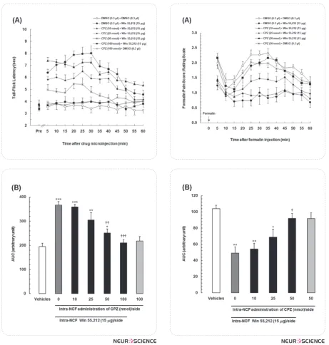
3.1. Effects of intra-NCF administration of capsaz-epine alone and in combination with WIN55,212-2 on TFLs in tail-flick test
To evaluate the effect of antagonizing TRPV1 re-ceptors within the NCF on antinociceptive response of WIN55,212-2 in tail-flick test as a model of acute pain, different doses of capsazepine (10, 25, 50 and 100 nmol/0.3 μl DMSO per side), a selective TRPV1 receptor antagonist, were microinjected into the NCF just before intra-NCF administration of WIN55,212-2 (15 µg/side). TFLs were measured at 5-min intervals during 60 min period. Fig.1A showed that intra-NCF administration of capsazepine could significantly at-tenuate the antinociceptive responses of WIN55,212-2 in a dose-dependent manner [treatment main ef-fect: F(6,481)=121.8, P<0.0001; time main efef-fect: F(12,481)=13.99, P<0.0001; treatment×time
interac-alone could not affect the baseline. One-way ANOVA followed by Newman-Keuls multiple comparison test also showed that there were significant differences in mean AUC values among the control and experimen-tal groups [F (6,43)=50.5, P<0.0001; Fig.1B]. Different doses of capsazepine dose-dependently decreased the AUC values as compared to mean AUC value in WIN-control animals that received solely WIN55,212-2 (15 µg/side) into the NCF.
3.2. Effects of intra-NCF administration of capsaz-epine alone and in combination with WIN55,212-2 on time-course of formalin-induced persistent pain behaviors
In the formalin test as a model of inflammatory persis-tent pain, to evaluate the role of TRPV1 receptors within the NCF in antinociceptive response of WIN55,212-2, different doses of capsazepine (10, 25 and 50 nmol/0.3 μl DMSO per side) were microinjected into the NCF just before intra-NCF administration of WIN55,212-2 (15 µg/side) and then the weighted pain scores were re-corded at 5-min blocks during a 60 min period. Fig. 2A showed that intra-NCF administration of capsazepine could significantly prevent the antinociceptive respons-es of WIN55,212-2 in a dose-dependent manner [treat-ment main effect: F(5,360)=37.76, P<0.0001; time main effect: F(11,360)=5.915, P<0.0001; treatment×time in-teraction: F(55,360)=3.612, P=0.0426]. On the other hand, there were no significant differences in formalin pain scores in all time set intervals between control (DMSO) group and animals that received only the high-est doses of capsazepine (50 nmol/side) into the NCF. Mean AUC calculated results in Fig. 2B revealed that different doses of capsazepine dose-dependently in-creased the AUC values as compared to that in WIN-control animals that received solely WIN55,212-2 ( 15 µg/side) into the NCF. Increase in AUC values in this figure are considered as a nociceptive index. One-way ANOVA followed by Newman-Keuls multiple compari-son test also showed that the antinociceptive response of WIN55,212-2 was completely suppressed by 50 nmol dose of capsazepine [F(5,35)=5.37, P=0.0012; Fig.2B].
4. Discussion
Summer 2011, Volume 2, Number 4
Basic and Clinical
Figure 1. (A) Time-dependent curves representing the effects of administration of different doses of capsazepine, a selec-tive TRPV1 receptor antagonist alone, and in combination with WIN55,212-2 (15 µg/side) into the nucleus cuneifor-mis (NCF) in the tail flick test. (B) The mean area under the curves (AUCs) was obtained from the time-response curves shown in A. In vehicle group, animals received DMSO (0.3 μl/side) into the NCF bilaterally. Data are represented as mean ± SEM for 6 rats.
* P<0.05; ** P<0.01; *** P<0.001 compared to DMSO control group
†† P<0.01; ††† P<0.001 compared to DMSO+Win55212,2 (Win control) group
Figure 2. (A) Time-course of formalin-induced pain be-haviors representing the effects of bilateral intra-nucleus cuneiformis (NCF) administration of different doses of cap-sazepine, a cannabinoid TRPV1 receptor antagonist, vehicle (DMSO; 0.3 µl/side) and WIN55,212-2 (15 µg/side) on pain-related behaviors in the formalin test. (B) Area under the curves (AUCs) was obtained from the time-course responses shown in A. Data are represented as mean ± SEM for 6 rats.
* P<0.05; ** P<0.01 compared to Vehicles group
pain-related behaviors in both tests.
Several lines of evidence indicate the existence of a cannabinoid pain modulatory system in different brain regions including the PAG (Hohmann et al., 2005; Mar-tin et al., 1999; Palazzo et al., 2001; Suplita, Richard, Farthing, Gutierrez, & Hohmann, 2005; Walker, Huang, Strangman & Tsou, 1999), RVM (Martin et al., 1998; Meng & Johansen, 2004; Meng, Manning, Martin, & Fields, 1998; Monhemius, Azami, Green, & Roberts, 2001), BLA (Hasanein et al., 2007) and NCF (Ebra-himzadeh & Haghparast, 2011). Our results, indicating the antinociceptive effects of intra-NCF administration of WIN55,212-2 in the tail-flick and formalin tests, are consistent with the results of investigations in these re-gions (Martin et al., 1995, 1998; Meng et al., 1998). The observed antinociceptive effect of WIN55,212-2 in the NCF was significantly antagonized by high doses of capsazepine in both tests. Attenuation of WIN55,212-2 effects by capsazepine has been reported at the level of spinal cord (Horvath et al., 2008). Pistis et al. (2004) also showed an antagonizing effect of capsazepine in an electrophysiological study in the amygdale (Pistis et al., 2004). Our results demonstrated that intra-NCF ad-ministration of capsazepine alone has no effect on the expression of pain-related behaviors in tail-flick and formalin tests. This is along with the results of study by Perkins and Campbell (1992) about the neutral ef-fect of capsazepine on nociception, when administered alone (Perkins & Campbell, 1992). In contrary, it has been shown that capsazepine and several other TRPV1 receptor antagonists affect noxious thermosensation (Gunthorpe et al., 2007; Tang et al., 2007). The pres-ent findings provide an evidence for a correlation be-tween antagonizing the TRPV1 receptors and attenua-tion of WIN55,212-2-mediated antinocicepattenua-tion in the NCF. Based on this correlation, a possible role could be suggested for TRPV1 receptors in NCF-mediated can-nabinoid-induced analgesia. To demonstrate the pres-ence and specific cell localization of cannabinoid and TRPV1 receptors in the NCF, other experiments like immunohistochemical and bimolecular analyses could be possible useful approaches. Also, for better elucidat-ing, the possible role of cannabinoids and vanilloids in the NCF-mediate analgesia, single unit extracellular re-cording experiments can be designed to measure the cell activity in the NCF in response to chemical (formalin-induced persistent pain) or thermal stimuli before and after pharmacological manipulation of cannabinoid and
ported by the grant (No. 88-301-A) from Neuroscience Research Center, Shahid Beheshti University of Medi-cal Sciences, Tehran, Iran.
References
Bisogno, T., Hanu , L., De Petrocellis, L., Tchilibon, S., Ponde, D. E., Brandi, I. (2001). Molecular targets for cannabidiol and its synthetic analogues: effect on vanilloid VR1 receptors and on the cellular uptake and enzymatic hydrolysis of ananda-mide. British Journal of Pharmacology, 134(4), 845-52.
Bloom, A. S., Dewey, W. L., Harris, L. S., & Brosius, K. K. (1977). 9-nor-9beta-hydroxyhexahydrocannabinol, a cannabinoid with potent antinociceptive activity: comparisons with mor-phine. Journal of Pharmacology and Experimental Thera-peutics, 200(2), 263-70.
Caterina, M. J., Schumacher, M. A., Tominaga, M., Rosen, T. A., Levine, J. D., & Julius, D. (1997). The capsaicin receptor: a heat-activated ion channel in the pain pathway. Nature, 389(6653), 816-24.
Coderre, T. J., Fundytus, M. E., McKenna, J. E., Dalal, S., & Melzack, R. (1993). The formalin test: a validation of the weighted-scores method of behavioural pain rating. Pain, 54(1), 43-50.
Cristino, L., De Petrocellis, L., Pryce, G., Baker, D., Guglielmot-ti, V., & Di Marzo, V. (2006). Immunohistochemical localiza-tion of cannabinoid type 1 and vanilloid transient receptor potential vanilloid type 1 receptors in the mouse brain. Neu-roscience, 139(4), 1405-15.
Davis, J. B., Gray, J., Gunthorpe, M. J., Hatcher, J. P., Davey, P. T., Overend, P. (2000). Vanilloid receptor-1 is essential for in-flammatory thermal hyperalgesia. Nature, 405(6783), 183-87.
Dubuisson, D., & Dennis, S. G. (1978). The formalin test: a quantitative study of the analgesic effects of morphine, meperidine, and brain stem stimulation in rats and cats. Pain, 4, 161-74.
Ebrahimzadeh, M., & Haghparast, A. (2011). Analgesic effects of cannabinoid receptor agonist WIN55,212-2 in the nucleus cuneiformis in animal models of acute and inflammatory pain in rats. Brain Research, 1420,19-28.
Gioia, M., & Bianchi, R. (1987). The cytoarchitecture of the nu-cleus cuneiformis. A Nissl and Golgi study. Journal of anat-omy, 155, 165-76.
Summer 2011, Volume 2, Number 4
Basic and Clinical
Haghparast, A., & Ahmad-Molaei, L. (2009). Effects of electro-lytic lesion of dorsolateral periaqueductal gray on analgesic response of morphine microinjected into the nucleus cunei-formis in rat. Neuroscience letters, 451(2), 165-69.
Haghparast, A., Gheitasi, I. P., & Lashgari, R. (2007). Involve-ment of glutamatergic receptors in the nucleus cuneiformis in modulating morphine-induced antinociception in rats. Europian Jurnal of Pain, 11(8), 855-62.
Haghparast, A., Ordikhani-Seyedlar, M., & Ziaei, M. (2008). Electrolytic lesion of the nucleus raphe magnus reduced the antinociceptive effects of bilateral morphine microinjected into the nucleus cuneiformis in rats. Neuroscience letters, 438(3), 351-55.
Haghparast, A., Soltani-Hekmat, A., Khani, A., & Komaki, A. (2007). Role of glutamatergic receptors located in the nucleus raphe magnus on antinociceptive effect of morphine micro-injected into the nucleus cuneiformis of rat. Neuroscience let-ters, 427(1), 44-9.
Hasanein, P., Parviz, M., Keshavarz, M., & Javanmardi, K. (2007). CB1 receptor activation in the basolateral amygdala produces antinociception in animal models of acute and ton-ic nocton-iception. Clinton-ical and Experimental Pharmacology and Physiology, 34(5 6), 439-49.
Heinzen, E. L., & Pollack, G. M. (2004). Pharmacodynamics of morphine-induced neuronal nitric oxide production and an-tinociceptive tolerance development. Brain research, 1023(2), 175-84.
Hohmann, A. G., Suplita, R. L., Bolton, N. M., Neely, M. H., Fegley, D., Mangieri, R. (2005). An endocannabinoid mecha-nism for stress-induced analgesia. Nature, 435(7045), 1108-12.
Horvath, G., Kekesi, G., Nagy, E., & Benedek, G. (2008). The role of TRPV1 receptors in the antinociceptive effect of anan-damide at spinal level. Pain, 134(3), 277-84.
Jacob, J. J., Ramabadran, K., & Campos-Medeiros, M. (1981). A pharmacological analysis of levonantradol antinociception in mice. Jurnal of Clinical Pharmacology, 21(8 suppl), 327-33S.
Jeske, N. A., Patwardhan, A. M., Gamper, N., Price, T. J., Ako-pian, A. N., & Hargreaves, K. M. (2006). Cannabinoid WIN 55,212-2 regulates TRPV1 phosphorylation in sensory neu-rons. Journal of Biological Chemistry, 281(43), 32879-90.
Kelly, S., & Chapman, V. (2002a). Effects of peripheral nerve in-jury on functional spinal VR1 receptors. Neuroreport, 13(9), 1147-50.
Kelly, S., & Chapman, V. (2002b). Spinal administration of cap-sazepine inhibits noxious evoked responses of dorsal horn neurons in non-inflamed and carrageenan inflamed rats. Brain research, 935(1-2), 103-8.
Maione, S., Bisogno, T., de Novellis, V., Palazzo, E., Cristino, L., Valenti, M. (2006). Elevation of endocannabinoid levels in the ventrolateral periaqueductal grey through inhibition of fatty acid amide hydrolase affects descending nociceptive pathways via both cannabinoid receptor type 1 and transient receptor potential vanilloid type-1 receptors. Journal of Phar-macology and Experimental Therapeutics, 316(3), 969-82.
Manning, B. H., & Franklin, K. B. J. (1998). Morphine analgesia in the formalin test: reversal by microinjection of quaternary naloxone into the posterior hypothalamic area or periaque-ductal gray. Behavioural brain research, 92(1), 97-102.
Martin, W. J., Coffin, P. O., Attias, E., Balinsky, M., Tsou, K., & Walker, J. M. (1999). Anatomical basis for cannabinoid-induced antinociception as revealed by intracerebral micro-injections. Brain research, 822(1-2), 237-42.
Martin, W. J., Patrick, S. L., Coffin, P. O., Tsou, K., & Walker, J. M. (1995). An examination of the central sites of action of cannabinoid-induced antinociception in the rat. Life sciences, 56(23-24), 2103-9.
Martin, W. J., Tsou, K., & Walker, J. M. (1998). Cannabinoid receptor-mediated inhibition of the rat tail-flick reflex after microinjection into the rostral ventromedial medulla. Neuro-science letters, 242(1), 33-6.
Meng, I., & Johansen, J. (2004). Antinociception and modula-tion of rostral ventromedial medulla neuronal activity by local microinfusion of a cannabinoid receptor agonist. Neu-roscience, 124(3), 685-93.
Meng, I. D., Manning, B. H., Martin, W. J., & Fields, H. L. (1998). An analgesia circuit activated by cannabinoids. Na-ture, 395(6700), 381-83.
Mezey, Toth, Z. E., Cortright, D. N., Arzubi, M. K., Krause, J. E., Elde, R. (2000). Distribution of mRNA for vanilloid recep-tor subtype 1 (VR1), and VR1-like immunoreactivity, in the central nervous system of the rat and human. Proceedings of the National Academy of Sciences of the United States of America, 97(7), 3655-60.
Monhemius, R., Azami, J., Green, D., & Roberts, M. (2001). CB1 receptor mediated analgesia from the Nucleus Reticularis Gi-gantocellularis pars alpha is activated in an animal model of neuropathic pain. Brain research, 908(1), 67-74.
Ohkubo, T., Shibata, M., & Takahashi, H. (1993). The analge-sia induced by intrathecal injection of ruthenium red. Pain, 54(2), 219-21.
Palazzo, E., de Novellis, V., Marabese, I., Cuomo, D., Rossi, F., Berrino, L. (2002). Interaction between vanilloid and gluta-mate receptors in the central modulation of nociception. Eu-ropean journal of pharmacology, 439(1-3), 69-75.
Palazzo, E., Marabese, I., De Novellis, V., Oliva, P., Rossi, F., Berrino, L. (2001). Metabotropic and NMDA glutamate re-ceptors participate in the cannabinoid-induced antinocicep-tion. Neuropharmacology, 40(3), 319-26.
Paxinos, G., & Watson, C. R. (2005). The Rat Brain in Stereotaxic Coordinates. (5th ed.). San Diego: Elsevier Academic Press.
Perkins, M., & Campbell, E. (1992). Capsazepine reversal of the antinociceptive action of capsaicin in vivo. British journal of pharmacology, 107(2), 329-33.
Rezvznipour, I. (2006). The Role of GABAA Receptor Inhibitor on Morphine Antinociception Action in Cuneiformis Nucle-us. International Journal of Pharmacology, 2(4), 400-5.
Roberts, J. C., Davis, J. B., & Benham, C. D. (2004). [3H] Res-iniferatoxin autoradiography in the CNS of wild-type and TRPV1 null mice defines TRPV1 (VR-1) protein distribution. Brain research, 995(2), 176-83.
Sanchez, J., Krause, J., & Cortright, D. (2001). The distribution and regulation of vanilloid receptor VR1 and VR1 5'splice variant RNA expression in rat. Neuroscience, 107(3), 373-81.
Suplita, I., Richard, L., Farthing, J. N., Gutierrez, T., & Hohm-ann, A. G. (2005). Inhibition of fatty-acid amide hydrolase en-hances cannabinoid stress-induced analgesia: sites of action in the dorsolateral periaqueductal gray and rostral ventro-medial medulla. Neuropharmacol, 49(8), 1201-9.
Tang, L., Chen, Y., Chen, Z., Blumberg, P. M., Kozikowski, A. P., & Wang, Z. J. (2007). Antinociceptive pharmacology of N-(4-chlorobenzyl)-N -(4-hydroxy-3-iodo-5-methoxyben-zyl) thiourea, a high-affinity competitive antagonist of the transient receptor potential vanilloid 1 receptor. Journal of Pharmacology and Experimental Therapeutics, 321(2), 791-98.
Toth, A., Boczan, J., Kedei, N., Lizanecz, E., Bagi, Z., Papp, Z. (2005). Expression and distribution of vanilloid receptor 1 (TRPV1) in the adult rat brain. Mol. Brain Res, 135(1-2), 162-68.
Walker, J. M., Huang, S. M., Strangman, N. M., Tsou, K., & Sa udo-Pe a, M. (1999). Pain modulation by release of the en-dogenous cannabinoid anandamide. Proceedings of the Na-tional Academy of Sciences of the United States of America, 96(21), 12198-203.