abnormalities of myocardial noradrenergic
function in Lewy body diseases
David S. Goldstein, … , Graeme Eisenhofer, Yehonatan
Sharabi
JCI Insight. 2019;
4(16)
:e130441.
https://doi.org/10.1172/jci.insight.130441
.
Graphical abstract
Clinical Medicine
Cardiology
Neuroscience
Find the latest version:
Conflict of interest: The authors have declared that no conflict of interest exists.
Copyright: © 2019, American Society for Clinical Investigation.
Submitted: May 20, 2019
Accepted: July 16, 2019
Published: August 22, 2019.
Reference information:JCI Insight. 2019;4(16):e130441.
https://doi.org/10.1172/jci. insight.130441.
Computational modeling reveals multiple
abnormalities of myocardial noradrenergic
function in Lewy body diseases
David S. Goldstein,1 Mark J. Pekker,2 Graeme Eisenhofer,3 and Yehonatan Sharabi4
1Autonomic Medicine Section (formerly Clinical Neurocardiology Section), Clinical Neurosciences Program, Division of
Intramural Research, National Institute of Neurological Disorders and Stroke (NINDS), National Institutes of Health, Bethesda, Maryland, USA. 2Mathematical Sciences, University of Alabama at Huntsville, Huntsville, Alabama, USA. 3Institute of Clinical Chemistry and Laboratory Medicine and Department of Medicine, Technische Universität Dresden,
Dresden, Germany. 4Tel Aviv University Sackler Faculty of Medicine and Chaim Sheba Medical Center, Tel HaShomer, Israel.
Introduction
The movement disorder in Parkinson disease (PD) results from depletion of the catecholamine dopamine in the brain’s nigrostriatal system (1). PD also entails profound deficiency of the closely related catechol-amine norepinephrine (NE) in the heart (2). Other Lewy body diseases — pure autonomic failure (PAF) and dementia with Lewy bodies — also involve severely decreased myocardial NE contents (3).
The cardiac sympathoneural lesion in these diseases probably is important clinically. Thus, neuroim-aging evidence of cardiac noradrenergic deficiency in PD is associated with cognitive impairment (4), exercise intolerance (5), olfactory dysfunction (6), rapid eye movement behavior disorder (7), visual hallu-cinations (8), falls from neurogenic orthostatic hypotension (9), fatigue (10), and shortened survival (11).
One might presume that the myocardial NE depletion in these disorders directly and solely reflects loss BACKGROUND. Lewy body diseases, a family of aging-related neurodegenerative disorders, entail loss of the catecholamine dopamine in the nigrostriatal system and equally severe deficiency of the closely related catecholamine norepinephrine in the heart. The myocardial noradrenergic lesion is associated with major nonmotor symptoms and decreased survival. Numerous mechanisms determine norepinephrine stores, and which of these are altered in Lewy body diseases has not been examined in an integrated way. We used a computational modeling approach to assess comprehensively pathways of cardiac norepinephrine synthesis, storage, release, reuptake, and metabolism in Lewy body diseases. Application of a potentially novel kinetic model identified a pattern of dysfunctional steps contributing to norepinephrine deficiency. We then tested predictions from the model in a new cohort of Parkinson disease patients.
METHODS. Rate constants were calculated for 17 reactions determining intraneuronal
norepinephrine stores. Model predictions were tested by measuring postmortem apical ventricular concentrations and concentration ratios of catechols in controls and patients with Parkinson disease.
RESULTS. The model identified low rate constants for 3 types of processes in the Lewy body group: catecholamine biosynthesis via tyrosine hydroxylase and aromatic l-amino acid decarboxylase, vesicular storage of dopamine and norepinephrine, and neuronal norepinephrine reuptake via the cell membrane norepinephrine transporter. Postmortem catechols and catechol ratios confirmed this triad of model-predicted functional abnormalities.
CONCLUSION. Denervation-independent impairments of neurotransmitter biosynthesis, vesicular sequestration, and norepinephrine recycling contribute to the myocardial norepinephrine deficiency attending Lewy body diseases. A proportion of cardiac sympathetic nerves are “sick but not dead,” suggesting targeted disease modification strategies might retard clinical progression.
of sympathetic noradrenergic nerves; however, studies using immunoreactive tyrosine hydroxylase (TH) as a marker of myocardial catecholaminergic innervation have noted about a 75% average decrease in PD (12–17), whereas, as shown here, the extent of loss of NE is 95%–99% (2, 3). The greater magnitude of neurotransmit-ter depletion than of loss of sympathetic noradrenergic innervation suggests the occurrence of abnormalities in residual nerves that are dysfunctional but extant. We call this the “sick but not dead” phenomenon.
There are many such potential abnormalities (Figure 1), including decreased vesicular uptake of cyto-plasmic catecholamines via the type 2 VMAT; (18) increased vesicular permeability (19); decreased axo-nal transport of vesicles or vesicle-associated proteins (20); decreased activities of the enzymes TH (21), LAAAD (22), and vesicular DBH (23); increased exocytotic release of vesicular NE (24); and decreased neuronal NE recycling via the U1 process mediated by the cell membrane NE transporter (NET) (25).
Previous studies and kinetic models (26) have not provided a cohesive, comprehensive view of overall cardiac catecholaminergic functioning in Lewy body diseases. In the present study we used a potentially novel systems biology approach by constructing and applying a detailed computational model that enabled a more complete picture.
A rich fund of in vivo neurochemical, neuroimaging, and tracer kinetic data has accumulated over many years about cardiac noradrenergic function in healthy humans (27) and in a variety of clinical disor-ders (28), and postmortem studies have provided data about myocardial tissue contents of NE, dopamine, and other compounds of interest in Lewy body diseases (2, 3). We exploited this information to assess for the first time to our knowledge all the major known pathways of cardiac catecholamine synthesis, release, recycling, and metabolism simultaneously, with the goal of identifying the intraneuronal functional abnor-malities contributing to myocardial noradrenergic deficiency in Lewy body diseases. We hypothesized that in addition to sympathetic denervation, a multiplicity of functional abnormalities in residual nerves work together to cause the dramatic loss of myocardial NE stores that characterizes Lewy body diseases.
conditions were used to calculate rate constants for each of the intraneuronal reactions in controls and patients with Lewy body disease (see Supplemental Appendix 1; supplemental material available online with this article; https://doi.org/10.1172/jci.insight.130441DS1).
We tested predictions from the model using data about postmortem concentrations and ratios of apical left ventricular catechols in cohorts of autopsy-proven patients with PD and control subjects.
Results
The Lewy body disease group had drastically decreased myocardial tissue contents of NE (by 96% from controls) and dopamine (by 95%) (Table 1 and Table 2; individual data with descriptive statistics are in Supplemental Table 1). There were smaller proportionate decreases in amounts of other catechols. The Lewy body disease group also had severely decreased rates of cardiac spillover of NE (by 86% from con-trol), DHPG (the main neuronal metabolite of NE) (by 97%), DOPAC (the main neuronal metabolite of dopamine) (by 84%), and DOPA (the precursor of the catecholamines) (by 98%) (individual data with descriptive statistics are in Supplemental Table 2).
Calculated rate constants in controls. Based on inputs for the model (Table 3, Table 4, Table 5, and Table
6), in controls the calculated rate constant for TH (kTH) was 1.9% of that for LAAAD (Figure 1 and Table 7). kVMAT_DA was about 7 times kMAO_DA, and kVMAT_NE was about 11 times kMAO_NE. The rate constant for neuronal uptake of NE from the extracellular fluid (kU1) was about 11 times that for loss of extracellular fluid NE from the tissue (kNE_Loss).
Calculated rate constants in patients with Lewy body disease. Compared with the control group, the Lewy body
disease group had lower values for kU1 (2% of control), kLAAAD (8%), kTH (30%), kNE_Loss (19%), kMAO_DA (27%), kVMAT_NE (28%), kMAO_NE (35%), and kVMAT_DA (74%), with higher values for kDBH (320%), kLeak_DA (208%), kLeak_NE (205%), and kNE_Release (141%) (Figure 2 and Table 7).
Across the series of reactions from TH to LAAAD and from LAAAD to VMAT_DA, the product of the percentages of control was 1.9%, meaning a 98.1% decrease in synthesis and vesicular uptake of cytoplasmic dopamine.
Curves showing amounts of reactants as functions of isolated decreases in rate constants failed to generate the pattern of abnormalities of reactants observed in the Lewy body group (Figure 3). That is, no single abnormality was sufficient to reproduce the observed differences between the control and Lewy body groups.
Table 1. Spillover rates and (mean values ± SEM) in control subjects and patients with Lewy body diseases
Rate (pmol/min) Control Lewy P Lewy vs. control
Myo. DHPG SO 733 ± 51 (45) 38 ± 25 (13) <0.00001 Myo. NE SO 99 ± 9 (45) 19 ± 4 (13) 0.00001 Myo. DOPA SO 219 ± 28 (45) 35 ± 7 (11) 0.00210 Myo. DOPAC SO 180 ± 34 (38) 4 ± 15 (11) 0.00773
Numbers in parentheses are numbers of subjects. Individual data and descriptive statistics are in Supplemental Tables 1 and 2. SO, spillover; Myo., myocardial.
Table 2. Myocardial catechol concentrations (mean values ± SEM) in control subjects and patients with Lewy body diseases
Concentration (pmol/mg wet weight) Control Lewy P Lewy vs. control
Myo. NE 2.11 ± 0.24 (26) 0.045 ± 0.013 (28) <0.00001 Myo. DA 0.140 ± 0.034 (26) 0.0031 ± 0.0006 (22) <0.00001 Myo. DHPG 0.111 ± 0.035 (26) 0.025 ± 0.017 (21) <0.00001 Myo. DOPAC 0.046 ± 0.010 (25) 0.010 ± 0.003 (26) 0.00021 Myo. DOPA 0.26 ± 0.06 (26) 0.21 ± 0.03 (28) ns
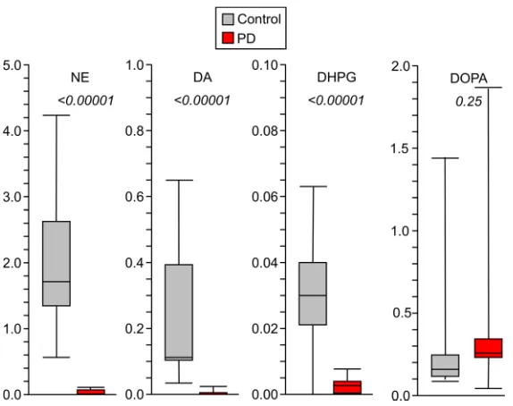
Testing model predictions. In new subject cohorts of PD patients and controls, myocardial NE content
was decreased by 99% in the PD group (Figure 4; individual data with descriptive statistics are in Supple-mental Table 3).
Myocardial concentration ratios of DHPG/NE in the PD group averaged 9.8 times, DOPAC/NE 11.2 times, and DOPAC/DA 14.8 times control (P = 0.00002, P < 0.00001, P = 0.0004; Figure 5 and Supplemental Table 3). Concentration ratios of DOPA/DOPAC, Cys-DOPA/DOPAC, and (DOPA + Cys-DOPA)/DOPAC in the PD group averaged 5.1, 3.5, and 5.0 times control (P = 0.002, P = 0.0004, P = 0.002), respectively. The mean value for (DOPA + Cys-DOPA) adjusted for LAAAD activity in the PD group averaged 30% that in the controls (P = 0.004; Figure 5).
Myocardial epinephrine in the PD group averaged 14.2% that in the controls (P < 0.0001; Supple-mental Table 3).
Discussion
“Sick but not dead.” As reported recently (3) and shown here, Lewy body diseases entail profound cardiac
NE depletion. The remarkably severe extent of this deficiency (99% in the present study) is greater than can be accounted for by sympathetic denervation alone (about 75% based on immunoreactive TH in left ventricular myocardial tissue) (12–17). How can there be greater loss of a neurotransmitter than of the Table 3. Reactant amounts under equilibrium conditions in control subjects
Reactant Parameter Value Range
1 TYRc TYRc amount 8747
2 DOPAc DOPAc amount 142 105–200
3 DAc DAc amount 0.6
4 DOPACc DOPACc amount 25 10–75
5 DAv DAv amount 76 30–110
6 NEv NEv amount 1150 885–1771
7 NEe NEe amount 0.14
8 NEc NEc amount 2.1
9 DHPGc DHPGc amount 60 47–165
All reactant amounts are in nanomoles. Ranges based on 25% and 75% quartiles of the empirical median values. DHPGc, cytoplasmic DHPG.
Table 4. Reaction rates under equilibrium conditions in control subjects
Reaction Parameter Value Range
1 TYR_Uptake TYR uptake rate to TYRc 56 34–89 2 TYR_Loss TYRc loss rate to ECF 54 33–87 3 TH TYRc conversion rate to DOPAc 1.8 1.1–2.8 4 LAAAD DOPAc conversion rate to DAc 1.5 1.2–1.8 5 DOPAc_Loss DOPAc exit rate from the model 0.22 0.15–0.33 6 VMAT_DA DAc vesicular uptake rate 2.2 1.8–2.7 7 Leak_DA DAv loss rate to cytoplasm 0.87 0.66–1.08 8 DBH DAv conversion rate to NEv 1.4 1.4–3.6 9 MAO_DA DAc conversion rate to DOPACc 0.18 0.11–0.29 10 DOPACc_Loss DOPACc exit rate from the model 0.18 0.11–0.29 11 VMAT_NE Vesicular uptake rate of NEc 14 11–22 12 Leak_NE NEv loss rate to cytoplasm 13 10–20 13 NE_Release NEv release rate to ECF 2.6 1.4–3.7 14 U1 NEe reuptake rate to NEc 2.4 1.3–3.5 15 MAO_NE NEc conversion rate to DHPGc 1.2 0.8–1.8 16 DHPGc_Loss DHPGc exit rate from the model 1.2 0.8–1.8 17 NEe_Loss NEe exit rate from the model 0.21 0.11–0.29
nerves that contain the neurotransmitter? Based on the results of this study, the resolution of this apparent paradox is substantial abnormalities in the synthesis, storage, and recycling of NE in residual nerves that are dysfunctional but extant, i.e., “sick but not dead.”
Reasonableness of the kinetic model. Review of the literature indicates that the comprehensive kinetic model
we developed was reasonable. First, TH is well known to be the rate-limiting enzyme in catecholamine bio-synthesis (29), and in the present study, the calculated rate constant for TH was about one-fiftieth that for the next enzyme in the synthetic cascade, LAAAD. Second, vesicular uptake is the dominant mode of disposition of cytoplasmic catecholamines (30), and the model-calculated rate constants for vesicular uptake of cytoplas-mic dopamine and NE each was about 12 times those for MAO acting on these catecholamines. Third, about 90% of released NE is removed by neuronal reuptake (28), and in the model the rate constant for U1 was 11.5 times the rate constant for loss of extracellular NE from the tissue. Fourth, under resting conditions most of the turnover of catecholamines occurs not by release and escape of neuronal reuptake but by leakage from the vesicles into the cytoplasm and enzymatic deamination catalyzed by MAO (30), and the calculated rate constant for NE leakage was about 5 times that of exocytotic NE release.
There was a huge range of values for the model-generated rate constants — from about 0.00020/min for kTH to almost 20/min for kU1. This span is also reasonable, as follows. The value for kTH in the mod-el agreed wmod-ell with that calculated from data published by Nagatsu, Levitt, and Udenfriend in their initial Table 5. Reactant amounts under equilibrium conditions in Lewy body disease patients
Reactant Parameter Value Range
1 TYRc TYRc amount 2239
2 DOPAc DOPAc amount 114 61–182
3 DAc DAc amount 0.05
4 DOPACc DOPACc amount 5.5 4.3–11.0
5 DAv DAv amount 1.7 1.3–2.6
6 NEv NEv amount 25 12–65
7 NEe NEe amount 0.14
8 NEc NEc amount 0.3
9 DHPGc DHPGc amount 14 4–40
All reactant amounts are in nanomoles. Ranges based on 25% and 75% quartiles of the empirical median values.
Table 6. Reaction rates under equilibrium conditions in Lewy body disease patients
Reaction Parameter Value Range
1 TYR_Uptake TYR uptake rate to TYRc 14.2 0–43 2 TYR_Loss TYRc loss rate to ECF 14.1 0–43 3 TH TYRc conversion rate to DOPAc 0.14 0–0.42 4 LAAAD DOPAc conversion rate to DAc 0.10 0–0.31 5 DOPAc_Loss DOPAc exit rate from the model 0.035 0.014–0.040 6 VMAT_DA DAc vesicular uptake rate 0.14 0–0.42 7 Leak_DA DAv loss rate to cytoplasm 0.04 0–0.12 8 DBH DAv conversion rate to NEv 0.10 0.05–0.26 9 MAO_DA DAc conversion rate to DOPACc 0.004 0–0.012 10 DOPACc_Loss DOPACc exit rate from the model 0.004 0–0.012 11 VMAT_NE Vesicular uptake rate of NEc 0.56 0.27–1.47 12 Leak_NE NEv loss rate to cytoplasm 0.57 0.28–1.51 13 NE_Release NEv release rate to ECF 0.08 0.03–0.11 14 U1 NEe reuptake rate to NEc 0.04 0.02–0.06 15 MAO_NE NEc conversion rate to DHPGc 0.06 0.01–0.10 16 DHPGc_Loss DHPGc exit rate from the model 0.06 0.01–0.10 17 NEe_Loss NEe exit rate from the model 0.04 0.01–0.05
description of TH as the rate-limiting enzyme in catecholamine biosynthesis (29). Regarding U1 we previous-ly estimated a much lower rate constant, 0.55/min, based on the kinetics of the cardiac sympathetic imaging agent, 18F-dopamine (31). The higher rate constant in the present model could reflect the use of an assumed NE amount of 0.14 nmol in the extracellular fluid. This was calculated indirectly from a published micro-dialysate NE concentration in rat myocardium (0.001 nmol/mL) (32), and micromicro-dialysate NE could have substantially underestimated NE at U1 sites, in which case kU1 in the model would be overestimated. Even so, the literature indicates a 2750-fold (calculated as 0.55/0.0002) range between kTH and kU1.
Computational modeling reveals multiple functional abnormalities. Accounting for the pattern of myocardial
catecholaminergic abnormalities found in Lewy body diseases required multiple alterations in values for rate constants. For instance, as shown in Figure 3, an isolated decrease in the rate constant for VMAT act-ing on cytoplasmic dopamine (kVMAT_DA) predicted increased DOPACc without a change in DOPAc, whereas patients with Lewy body disease showed severely decreased rates of cardiac DOPA spillover (Table 1 and Table 2).
TH, LAAAD, and vesicular uptake of DOPACc occur in a series (Figure 1). The calculated rate con-stants for these processes in the Lewy body disease group were 30%, 8%, and 74% of control, respectively. The series arrangement of these reactions (thick green arrows in Figure 1) predicted a 98.1% decrease in tissue dopamine content in the Lewy body disease group, which agrees with the empirical value of 98% (1 – [0.0031/0.140]; Table 2).
The calculated rate constant for LAAAD in the Lewy body group was only 8% of that in the controls. Based on postmortem patterns of catechol concentrations, PD also involves markedly decreased LAAAD activity in the brain’s nigrostriatal system (33, 34). LAAAD therefore seems a promising target for gene enhancement therapy (35).
NE synthesis in sympathetic nerves requires DBH, the intravesicular enzyme catalyzing the conversion of dopamine to NE. The calculated rate constant for DBH in the Lewy body disease group was not lower than in the controls. This finding indicates that the functional abnormalities in cardiac sympathetic nerves in Lewy body diseases are not generalized to all aspects of catecholaminergic function. Similarly, the cal-culated rate constant for NE release was not decreased in the Lewy body group. The low empirical rate of cardiac NE spillover in Lewy body diseases (19% of control, Table 1) seems to reflect the depletion of releasable NE stores.
Table 7. Calculated rate constants (in per minute units) for the model of the cardiac sympathetic nervous system in healthy humans and patients with Lewy body disease
Rate constant Process Control Lewy
1. kTYR_Uptake → TYR (cyto.) 56 14 2. kTYR_Loss TYR (cyto.) → 0.0062 0.0063 3. kTH TYR (cyto.) → DOPA (cyto.) 0.00020 0.000062 4. kLAAAD DOPA (cyto.) → DA (cyto.) 0.011 0.00091 5. kDOPA_Loss DOPA (cyto.) → 0.0016 0.00031 6. kMAO_DA DA (cyto.) → DOPAC (cyto.) 0.30 1.10 7. kVMAT_DA DA (cyto.) → DA (vesicles) 3.75 2.79 8. kDBH DA (vesicles) → NE (vesicles) 0.018 0.058 9. kLeak_DA DA (vesicles) → DA (cyto.) 0.011 0.024 10. kNE_Release NE (vesicles) →NE (ECF) 0.0023 0.0032 11. kLeak_NE NE (vesicles) → NE (cyto.) 0.011 0.023 12. kVMAT_NE NE (cyto.) → NE (vesicles) 6.72 1.85 13. kMAO_NE NE (cyto.)→ DHPG (cyto.) 0.56 0.20 14. kU1 NE (ECF) → NE (cyto.) 17.1 0.29 15. kDOPAC_Loss DOPAC (cyto.) → 0.0072 0.00073 16. kDHPG_Loss DHPG (cyto.) → 0.020 0.0043 17. kNE_Loss NE (ECF) → 1.49 0.28
One may ask whether the obtained results would differ if, given the relatively high cytoplasmic concen-tration of tyrosine as substrate, TH were saturated under physiological conditions. The proportionate dif-ference between the Lewy body and control groups in model-generated rate constants assuming first-order kinetics for TH was identical to the difference assuming enzyme saturation (zero-order kinetics). That is, after taking denervation into account, the same large decrease in TH activity was found in the Lewy body group regardless of enzyme saturation.
In the model the rate constant for vesicular leakage was set at 1.14% per minute in controls, based on previously published data (28); this value was increased in the Lewy body disease group because of previously published accelerated loss of myocardial 18F-dopamine–derived radioactivity (36). Increased sympathoneural NE turnover could be from decreased vesicular uptake or from increased vesicular leak-age (2), and the model cannot separate these possibilities. For instance, the decrease in vesicular storleak-age of dopamine in the Lewy body group could have reflected a 25.4% drop in the rate constant for vesicular uptake and 208% increase in the rate constant for leakage (as in Figure 1) but could also have reflected a 53.2% drop (25.4% × 208%) in vesicular uptake and no change in the rate constant for vesicular leakage. Experiments on vesicles isolated from striatal tissue from patients with PD have found markedly decreased vesicular uptake, with evidence for both decreased numbers of VMAT molecules and abnormal transport function of the protein itself (37).
The predominant mechanism of inactivation of released NE is reuptake (U1) mediated by the NET. U1 activity has been reported to be decreased in Lewy body diseases (25, 38); however, previous studies have not examined NET activity after taking denervation into account. The present kinetic model yield-ed a value for the U1 rate constant (kU1) in residual sympathetic neurons in Lewy body diseases that was only about 2% of control.
Empirical testing of model predictions. Predictions from the model were assessed by myocardial levels of
group these averaged 5.1, 3.5, and 5.0 times control, respectively. TH activity in the residual sympathetic nerves in the PD group was estimated to be decreased by about 70% after taking denervation into account. An indirect estimate of U1 activity, the myocardial epinephrine concentration, was decreased to 14% of control; however, this could reflect denervation, NET dysfunction, or a combination of these abnormali-ties. In general, the new empirical data fit with the concept of attenuated catecholamine biosynthesis, vesic-ular storage, and NE recycling in residual myocardial sympathetic nerves in Lewy body diseases.
A common cause? Is it possible that there is a single common cause for the pattern of functional abnormalities
described here? Oxidative stress or decreased mitochondrial energy generation would seem likely culprits (39, 40). For LAAAD to catalyze dopamine synthesis from DOPA, however, requires neither oxygen nor energy, yet the calculated rate constant for LAAAD was substantially decreased in the Lewy body disease group. More gen-erally, widespread oxidative stress or deficient mitochondrial energy generation would not account easily for the syndromic nature of Lewy body diseases or the relatively selective, profound catecholamine deficiencies found in the putamen and heart compared with other regions receiving catecholaminergic innervation.
A specific unifying mechanism may be harmful interactions between the catecholaldehyde DOPAL and the protein α-synuclein (AS). DOPAL is an obligate intermediate in neuronal dopamine metabolism (Figure 1). DOPAL inhibits activities of both TH (21) and LAAAD (41). DOPAL also potently oligomerizes AS (41), converting the protein to oligomeric forms that impede vesicular functions (19), and AS inhibits LAAAD (22). Moreover, DOPAL forms quinone-protein adducts with other proteins involved in catecholaminergic functions, including TH, LAAAD, and VMAT2, probably via spontaneous oxidation to DOPAL-quinone (41). Lewy body diseases all feature AS deposition in sympathetic noradrenergic nerves (42), and putamen DOPAL is built up in PD (43). Nevertheless, accumulation of neither DOPAL nor AS in catecholaminergic neurons has been shown to be pathogenic, as opposed to both being nonpathogenic biomarkers of the disease process.
Implications. The present findings based on application of a potentially novel computational modeling
approach indicate that, rather than there being a single pathogenetic mechanism underlying cardiac NE defi-ciency, neuronal loss, there are multiple functional abnormalities in extant neurons in Lewy body diseases. Both decreased innervation and neuronal dysfunctions in residual neurons that are “sick but not dead” seem to contribute to the dramatic NE depletion found in the heart in PD and other Lewy body diseases.
It is reasonable to propose that an analogous phenomenon applies in central catecholaminergic neurons. Thus, there is evidence for denervation-independent abnormalities in vesicular storage, LAAAD activity, and ALDH activity in putamen tissue from patients with parkinsonian synucleinopathies (33, 36, 43).
Although the kinetic model presumed a balance of rates of gain and loss for each of the reactants, this equilibrium does not imply stability. On the contrary, the kinetic model does not involve negative feedback regulation, i.e., no homeostasis. Any perturbation would be unopposed. A systems approach involving compensatory negative feedback and progressively declining homeostatic capacity seems required to model accurately the period of preclinical or prodromal disease.
The finding that a proportion of catecholaminergic neurons are “sick but not dead” offers hope that a disease modification strategy might slow or prevent the progression of catecholaminergic neurodegener-ation in this family of disorders. Understanding about how catecholaminergic neurons are dysfunctional may incite rational, mechanism-directed treatment. The “sick but not dead” concept also supports the pos-sibility that one can develop pathophysiological biomarkers to detect the disease process in an early phase.
Methods
Subjects. Data from previous publications were based on (a) transcardiac kinetic studies of
tracer-la-beled catecholamines during cardiac catheterization in 45 control subjects and 13 patients with Lewy body diseases (7 PAF, 6 PD with orthostatic hypotension, 64 ± 4 years, 9 men) (28, 38) and (b) post-mortem studies of apical myocardial catechols in 23 control subjects and 8 patients with neurogenic orthostatic hypotension in the setting of a Lewy body disease (5 PD, 3 PAF) (2, 3). Data about rates of reactions in controls were obtained from a previously published study by Eisenhofer et al. in 1996 (28), except for a minor adjustment in the calculated rate of NE production to obey conservation of mass. Rates of reactions in patients with Lewy body disease were from publications about the rate of appearance of endogenous NE in coronary sinus plasma (spillover) and arterial-coronary sinus differences in plasma concentrations of DHPG, DOPAC, and DOPA (3, 38). Data about myocardial tissue amounts of catechols in the control and patient groups were obtained from publications about concentrations of these catechols as well as Cys-DOPA, a product of spontaneous oxidation of cyto-plasmic dopamine (2).
Kinetic model. A multicompartment model was constructed depicting 17 reactions involving 9 reactants
in cardiac sympathetic nerves (Figure 1 and Table 2). Tables 3 and 4 list reaction rates and amounts in the control and Lewy body nOH groups.
Testing model predictions. To test predictions from the pattern of rate constants obtained by applying the
kinetic model to previously published data, we used empirical data about myocardial catechols and catechol ratios in cohorts of patients with PD (n = 11) and controls (n = 11) of similar age. Coded samples of myocar-dial tissue from patients with PD and control subjects were received from the Banner Sun Health Research Institute (Sun City, Arizona, USA) under a Material Transfer Agreement and assayed for catechol contents (2). The investigators and assay personnel were blinded to the postmortem neuropathological diagnosis.
We reported previously on myocardial concentration ratios of DHPG/NE, DOPAC/NE, and DOPAC/ DA and their relationships to vesicular sequestration of cytoplasmic catecholamines in PD (2). Loss of sym-pathetic noradrenergic innervation alone would not be expected to alter these ratios. If there were a vesicular storage defect in residual myocardial sympathetic nerves, with a shift from vesicular sequestration to oxidative deamination of cytoplasmic catecholamines, then values for these ratios would be increased (Figure 1). If there were decreased LAAAD activity in residual cardiac sympathetic nerves, DOPA and Cys-DOPA, which are proximal to the LAAAD step, would be built up with respect to DOPAC. If TH activity were decreased in residual sympathetic neurons, myocardial endogenous DOPA and Cys-DOPA levels would be decreased; however, because decreased LAAAD activity would increase levels of both compounds, to assess TH activity, it was necessary to adjust DOPA and Cys-DOPA levels for the decrease in LAAAD activity. Myocardial epinephrine content is derived substantially from neuronal uptake of epinephrine from the coronary arterial blood (44). If there were decreased NET activity in residual cardiac sympathetic neurons in Lewy body dis-eases, myocardial epinephrine content would therefore be decreased.
Statistics. Mean values were expressed ± 1 SEM. A P value less than 0.05 defined statistical significance.
Mean values for catechol content and catechol ratios in the Lewy body disease patients and controls were compared by independent-means t tests conducted on log-transformed data.
Study approval. For all the subjects, written informed consent was obtained before participation in
pro-tocols approved by the Institutional Review Board of the NINDS, or else the next of kin gave written informed consent for postmortem tissue harvesting for research purposes.
Author contributions
DSG performed the literature search, created the figures, designed the study, collected data, analyzed and interpreted data, and wrote the manuscript. MJP performed the literature search, designed the study,
ana-Figure 5. Box-and-whisker plots for postmortem indices of sympathetic intraneuronal functions in con-trols and PD patients. Highest, third quartile, median, second quartile, and lowest values are shown. Numbers in italics are P values for independent-means t tests conducted on log-transformed data comparing the control and PD groups. DHPG/NE and DOPAC/NE ratios provided inverse indices of VMAT2 activity. The sum of Cys-DOPA and DOPA, divided by the sum of DA and its metabolites DOPAC and 3,4-dihydroxyphenylethanol, provided an inverse index of LAAAD activity. The sum of Cys-DOPA and DOPA, adjusted for LAAAD activity, provided an index of TH activity. The results indicate decreased VMAT2, LAAAD, and TH activities in PD (shown in red) compared with controls (shown in gray). DHPG/NE: control, n = 11; PD, n = 7. DOPAC/NE: control,
n = 11; PD, n = 11. Inv, Index of LAAD: control, n = 11; PD, n = 11. Index of TH: control, n = 11; PD, n = 11. DOPAC,
lyzed and interpreted data, and wrote the manuscript. GE performed the literature search, data interpreta-tion, and writing of the manuscript. YS performed the literature search, designed the study, analyzed and interpreted data, and wrote the manuscript.
Acknowledgments
The research reported here was supported by the Division of Intramural Research, NINDS.
Address correspondence to: David S. Goldstein, Autonomic Medicine Section (formerly Clinical Neuro-cardiology Section), CNP/DIR/NINDS/NIH, 9000 Rockville Pike MSC-1620, Building 10 Room 8N260, Bethesda, Maryland 20892-1620, USA. Phone: 301.675.1110; Email: [email protected].
1. Ehringer H, Hornykiewicz O. [Distribution of noradrenaline and dopamine (3-hydroxytyramine) in the human brain and their behavior in diseases of the extrapyramidal system]. Klin Wochenschr. 1960;38:1236–1239.
2. Goldstein DS, Sullivan P, Holmes C, Miller GW, Sharabi Y, Kopin IJ. A vesicular sequestration to oxidative deamination shift in myocardial sympathetic nerves in Parkinson’s disease. J Neurochem. 2014;131(2):219–228.
3. Goldstein DS, Sharabi Y. The heart of PD: Lewy body diseases as neurocardiologic disorders. Brain Res. 2019;1702:74–84. 4. Kim JS, et al. Cardiac sympathetic denervation and its association with cognitive deficits in Parkinson’s disease. Parkinsonism
Relat Disord. 2009;15(9):706–708.
5. Nakamura T, et al. Lowered cardiac sympathetic nerve performance in response to exercise in Parkinson’s disease. Mov Disord. 2010;25(9):1183–1189.
6. Goldstein DS, et al. Biomarkers to detect central dopamine deficiency and distinguish Parkinson disease from multiple system atrophy. Parkinsonism Relat Disord. 2008;14(8):600–607.
7. Kim JS, et al. Orthostatic hypotension and cardiac sympathetic denervation in Parkinson disease patients with REM sleep behavioral disorder. J Neurol Sci. 2016;362:59–63.
8. Oka H, et al. Impaired cardiovascular autonomic function in Parkinson’s disease with visual hallucinations. Mov Disord. 2007;22(10):1510–1514.
9. Romagnolo A, et al. Cardiovascular autonomic neuropathy and falls in Parkinson disease: a prospective cohort study. J Neurol. 2019;266(1):85–91.
10. Nakamura T, Hirayama M, Hara T, Hama T, Watanabe H, Sobue G. Does cardiovascular autonomic dysfunction contribute to fatigue in Parkinson’s disease? Mov Disord. 2011;26(10):1869–1874.
11. Goldstein DS, Holmes C, Sharabi Y, Wu T. Survival in synucleinopathies: A prospective cohort study. Neurology. 2015;85(18):1554–1561.
12. Amino T, Orimo S, Itoh Y, Takahashi A, Uchihara T, Mizusawa H. Profound cardiac sympathetic denervation occurs in Parkin-son disease. Brain Pathol. 2005;15(1):29–34.
13. Dickson DW, et al. Evidence that incidental Lewy body disease is pre-symptomatic Parkinson’s disease. Acta Neuropathol. 2008;115(4):437–444.
14. Orimo S, et al. Axonal alpha-synuclein aggregates herald centripetal degeneration of cardiac sympathetic nerve in Parkinson’s disease. Brain. 2008;131(Pt 3):642–650.
15. Fujishiro H, et al. Cardiac sympathetic denervation correlates with clinical and pathologic stages of Parkinson’s disease. Mov
Disord. 2008;23(8):1085–1092.
16. Ghebremedhin E, Del Tredici K, Langston JW, Braak H. Diminished tyrosine hydroxylase immunoreactivity in the cardiac con-duction system and myocardium in Parkinson’s disease: an anatomical study. Acta Neuropathol. 2009;118(6):777–784.
17. Takahashi M, et al. Quantitative correlation between cardiac MIBG uptake and remaining axons in the cardiac sympathetic nerve in Lewy body disease. J Neurol Neurosurg Psychiatry. 2015;86(9):939–944.
18. Goldstein DS, Holmes C, Kopin IJ, Sharabi Y. Intra-neuronal vesicular uptake of catecholamines is decreased in patients with Lewy body diseases. J Clin Invest. 2011;121(8):3320–3330.
19. Plotegher N, et al. DOPAL derived alpha-synuclein oligomers impair synaptic vesicles physiological function. Sci Rep. 2017;7:40699.
20. Volpicelli-Daley LA. Effects of α-synuclein on axonal transport. Neurobiol Dis. 2017;105:321–327.
21. Mexas LM, Florang VR, Doorn JA. Inhibition and covalent modification of tyrosine hydroxylase by 3,4-dihydroxyphenylacet-aldehyde, a toxic dopamine metabolite. Neurotoxicology. 2011;32(4):471–477.
22. Tehranian R, Montoya SE, Van Laar AD, Hastings TG, Perez RG. Alpha-synuclein inhibits aromatic amino acid decarboxylase activity in dopaminergic cells. J Neurochem. 2006;99(4):1188–1196.
23. Nagatsu T, Sawada M. Biochemistry of postmortem brains in Parkinson’s disease: historical overview and future prospects. J
Neural Transm Suppl. 2007;(72):113–120.
24. Meredith IT, Eisenhofer G, Lambert GW, Dewar EM, Jennings GL, Esler MD. Cardiac sympathetic nervous activity in congestive heart failure. Evidence for increased neuronal norepinephrine release and preserved neuronal uptake. Circulation. 1993;88(1):136–145.
25. Polinsky RJ, Goldstein DS, Brown RT, Keiser HR, Kopin IJ. Decreased sympathetic neuronal uptake in idiopathic orthostatic hypotension. Ann Neurol. 1985;18(1):48–53.
26. Sasidharakurup H, Melethadathil N, Nair B, Diwakar S. A systems model of Parkinson’s disease using biochemical systems theory. OMICS. 2017;21(8):454–464.
28. Eisenhofer G, et al. Cardiac sympathetic nerve function in congestive heart failure. Circulation. 1996;93(9):1667–1676. 29. Nagatsu T, Levitt M, Udenfriend S. Tyrosine hydroxylase. The initial step in norepinephrine biosynthesis. J Biol Chem.
1964;239:2910–2917.
30. Eisenhofer G, Kopin IJ, Goldstein DS. Catecholamine metabolism: a contemporary view with implications for physiology and medicine. Pharmacol Rev. 2004;56(3):331–349.
31. Goldstein DS, Katzper M, Linares OA, Kopin IJ. Kinetic model for the fate of the sympathoneural imaging agent 6-[18F]flu-orodopamine in the human heart: a novel means to assess cardiac sympathetic neuronal function. Naunyn-Schmiedeberg’s Arch
Pharmacol. 2002;365:38–49.
32. Gilinsky MA, Faibushevish AA, Lunte CE. Determination of myocardial norepinephrine in freely moving rats using in vivo microdialysis sampling and liquid chromatography with dual-electrode amperometric detection. J Pharm Biomed Anal. 2001;24(5-6):929–935.
33. Goldstein DS, Sullivan P, Holmes C, Mash DC, Kopin IJ, Sharabi Y. Determinants of denervation-independent depletion of putamen dopamine in Parkinson’s disease and multiple system atrophy. Parkinsonism Relat Disord. 2017;35:88–91.
34. Ciesielska A, et al. Depletion of AADC activity in caudate nucleus and putamen of Parkinson’s disease patients; implications for ongoing AAV2-AADC gene therapy trial. PLoS One. 2017;12(2):e0169965.
35. Mittermeyer G, et al. Long-term evaluation of a phase 1 study of AADC gene therapy for Parkinson’s disease. Hum Gene Ther. 2012;23(4):377–381.
36. Goldstein DS, et al. Deficient vesicular storage: a common theme in catecholaminergic neurodegeneration. Parkinsonism Relat
Disord. 2015;21(9):1013–1022.
37. Pifl C, et al. Is Parkinson’s disease a vesicular dopamine storage disorder? Evidence from a study in isolated synaptic vesicles of human and nonhuman primate striatum. J Neurosci. 2014;34(24):8210–8218.
38. Goldstein DS, Holmes C, Li ST, Bruce S, Metman LV, Cannon RO. Cardiac sympathetic denervation in Parkinson disease. Ann
Intern Med. 2000;133(5):338–347.
39. Perfeito R, Cunha-Oliveira T, Rego AC. Reprint of: revisiting oxidative stress and mitochondrial dysfunction in the pathogenesis of Parkinson disease-resemblance to the effect of amphetamine drugs of abuse. Free Radic Biol Med. 2013;62:186–201. 40. Schapira AH, Jenner P. Etiology and pathogenesis of Parkinson’s disease. Mov Disord. 2011;26(6):1049–1055.
41. Jinsmaa Y, Sharabi Y, Sullivan P, Isonaka R, Goldstein DS. 3,4-Dihydroxyphenylacetaldehyde-Induced protein modifications and their mitigation by N-acetylcysteine. J Pharmacol Exp Ther. 2018;366(1):113–124.
42. Isonaka R, Sullivan P, Jinsmaa Y, Corrales A, Goldstein DS. Spectrum of abnormalities of sympathetic tyrosine hydroxylase and alpha-synuclein in chronic autonomic failure. Clin Auton Res. 2018;28(2):223–230.
43. Goldstein DS, et al. Determinants of buildup of the toxic dopamine metabolite DOPAL in Parkinson’s disease. J Neurochem. 2013;126(5):591–603.