R E S E A R C H A R T I C L E
Open Access
Reduced resting-state brain functional
network connectivity and poor regional
homogeneity in patients with CADASIL
Jingjing Su
1†, Shiyu Ban
2†, Mengxing Wang
2,3, Fengchun Hua
4, Liang Wang
5, Xin Cheng
5, Yuping Tang
5,
Houguang Zhou
6, Yu Zhai
1, Xiaoxia Du
2*and Jianren Liu
1*Abstract
Background:Cerebral autosomal dominant arteriopathy with subcortical infarcts and leukoencephalopathy (CADASIL) manifests principally as a suite of cognitive impairments, particularly in the executive domain. Executive functioning requires the dynamic coordination of neural activity over large-scale networks. It remains unclear whether changes in resting-state brain functional network connectivity and regional homogeneities (ReHos) underly the mechanisms of executive dysfunction evident in CADASIL patients.
Methods:In this study, 22 CADASIL patients and 44 matched healthy controls underwent resting-state functional magnetic resonance imaging (fMRI). Independent component analysis (ICA) was used to measure functional brain network connectivity, and ReHos were calculated to evaluate local brain activities. We used seed-based functional connectivity (FC) analyses to determine whether dysfunctional areas (as defined by ReHos) exhibited abnormal FC with other brain areas. Relationships among the mean intra-network connectivity z-scores of dysfunctional areas within functional networks, and cognitive scores were evaluated using Pearson correlation analyses.
Results:Compared to the controls, CADASIL patients exhibited decreased intra-network connectivity within the bilateral lingual gyrus (LG) and the right cuneus (CU) (thus within the visual network [VIN)], and within the right precuneus (Pcu), inferior frontal gyrus (IFG), and precentral gyrus (thus within the frontal network [FRN]). Compared to the controls, patients also exhibited significantly lower ReHos in the right precuneus and cuneus (Pcu/CU), visual association cortex, calcarine gyri, posterior cingulate, limbic lobe, and weaker FC between the right Pcu/CU and the bilateral parahippocampal gyrus (PHG), and between the right Pcu/CU and the right postcentral gyrus. Notably, the mean connectivity z-scores of the bilateral LG and the right CU within the VIN were positively associated with compromised attention, calculation and delayed recall as revealed by tests of the various cognitive domains explored by the Mini-Mental State Examination.
(Continued on next page)
© The Author(s). 2019Open AccessThis article is distributed under the terms of the Creative Commons Attribution 4.0 International License (http://creativecommons.org/licenses/by/4.0/), which permits unrestricted use, distribution, and reproduction in any medium, provided you give appropriate credit to the original author(s) and the source, provide a link to the Creative Commons license, and indicate if changes were made.
* Correspondence:xxdu@phy.ecnu.edu.cn;liujr021@sjtu.edu.cn
†Jingjing Su and Shiyu Ban are authors have contributed equally to this work
as co-first authors.
2Shanghai Key Laboratory of Magnetic Resonance and Department of
Physics, School of Physics and Materials Science, East China Normal University, 3663 North Zhongshan Road, Shanghai 200062, People’s Republic of China
1Department of Neurology, Shanghai Ninth People’s Hospital, Shanghai Jiao
Tong University School of Medicine, 639 Zhizaoju Road, Shanghai 200011, People’s Republic of China
(Continued from previous page)
Conclusions:The decreases in intra-network connectivity within the VIN and FRN and reduced local brain activity in the posterior parietal area suggest that patients with CADASIL may exhibit dysfunctional visuomotor behaviors (a hallmark of executive function), and that all visual information processing, visuomotor planning, and movement execution may be affected.
Keywords:Functional network connectivity, Regional homogeneity, CADASIL, Resting-state fMRI, Visuomotor behaviors
Introduction
Cerebral autosomal dominant arteriopathy with subcor-tical infarcts and leukoencephalopathy (CADASIL) is the most common form of hereditary, subcortical vascular dementia; the condition is attributable to pathogenic mutations in the NOTCH3 gene of chromosome 19. Usually, the earliest signs are recurrent strokes and cog-nitive deficits dominated by early impairment of execu-tive functions, and it is commonly associated with
deficits in attention and memory [1,2]. Such dysfunction
was present in all subjects aged 35–73 years in a study
that recruited 42 consecutive, symptomatic CADASIL
patients [2]. The cognitive impairments including
execu-tive abnormalities in CADASIL are mainly characterised by frontal-like symptoms such as poor attention, persev-erations and apathy, and memory impairment associated with pyramidal signs, gait difficulties, pseudobulbar
palsy, and sphincter incontinence [3]. Cognitive decline
increases with age, and is associated with progressive de-terioration of instrumental activities, visuospatial
abil-ities, visual memory, and reasoning [2]. After the age of
60 years, significant deficits in all cognitive domains are evident, severely compromising independent daily living
and even causing death [2]. No effective treatment is
known [1]. Thus, an understanding of the mechanisms
underlying executive dysfunction in patients with this devastating disorder will increase our understanding of this condition and pave the way for new therapeutic trials.
Age, male sex, active smoking, and systolic blood pres-sure are strong independent risk factors for clinical de-terioration (including executive deficits) in patients with
CADASIL [4–7]. Of these, smoking and systolic blood
pressure are modifiable [6,7]. Quantitative magnetic
res-onance imaging (MRI) has revealed close associations between measured parameters and the executive func-tions of patients with CADASIL. Both the overall lacu-nar lesional burden and extent of brain atrophy, as reflected by the normalized brain volume yielded by structural image evaluation using normalization of atro-phy software, significantly influence the severity of ex-ecutive performance deterioration and cognitive decline
as assessed by executive scores [4,8–10]. The global
cor-tical atrophy scale (another measure of brain volume
change) was developed by Pasquier to rate the extent of atrophy in 13 brain MRI regions on a visual scale, and
predict poor executive performance [11]. The corpus
callosum area as determined by three-dimensional (3D)-T1 MRI sequences (using well-validated methodology) is independently correlated with reaction time during the performance of a simple task, and can serve as a proxy of early cognitive and behavioral changes in patients
with CADASIL [12, 13]. Diffusion tensor imaging (DTI)
yields useful information on the extent of white matter (WM) tract damage; the cingulum bundle seems to be important in terms of maintaining the anteroposterior connections necessary for executive functioning in
non-demented CADASIL patients [14]. Furthermore, DTI
ab-normalities in white and deep gray matter that appear normal using other imaging modalities have been de-tected in nondemented CADASIL patients; these abnor-malities correlated particularly strongly with executive
function [15]. Thus, it may be that widespread WM
damage explains the executive dysfunction apparent dur-ing the early stage of CADASIL; this suggestion is (at least in part) consistent with pathological data derived from post-mortem brains. Extensive pathological WM lesions were evident in all brain regions, particularly the frontal motor cortex, suggesting the disruption of either the cortico-cortical or cortico-subcortical networks of frontal lobe WM, which may explain the observed motor
deficits and executive dysfunctions [16].
Brain functional MRI (fMRI) has shed light on the causal mechanisms of executive dyscognition in CADA-SIL patients. A study evaluating the association be-tween resting-state functional network connectivity and cognitive performance indicated that the status of two frontoparietal components correlated with executive
performance [17]. Our recent resting-state amplitude of
low-frequency fluctuation (ALFF) analyses revealed negative associations between the ALFF values of the left cerebellar anterior and posterior lobes, and
execu-tive function scores [18]. An event-related Go/No-go
task study on CADASIL patients revealed lower blood oxygen level-dependent (BOLD) effects in the alerting network and in areas involved in executive functions, possibly reflecting hemodynamic responses that
Thus, although the scores on various indices appear to correlate with the extent of executive disability in CADA-SIL patients, it is currently considered that human execu-tive functions are mediated by the dynamic interplay of
large-scale brain networks [20]. However, it remains largely
unknown whether changes in resting-state brain functional network connectivity and regional homogeneities (ReHos) can provide novel insights into the executive dysfunction mechanisms in play in CADASIL patients. Resting-state functional network connectivity evaluated via independent component analysis (ICA) has been used to define several,
intrinsic functional networks [17]. In addition, ReHos can
be used to identify local features of spontaneous brain activ-ity when the Kendall coefficient of concordance (KCC) is employed during synchrony evaluation of BOLD time series. The intra-network connectivity within brain net-works and ReHos have been shown to correlate with be-havioral measures in both healthy subjects and those with
neuropsychiatric conditions [21]. Based on the above
men-tioned abnormalities in executive behaviors and brain
structure [6–15], we hypothesized that both brain
func-tional network connectivity and ReHos would be altered in CADASIL patients. To this end, we used resting-state fMRI to explore changes in brain functional networks as assessed by ICA and local ReHos.
Methods Participants
We recruited 22 patients with CADASIL (13 males and 9 fe-males) from 11 families; all patients were referred to the
De-partment of Neurology at Shanghai Ninth People’s Hospital,
Shanghai Jiao Tong University School of Medicine between May 2015 and August 2017. The probands for each family were selected based on the presence of recurrent stroke, vas-cular dementia, and leukoencephalopathy; all were eventu-ally geneticeventu-ally diagnosed with CADASIL. Subsequently, other family members underwent genetic screening, and those with genetic diagnoses of CADASIL were also in-cluded. Of the 22 cases, 4 were asymptomatic whereas the remaining 18 suffered from stroke, headache, and cognitive impairments. The exclusion criteria were any other neuro-degenerative disorder or severe untreated depression or anx-iety. Forty-four healthy matched controls (26 males and 18 females) served as the control group. None had a history of stroke, headache, or cognitive impairment; their family members lacked any history of cerebrovascular disease and did not exhibit vascular disease risk factors. All neurological and psychiatric diseases were excluded based on clinical examination and MRI evaluation; lacunae and WM lesions were absent.
Clinical assessment
We recorded sex; age; family medical history; any history of stroke, transient ischemic attack (TIA), or headache;
and vascular disease risk factors (e.g., hypertension, dia-betes, coronary heart disease, hyperlipidemia, and
smok-ing). Cognitive scores on the Mini-Mental State
Examination (MMSE) and Montreal Cognitive Assess-ment (MoCA) were noted. MMSE and MoCA allowed the assessments of different cognitive domains. The three-word delayed recall test from MMSE was used to measure the degree of cognitive impairment and assess the domain of episodic memory. In the delayed recall task, the research staff read out loud 3 unrelated words to the subjects and the subjects were informed to be asked to recall the words later. Its scores ranged from 0 to 3, reflecting the number of words correctly recalled.
A score≤1 identified the participants with poor delayed
recall [22, 23]. Furthermore, a neurologist evaluated the
neurological deficits of patients using the National Insti-tute of Health Stroke Scale (NIHSS) and the modified Rankin scale (mRs). State depression and anxiety were assessed using the Hamilton Depression Scale (HAMD) and the Hamilton Anxiety Scale (HAMA), respectively.
MRI
MRI scans were performed at the East China Normal Uni-versity using a 3.0-T Siemens Trio Tim system fitted with a 12-channel head coil. Head movements were minimized using custom-fitted foam pads. The structural MRI scan in-cluded T1- and T2-weighted and FLAIR imaging. The FLAIR sequence parameters were repetition time of 9000
ms; echo time of 93 ms; field-of-view of 199 × 220 mm2; 30
slices; and slice thickness of 3.5 mm. The parameters of the T2-weighted imaging were as follows: turber spin echo dark fluid sequence, repetition time of 5500 ms; echo time of 83
ms; field-of-view of 220 × 220 mm2; 35 slices; and slice
thickness of 3 mm. The resting-state fMRI images were ac-quired using a T2*-weighted gradient-echo, echo-planar, and imaging pulse sequence with the following parameters: repetition time of 2000 ms; echo time of 30 ms; flip angle of
90°; field-of-view of 220 × 220 mm2; matrix size of 64 × 64;
33 slices; slice thickness of 3.5 mm; and 210 volumes. Whole-brain anatomical volume was obtained using a high-resolution, T1-weighted, 3D, magnetization-prepared, rapid-acquisition, gradient-echo pulse sequence with the following parameters: repetition time of 2530 ms; echo time
of 2.34 ms; field-of-view of 256 × 256 mm2; 192 slices; slice
thickness of 1 mm; and flip angle of 7°.
Assessments of lacunae and WM lesions
The presence of lacunae was assessed using 3D, T1 im-ages. Hypointense lesions on T1-weighted imaging with a signal identical to cerebrospinal fluid, sharp delinea-tion, and diameter > 2 mm were selected as previously
reported [24]. The number of lacunae was thereafter
intensity on FLAIR sequences. The severity of WM hyperintensity was assessed visually on axial FLAIR
im-ages according to the modified Fazekas scale [25], the
most widely used scale to describe WM hyperintensity severity. This scale divided WM hyperintensities into periventricular and deep categories. Periventricular WM hyperintensities were graded according to the following patterns: 0 = absent, 1 = caps or a pencil-thin lining, 2 = a smooth halo, and 3 = irregular WM hyperintensities ex-tending into the deep WM. Deep WM hyperintensities were graded according to the following patterns: 0 = ab-sent or single punctate foci, 1 = multiple punctate foci, 2 = beginning confluence of foci, and 3 = large fused foci. The total scores were acquired by adding the periven-tricular and deep WM hyperintensity scores.
Resting-state fMRI data preprocessing
The resting-state fMRI data were preprocessed using the Data Processing and Analyses of Brain Imaging software (a newly developed toolbox), and loaded into statistical,
parametric mapping software ver. 12 (http://www.fil.ion.
ucl.ac.uk/spm/software/spm12) [26]. Thus, manual ma-nipulations prior to data analysis were minimized. The preprocessing steps featured slice-timing correction, re-alignment of functional data to the first images, and co-registration of functional and structural images. All data were spatially normalized to the standard Montreal Neurological Institute space. Functional images were subjected to Gaussian spatial smoothing (6 mm full-width at half-maximum). Subjects exhibiting head move-ments > 2 mm were excluded from the analyses, and sig-nals from cerebral WM and cerebrospinal fluid were removed using a general linear model.
Data analyses
ICA and resting-state networks analyses
We used ICA to preprocess data using the Group ICA
of fMRI toolbox (GIFT 4.0a,http://icatb.sourceforge.net/
), which runs an Infomax algorithm. The preprocessed group data were decomposed into 31 spatial independ-ent componindepend-ents (ICs). The data were concatenated and reduced using two-stage principal component analysis (PCA) and ICs, and then calculated using the Infomax algorithm. The GICA-3 back-reconstruction step was used to separate single-subject components from the set of aggregate components calculated in the previous step. Finally, for all subjects, the acquired spatial component maps were converted into z-score maps.
The correlation analyses were performed between the 31 spatial ICs and 7 resting-state networks using
Pear-son correlation as previous study had demonstrated [27].
The brain networks were divided based on their anatom-ical and functional properties and included basal ganglia, auditory, sensorimotor, visual, default-mode, attentional,
and frontal networks. We then obtained a matrix of Pearson correlation coefficients and applied a threshold
to the correlation coefficients at r> 0.4, indicating that
the ICs belonged to the corresponding brain networks.
ReHo analyses
ReHo analyses were based on the preprocessed data de-scribed above. Individual ReHo maps were generated by calculating the KCC concordance of each time series to
those of its 26 nearest neighbors for each voxel [21]. To
eliminate any effect of individual differences, the ReHo of each voxel was converted into a z-score by subtract-ing the average ReHo value and dividsubtract-ing the value thus obtained by the standard deviation of the whole-brain ReHo map, yielding a standard ReHo value.
Seed-based functional connectivity (FC) analyses
We used seed-based FC analyses to explore whether dys-functional areas (as revealed by ReHos) exhibited
abnor-mal FC with other brain areas. The former
(dysfunctional) brain areas served as the seed regions. The mean time series of each seed region was correlated with the time series of each whole-brain voxel, yielding FC maps that were converted into z-score maps using the Fisher Z-transformation.
Correlational analyses
Individual mean ICA z-scores and ReHo z-scores for the surviving CADASIL clusters were extracted. Associa-tions between the mean z-scores for ICA and ReHo in the significantly altered brain regions and the clinical measures, including the MMSE, MoCA, NIHSS, mRs, HAMD, and HAMA, were assessed. In order to explore different cognitive domains in the MMSE and MoCA, we used subanalyses to detect specific correlations with mean ICA z-scores, and ReHo z-scores of the surviving CADASIL clusters.
Statistical analyses
Maps showing significant differences (i.e., ICA, ReHos, and seed-based FC maps of the 22 CADASIL patients and 44 controls) were compared using voxel-wise
two-sample t-tests, with age and sex as covariates, after
ap-plying a brain mask. To deal with the issue of multiple comparisons, the aforementioned statistical maps were
assigned thresholds at p< 0.001 (thus at the voxel level),
and family wise errors (FWE) were corrected to a p
-value < 0.05 at the cluster level. Then the surviving clus-ters were analyzed. Pearson correlation analyses were used to identify correlations with clinical scores. The clinical data of the two groups were compared using
Pearson’s chi-square test to explore possible sex-based
differences, and the independent-samplest-test was used
and HAMA scores. P< 0.05 was considered statistically significant.
Results Clinical data
The demographic and clinical data of the CADASIL
pa-tients and control groups were listed in Table1. Sex, age
at the time of the visit, and the median depression and anxiety symptom scores did not differ between the groups. However, there were differences in the cognitive scores on the MMSE and MoCA. The different sub-scores according to the different cognitive domains of MMSE and MoCA in CADASIL patients had also been
showed in Tables2and3.
ICA
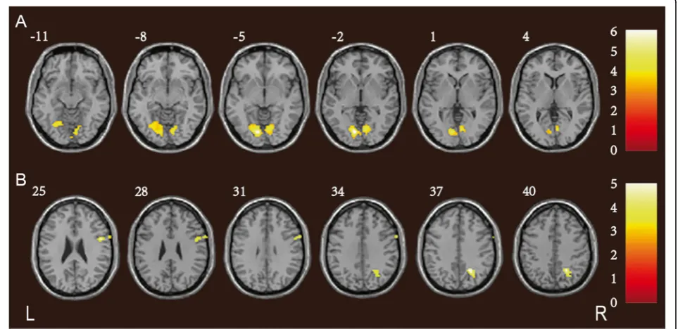
Compared to the controls, patients with CADASIL ex-hibited decreased intra-network connectivity in the bilat-eral lingual gyrus (LG) and the right cuneus (CU) of the visual network (VIN), and within the right precuneus (Pcu), inferior frontal gyrus (IFG), and precentral gyrus
of the frontal network (FRN) (Fig.1; Table4).
ReHos and seed-based FC
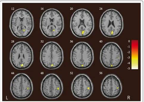
Compared to the controls, patients with CADASIL ex-hibited significantly lower ReHos in the right precuneus and cuneus (Pcu/CU), visual association cortex, calcarine
gyri, posterior cingulate and limbic lobe (Fig.2; Table5).
We compared the FC of the right Pcu/CU to those of other brain regions in the CADASIL and control groups. Compared to the controls, the former group exhibited weaker FC between the right Pcu/CU and the bilateral
parahippocampal gyrus (PHG), and between the right
Pcu/CU and the right postcentral gyrus (Fig.3; Table5).
Correlations with clinical scores
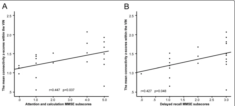
The mean connectivity z-scores of the bilateral LG and the right CU within the VIN were positively correlated with
both of the attention and calculation (r= 0.447,p= 0.037),
and delayed recall (r= 0.427,p= 0.048) scores, as revealed
by the cognitive domains explored by the MMSE (Fig.4).
There was no correlation between the mean ReHo z-scores of the right Pcu/CU and clinical scores, or between the mean connectivity z-scores within the functional networks and any of the scores on the neurological deficits, depres-sion, or anxiety scales.
Discussion
ICA revealed reduced intra-network connectivity in the VIN and FRN of CADASIL patients, marked decreases in the ReHos of the right Pcu/CU, visual association cor-tex, calcarine gyri, posterior cingulate, limbic lobe, and weaker FC between the right Pcu/CU and other brain areas. Furthermore, the mean connectivity z-scores of the dysfunctional areas within the VIN were positively associated with the several cognitive domains of MMSE.
Human executive functions are not mediated by a sin-gle brain region, but rather, reflect the dynamic interplay
of multiple networks [28–32]. A hallmark of executive
function is the ability of rapidly arbitrary links between visual inputs, on the one hand, and actions and goals, on the other, by the use of the learned informations (e.g., applying the brakes of a car when a person is seen
ahead) [29]. This ability is termed arbitrary visuomotor
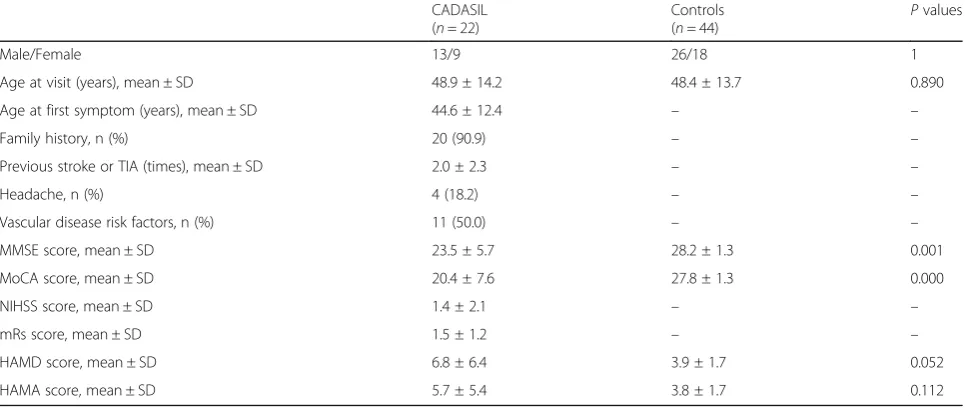
Table 1Demographic and clinical characteristics of CADASIL patients and controls
CADASIL (n= 22)
Controls (n= 44)
Pvalues
Male/Female 13/9 26/18 1
Age at visit (years), mean ± SD 48.9 ± 14.2 48.4 ± 13.7 0.890
Age at first symptom (years), mean ± SD 44.6 ± 12.4 – –
Family history, n (%) 20 (90.9) – –
Previous stroke or TIA (times), mean ± SD 2.0 ± 2.3 – –
Headache, n (%) 4 (18.2) – –
Vascular disease risk factors, n (%) 11 (50.0) – –
MMSE score, mean ± SD 23.5 ± 5.7 28.2 ± 1.3 0.001
MoCA score, mean ± SD 20.4 ± 7.6 27.8 ± 1.3 0.000
NIHSS score, mean ± SD 1.4 ± 2.1 – –
mRs score, mean ± SD 1.5 ± 1.2 – –
HAMD score, mean ± SD 6.8 ± 6.4 3.9 ± 1.7 0.052
HAMA score, mean ± SD 5.7 ± 5.4 3.8 ± 1.7 0.112
Values are means ± SDs or numbers with percentages
CADASILCerebral autosomal dominant arteriopathy with subcortical infarcts and leukoencephalopathy,SDStandard deviation,TIATransient ischemic attack,MMSE
mapping; large-scale brain networks engage in
reconfig-uration and dynamic integration and occipital–parietal–
frontal cortical and cortico-subcortical FC networks are involved. First, the visual and parietal regions coordinate with the sensorimotor and premotor areas. Second, the dorsal frontoparietal circuit links to the sensorimotor and frontostriatal networks. Finally, the cortico-cortical interhemisphere coordinates the bilateral sensorimotor
regions [20]. During this process, the posterior parietal
area, particularly the Pcu, plays a key role in arranging visuomotor planning and using of visual information in movement. The premotor areas serve as relays from the posterior parietal areas to the medial prefrontal cortices,
which play receptor roles [33]. The networks exhibit
temporal evolution, commencing with the processing of visual information, which is followed by emergence of a
visuomotor plan and then action [34]. We found that
the intra-network connectivity of the VIN and FRN were reduced in the patient group; the VIN and FRN lie in the occipital and frontoparietal areas, respectively, and include visual regions such as the LG and CU, motor areas such as the IFG and precentral gyrus, and the pos-terior parietal Pcu. We suggest that all visual input,
motor output, and visuomovement transformation may be interrupted in CADASIL patients, thereby affecting arbitrary visuomotor mapping (a form of acquired in-strumental behavior) in such patients.
Cortical network nodes are involved in arbitrary visuo-motor mapping. The Pcu and CU lie on both sides of the parieto-occipital fissure, in the posterior parietal and inferior occipital lobes, respectively. Resting-state fMRI has revealed three distinct FC patterns in the Pcu. The anterior Pcu is functionally connected with the postcen-tral and precenpostcen-tral gyri, the sensorimotor regions. The central Pcu connects with the dorsolateral and dorsome-dial prefrontal cortex (a cognitive/associative region) and the posterior Pcu with the adjacent, visual cortical
re-gions [35]. The FC data showed that the Pcu serves as a
bridge connecting visual perceptions with action during the visuomotor process, being involved in both visuo-motor planning and transmission of visual information
from the occipital lobe to sensorimotor regions [36–40].
Furthermore, the CU exhibited significant, contralateral visual selectivity when engaged in processing of visual
information [35]. Therefore, the Pcu/CU are considered
(respectively) the node and hub of the frontoparietal,
Table 2The different subscores of MoCA in CADASIL patients
Patients MoCA
Visuospatial/execution Naming Attention Language Abstraction Delayed recall Orientation
1 4 3 6 2 2 5 6
2 5 3 6 3 2 5 6
3 5 3 6 3 2 5 6
4 5 3 6 2 2 5 6
5 4 3 6 3 2 5 6
6 4 3 6 3 2 2 6
7 3 3 4 2 2 3 6
8 1 2 1 0 1 0 3
9 4 3 4 2 2 0 6
10 3 2 4 1 2 3 6
11 4 3 6 3 2 3 6
12 4 1 4 1 2 0 6
13 2 1 1 0 1 0 1
14 0 1 3 1 1 1 2
15 2 3 6 1 2 2 6
16 0 2 3 0 2 1 3
17 4 3 6 1 2 2 6
18 0 2 1 0 0 0 5
19 2 2 5 1 1 1 4
20 3 3 5 1 1 0 5
21 3 3 6 1 1 4 6
22 1 3 3 1 2 0 2
central executive network involved in arbitrary
visuo-motor mapping [41]. In the current study, we found that
the ReHos of the right Pcu/CU were decreased in CADASIL patients, suggesting that Pcu/CU-mediated executive cognition might be impaired. The functional changes in the Pcu/CU were similar to those of patients
with sporadic, subcortical vascular dementia (SVD) [41,
42], indicating that Pcu/CU dysfunction might explain
the executive impairments of both hereditary and spor-adic SVD.
We found that the intra-network connectivity of the right IFG (within the FRN) was also reduced in CADASIL patients. The IFG plays a vital role in implementing the
strategies of multi-component behaviors [43].
Magnetoen-cephalography revealed that the IFG connected directly with the visual and motor cortices when a visuomotor, precision, grip force task was underway. This network may effectively complement the traditional step-by-step processing of the occipital-parietal-premotor-motor
path-way during such tasks [44]. We found that two pathways
connecting the visual and motor cortices during visuo-motor behavior might be affected in CADASIL patients; the pathways involve the traditional frontoparietal nodes (the Pcu/CU) and the IFG.
We also found that patients exhibited weaker FC tween the right Pcu/CU and the bilateral PHG, and be-tween the right Pcu/CU and the right postcentral gyrus (the primary somatosensory area), than controls. Matyas et al. used the mouse whisker model to show that cor-tical motor control was driven by the primary somato-sensory cortex, which also delivered rapid negative
feedback during sensorimotor integration [45]. Similar
to the expansion of motor control into the sensory cor-tex, the PHG lies in the medial-temporal lobe and pro-cesses visual motion such as speed, acceleration, and the direction of hand movement, but only during a visuo-motor task. Thus, the PHG may engage in visuovisuo-motor Fig. 1Reduced intra-network connectivity in patients with CADASIL. The CADASIL group exhibited decreased activity of the bilateral LG and the
right CU within the VIN (a); and of the right Pcu, IFG, and precentral gyrus within the FRN (b), compared to the control group. Thet-values are
color-coded. CADASIL: cerebral autosomal dominant arteriopathy with subcortical infarcts and leukoencephalopathy; LG: lingual gyrus; CU: cuneus; VIN: visual network; Pcu: precuneus; IFG: inferior frontal gyrus; FRN: frontal network
Table 4Significant inter-group ICA differences between CADASIL patients and controls
Predominant cluster regions
Cluster size
Peak T value
MNI coordinates Cluster level P FWE-corr
x y z
CADASIL < controls in VIN
Bilateral LG 348 −6.19 −9 −78 −3 0.000
Right CU
CADASIL < controls in FRN
Right Pcu 59 −5.18 24 −57 36 0.002
Right IFG 67 −5.05 48 3 21 0.001
Right precentral gyrus
The surviving ICA clusters were assigned thresholds ofp< 0.001 and FWE-corrected top< 0.05 at the cluster level
Fig. 2Reductions in the ReHos in patients with CADASIL. The CADASIL group exhibited significantly lower ReHos in the right Pcu/CU, visual
association cortex, calcarine gyri, posterior cingulate and limbic lobe, compared to controls. Thet-values are color-coded. ReHo: regional
homogeneity; CADASIL: cerebral autosomal dominant arteriopathy with subcortical infarcts and leukoencephalopathy; Pcu/CU: precuneus and cuneus
Table 5Significant inter-group differences in ReHo and seed-based FC analysis between CADASIL patients and controls
Predominant cluster regions Cluster
size
Peak T value
MNI coordinates Cluster
level P FWE-corr
x y z
ReHo reduction in CADASIL patients
Right Pcu/CU 155 −5.33 6 −78 30 0.009
Right visual association cortex
Right calcarine gyri
Right posterior cingulate
Right limbic lobe
FC reductions in CADASIL patients (seed region: right Pcu/CU)
Right PHG 206 −5.62 24 −27 −3 0.005
Left PHG 171 −4.59 −18 −39 −12 0.011
Right postcentral gyrus 115 −4.14 33 −39 57 0.044
The ReHo clusters and seed-based FC that survived were assigned thresholds ofp< 0.001 and FWE-corrected top< 0.05 at the cluster level
integration [46,47]. In general, the poor FC between the Pcu/CU, on the one hand, and the postcentral gyrus and the PHG, on the other, suggest that both coordination and integration of arbitrary sensorimotor associations are dysfunctional in CADASIL patients.
We found positive associations between the mean con-nectivity z-scores of the LG and CU within the VIN, and the scores on different cognitive domains of the MMSE including those reflecting attention, calculation and de-layed recall. Patients with the most serious cognitive im-pairments may exhibit lower VIN connectivity. In a sense, such decreases render it easier to detect severe cognitive issues in patients with CADASIL.
Our work had certain limitations. First, we only stud-ied a small sample of CADASIL patients. Second, we only used the MMSE and MoCA to evaluate cognitive function. Future studies with larger sample sizes should focus on more detailed cognitive domains such as execu-tive function and processing speed.
ALFF and ReHos are two quantitative methods used to evaluate local spontaneous neuronal activity. ALFF re-flects the intensity of neuronal activity within a single voxel, and ReHos indicate the temporal similarity of neuronal activity between a single voxel and its
neigh-boring voxels [18, 21]. In our recent resting-state ALFF
analyses, we found that CADASIL exhibited lower ALFF Fig. 3Reductions in the FC between the right Pcu/CU and other brain areas in patients with CADASIL. The CADASIL group exhibited weaker FC
between the right Pcu/CU and both PHGs, and between the right Pcu/CU and right postcentral gyrus, compared to controls. Thet-values are
color-coded. FC: functional connectivity; Pcu/CU: precuneus and cuneus; CADASIL: cerebral autosomal dominant arteriopathy with subcortical infarcts and leukoencephalopathy; PHG: parahippocampal gyrus
Fig. 4Significant correlations between the mean connectivity z-scores of the LG and CU within the VIN and cognitive measures for surviving
clusters in the CADASIL group.aThe mean connectivity z-scores within the VIN and the attention and calculation MMSE subscores;r= 0.447,p=
0.037.bThe mean connectivity z-scores within the VIN and the delayed recall MMSE subscores;r= 0.427,p= 0.048. LG: lingual gyrus; CU: cuneus;
values in the right Pcu/CU [18]. In contrast, the present study revealed lower ReHos in the right Pcu/CU and other areas including visual association cortex, calcarine gyri, posterior cingulate, limbic lobe. More importantly, we also found that CADASIL exhibited decreased intra-network connectivity within the VIN and FRN. These results suggest that CADASIL may exhibit dysfunctional visuomotor behaviors, a hallmark of executive function.
Conclusions
In this study, we found aberrant resting-state brain func-tional network connectivity within the VIN and FRN, and lower ReHos, in patients with CADASIL. These may explain the arbitrary visuomotor behaviors of such pa-tients, reflecting (downward) transitions in visual stimuli, movement location, and execution response mapping. The work advances our understanding of the executive cognition architectures underlying CADASIL, and will aid in the development of therapeutic strategies.
Abbreviations
CADASIL:Cerebral autosomal dominant arteriopathy with subcortical infarcts and leukoencephalopathy; FC: Functional connectivity; fMRI: Functional magnetic resonance imaging; FRN: Frontal network; MMSE: Mini-Mental State Examination; Pcu/CU: Precuneus and cuneus; ReHo: Regional homogeneity; VIN: Visual network
Acknowledgements
We acknowledged the specialist editors with suitable professional knowledge who provided professional services for reviewing and correcting the English.
Authors’contributions
Study conception and design: JL and XD; acquisition of data: JS, FH, LW, XC, YT, HZ, YZ; analysis and interpretation of data: SB and MW; drafting the article: all authors; final approval of the version to be published: all authors.
Funding
This work was supported by the National Natural Science Foundation of China [No. 81271302 to J.R. Liu, No. 81571658 and 81201082 to X.X. Du], the Biomedicine Key Programme of the Shanghai Municipal Science and Technology Commission [No. 16411953100 to J.R. Liu], a science popularization project and research innovation project of the Shanghai Municipal Science and Technology Commission [No. 18dz2313603 and No.
14JC1404300 to J.R. Liu], the“Prevention and Control of Chronic Diseases
Project”of the Shanghai Hospital Development Center [No. SHDC12015310
to J.R. Liu], a project of Shanghai Municipal Education Commission-Gaofeng Clinical Medicine Grant Support [No. 20161422 to J.R. Liu], and the Clinical Research Project of Shanghai Jiao Tong University School of Medicine [No. DLY201614 to J.R. Liu].
The roles of the funding body in the design of the study: the National Natural Science Foundation of China; data collection: the Biomedicine Key Programme of the Shanghai Municipal Science and Technology Commission; data analysis: a science popularization project and research innovation project of the Shanghai Municipal Science and Technology Commission;
interpretation of data: the“Prevention and Control of Chronic Diseases
Project”of the Shanghai Hospital Development Center and a project of
Shanghai Municipal Education Commission-Gaofeng Clinical Medicine Grant Support; writing the manuscript: the Clinical Research Project of Shanghai Jiao Tong University School of Medicine.
Availability of data and materials
The datasets used and/or analysed during the current study are available from the corresponding author on reasonable request.
Ethics approval and consent to participate
The study was approved by the Independent Ethics Committee of the East
China Normal University on Human Research (Project No. HR 062–2018) and
Shanghai Ninth People’s Hospital (Project No. 2016–44-T1). All participants
provided written informed consent.
Consent for publication
Not applicable.
Competing interests
The authors declare that they have no competing interests.
Author details
1
Department of Neurology, Shanghai Ninth People’s Hospital, Shanghai Jiao Tong University School of Medicine, 639 Zhizaoju Road, Shanghai 200011, People’s Republic of China.2Shanghai Key Laboratory of Magnetic
Resonance and Department of Physics, School of Physics and Materials Science, East China Normal University, 3663 North Zhongshan Road, Shanghai 200062, People’s Republic of China.3College of Medical Imaging,
Shanghai University of Medicine & Health Sciences, 279 Zhouzhu Highway, Shanghai 201318, People’s Republic of China.4PET Center, Huashan Hospital,
Fudan University, 518 East Wuzhong Road, Shanghai 200235, People’s Republic of China.5Department of Neurology, Huashan Hospital, Fudan
University, 12 Middle Wulumuqi Road, Shanghai 200040, People’s Republic of China.6Department of Geriatrics Neurology, Huashan Hospital, Fudan
University, 12 Middle Wulumuqi Road, Shanghai 200040, People’s Republic of China.
Received: 1 August 2019 Accepted: 10 October 2019
References
1. Chabriat H, Joutel A, Dichgans M et al (2009) Cadasil. Lancet Neurol 8:643–653.
https://doi.org/10.1016/S1474-4422(09)70127-9
2. Buffon F, Porcher R, Hernandez K et al (2006) Cognitive profile in CADASIL. J
Neurol Neurosurg Psychiatry 77:175–180.https://doi.org/10.1136/jnnp.2005.068726
3. Chabriat H, Vahedi K, Iba-Zizen MT et al (1995) Clinical spectrum of
CADASIL: a study of 7 families. Cerebral autosomal dominant arteriopathy
with subcortical infarcts and leukoencephalopathy. Lancet 346:934–939.
https://doi.org/10.1016/s0140-6736(95) 91557-5
4. Jouvent E, Duchesnay E, Hadj-Selem F et al (2016) Prediction of 3-year
clinical course in CADASIL. Neurology 87:1787–1795.https://doi.org/10.
1212/WNL.0000000000003 252
5. Gunda B, Hervé D, Godin O et al (2012) Effects of gender on the phenotype of
CADASIL. Stroke 43:137–141.https://doi.org/10.1161/STROKEAHA.111.631028
6. Chabriat H, Hervé D, Duering M et al (2016) Predictors of clinical worsening
in cerebral autosomal dominant arteriopathy with subcortical infarcts and
leukoencephalopathy: prospective cohort study. Stroke 47:4–11.https://doi.
org/10.1161/STROKEAHA.115.010696
7. Ling Y, De Guio F, Duering M et al (2017) Predictors and clinical impact of
incident lacunes in cerebral autosomal dominant arteriopathy with
subcortical infarcts and leukoencephalopathy. Stroke 48:283–289.https://doi.
org/10.1161/STROKEAHA.116.015 750
8. Liem MK, van der Grond J, Haan J et al (2007) Lacunar infarcts are the main
correlate with cognitive dysfunction in CADASIL. Stroke 38:923–928.https://
doi.org/10.1161/01.STR.0000257968.24015.bf
9. Viswanathan A, Gschwendtner A, Guichard JP et al (2007) Lacunar lesions are
independently associated with disability and cognitive impairment in CADASIL.
Neurology 69:172–179.https://doi.org/10.1212/01.wnl.0000265221.05610.70
10. Peters N, Holtmannspötter M, Opherk C et al (2006) Brain volume changes in
CADASIL: a serial MRI study in pure subcortical ischemic vascular disease.
Neurology 66:1517–1522.https://doi.org/10.1212/01.wnl.0000216271.96364.50
11. Shi Y, Li S, Li W et al (2018) MRI lesion load of cerebral small vessel disease
and cognitive impairment in patients with CADASIL. Front Neurol 9:862. https://doi.org/10.3389/fneur.2018.00862
12. Jouvent E, Reyes S, De Guio F et al (2015) Reaction time is a marker of early
cognitive and behavioral alterations in pure cerebral small vessel disease. J
Alzheimers Dis 47:413–419.https://doi.org/10.3233/JAD-150083
13. Delorme S, De Guio F, Reyes S et al (2017) Reaction time is negatively
associated with Corpus callosum area in the early stages of CADASIL. AJNR
14. O'Sullivan M, Barrick TR, Morris RG et al (2005) Damage within a network of white matter regions underlies executive dysfunction in CADASIL. Neurology
65:1584–1590.https://doi.org/10.1212/01.wnl.0000184480.07394.fb
15. O'Sullivan M, Singhal S, Charlton R et al (2004) Diffusion tensor imaging of
thalamus correlates with cognition in CADASIL without dementia.
Neurology 62:702–707.https://doi.org/10.1212/01.wnl.0000113760.72706.d2
16. Craggs LJ, Yamamoto Y, Ihara M et al (2014) White matter pathology and
disconnection in the frontal lobe in cerebral autosomal dominant arteriopathy with subcortical infarcts and leukoencephalopathy (CADASIL).
Neuropathol Appl Neurobiol 40:591–602.https://doi.org/10.1111/nan.12073
17. Cullen B, Moreton FC, Stringer MS et al (2016) Resting state connectivity
and cognitive performance in adults with cerebral autosomal-dominant arteriopathy with subcortical infarcts and leukoencephalopathy. J Cereb
Blood Flow Metab 36:981–991.https://doi.org/10.1177/0271678X16636395
18. Su J, Wang M, Ban S et al (2019) Relationship between changes in
resting-state spontaneous brain activity and cognitive impairment in patients with
CADASIL. J Headache Pain 20:36.https://doi.org/10.1186/s10194-019-0982-3
19. Gavazzi G, Orsolini S, Salvadori E et al (2018) Functional magnetic resonance
imaging of inhibitory control reveals decreased blood oxygen level dependent effect in cerebral autosomal dominant arteriopathy with
subcortical infarcts and leukoencephalopathy. Stroke.https://doi.org/10.
1161/STROKEAHA.118.022923[Epub ahead of print]
20. Brovelli A, Badier JM, Bonini F et al (2017) Dynamic reconfiguration of
visuomotor-related functional connectivity networks. J Neurosci 37:839–853.
https://doi.org/10.1523/JNEUROSCI.1672-16.2016
21. Zang Y, Jiang T, Lu Y et al (2004) Regional homogeneity approach to fMRI
data analysis. Neuroimage 22:394–400.https://doi.org/10.1016/j.neuroimage.
2003.12.030
22. Bramell-Risberg E, Jarnlo GB, Elmståhl S (2012) Separate physical tests of lower
extremities and postural control are associated with cognitive impairment. Results from the general population study good aging in Skåne (GÅS-SNAC).
Clin Interv Aging 7:195–205.https://doi.org/10.2147/CIA.S31777
23. Marengoni A, Bandinelli S, Maietti E et al (2017) Combining gait speed and
recall memory to predict survival in late life: population-based study. J Am
Geriatr Soc 65:614–618.https://doi.org/10.1111/jgs.14705
24. Duering M, Righart R, Csanadi E et al (2012) Incident subcortical infarcts
induce focal thinning in connected cortical regions. Neurology 79:2025–
2028.https://doi.org/10.1212/WNL.0b013e3182749f39
25. Fazekas F, Chawluk JB, Alavi A et al (1987) MR signal abnormalities at 1.5 T in
Alzheimer’s dementia and normal aging. AJR Am J Roentgenol 149:351–356.
https://doi.org/10.2214/ajr.149.2.351
26. Yan CG, Wang XD, Zuo XN et al (2016) DPABI: data processing & analysis for
(resting-state) brain imaging. Neuroinformatics 14:339–351.https://doi.org/
10.1007/s12021-016-9299-4
27. Allen EA, Erhardt EB, Damaraju E et al (2011) A baseline for the multivariate
comparison of resting-state networks. Front Syst Neurosci 5:2.https://doi.
org/10.3389/fnsys.2011.00002
28. Braun U, Schäfer A, Walter H et al (2015) Dynamic reconfiguration of frontal
brain networks during executive cognition in humans. Proc Natl Acad Sci U
S A 112:11678–11683.https://doi.org/10.1073/pnas.1422487112
29. Eliassen JC, Souza T, Sanes JN (2003) Experience-dependent activation patterns in
human brain during visual-motor associative learning. J Neurosci 23:10540–10547
30. Chouinard PA, Goodale MA (2009) FMRI adaptation during performance of
learned arbitrary visuomotor conditional associations. Neuroimage 48:96–706.
https://doi.org/10.1016/j.neuroimage.2009.07.020
31. Madhavan R, Bansal AK, Madsen JR et al (2018) Neural interactions
underlying Visuomotor associations in the human brain. Cereb Cortex. https://doi.org/10.1093/cercor/bhy333[Epub ahead of print]
32. Ester EF, Sprague TC, Serences JT (2015) Parietal and frontal cortex encode
stimulus-specific mnemonic representations during visual working memory.
Neuron 87:893–905.https://doi.org/10.1016/j.neuron.2015.07.013
33. Brovelli A, Chicharro D, Badier JM et al (2015) Characterization of cortical networks
and corticocortical functional connectivity mediating arbitrary Visuomotor mapping.
J Neurosci 35:12643–12658.https://doi.org/10.1523/JNEUROSCI.4892-14.2015
34. Cappadocia DC, Monaco S, Chen Y et al (2017) Temporal evolution of target
representation, movement direction planning, and reach execution in
occipital-parietal-frontal cortex: an fMRI study. Cereb Cortex 27:5242–5260.
https://doi.org/10.1093/cercor/bhw304
35. Margulies DS, Vincent JL, Kelly C et al (2009) Precuneus shares intrinsic
functional architecture in humans and monkeys. Proc Natl Acad Sci U S A
106:20069–20074.https://doi.org/10.1073/pnas.0905314106
36. Grol MJ, de Lange FP, Verstraten FA et al (2006) Cerebral changes during
performance of overlearned arbitrary visuomotor associations. J Neurosci 26: 117–125.https://doi.org/10.1523/JNEUROSCI.2786-05.2006
37. Fernandez-Ruiz J, Goltz HC, DeSouza JF et al (2007) Human parietal“reach
region”primarily encodes intrinsic visual direction, not extrinsic movement
direction, in a visual motor dissociation task. Cereb Cortex 17:2283–2292.
https://doi.org/10.1093/cercor/bhl137
38. Culham JC, Valyear KF (2006) Human parietal cortex in action. Curr Opin
Neurobiol 16:205–212.https://doi.org/10.1016/j.conb.2006.03.005
39. Grol MJ, Toni I, Lock M et al (2009) Spatial representation of overlearned
arbitrary visuomotor associations. Exp Brain Res 192:751–759.https://doi.
org/10.1007/s00221-008-1653-9
40. Gertz H, Fiehler K (2015) Human posterior parietal cortex encodes the
movement goal in a pro−/anti-reach task. J Neurophysiol 114:170–183.
https://doi.org/10.1152/jn.01039.2014
41. Menon V (2011) Large-scale brain networks and psychopathology: a
unifying triple network model. Trends Cogn Sci 15:483–506.https://doi.org/
10.1016/j.tics.2011.08.003
42. Li C, Liu C, Yin X et al (2014) Frequency-dependent changes in the
amplitude of low-frequency fluctuations in subcortical ischemic vascular
disease (SIVD): a resting-state fMRI study. Behav Brain Res 274:205–210.
https://doi.org/10.1016/j.bbr.2014.08.019
43. Dippel G, Beste C (2015) A causal role of the right inferior frontal cortex in
implementing strategies for multi-component behaviour. Nat Commun 6:
6587.https://doi.org/10.1038/ncomms7587
44. Papadelis C, Arfeller C, Erla S et al (2016) Inferior frontal gyrus links visual
and motor cortices during a visuomotor precision grip force task. Brain Res
1650:252–266.https://doi.org/10.1016/j.brainres.2016.09.011
45. Matyas F, Sreenivasan V, Marbach F et al (2010) Motor control by sensory
cortex. Science 330:1240–1243.https://doi.org/10.1126/science.1195797
46. Tankus A, Fried I (2012) Visuomotor coordination and motor representation
by human temporal lobe neurons. J Cogn Neurosci 24:600–610.https://doi.
org/10.1162/jocn_a_00160
47. Sato N, Nakamura K (2003) Visual response properties of neurons in the
parahippocampal cortex of monkeys. J Neurophysiol 90:876–886.https://doi.
org/10.1152/jn.01089.2002
Publisher’s Note